65 years old female patient with diagnosis of supratentorial mass was operated. F-18 FDG PET/CT was performed for determination of primary tumor of the lesion which was assumed to be metastatic. However metabolic imaging revealed multiple lesions all over the body additionally the pathology revealed this exceptional case with synovial sarcoma as the primary diagnosis.
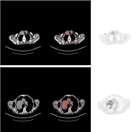
Figure 1. A 65 year old woman with the diagnosis of brain mass lesion was referred for FDG PET/CT imaging. The patient fasted for 10 hours before the imaging study and 7 mCi F-18 FDG was administered via venous line and waited for one hour. PET/CT imaging was performed in craniocaudal direction from head to feet with low dose noncontrast enhanced CT. The images showed postoperative changes in the brain region as well as mediastinal 100 mm (SUVmax=33.4) and left adrenal 36 mm (SUVmax=12.3) hypermetabolic mass lesions and bilateral thyroid nodules accumulating FDG (SUVmax=8.2)
sarcoma, FDG, PET/CT, brain
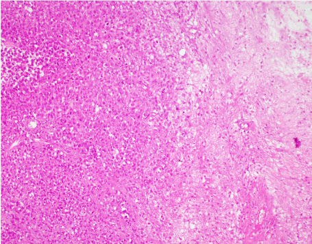
Figure 2. Biopsy results revealed snovial sarcoma obtained from the brain. Synovial sarcoma is a mesenchymal tumor however sarcoma arising from brain region is extremely rare and disseminated sarcoma in multiple regions has not been reported previously as far as we know. Previous case reports presented epithelioid sarcoma of the temporal space [1] which was also rare in the head and neck region. F-18 FDG PET/CT findings of an spinal cord mesenchymal chondrosarcoma with significant FDG uptake was also reported in a previous case report [2]. Another case report with brain and lung histiocytic sarcoma with F-18 FDG PET/CT findings showed hypermetabolism in the determined lesions of the patient [3]. The current case presented as a brain mass at first presentation and F-18 FDG PET/CT demonstrated additional multiple hypermetabolic mass lesions all over the body. Unfortunately, the brain lesion was operated at the time of imaging thus could not be characterized. This case report determines the first case of disseminated synovial sarcoma as shown by FDG PET/CT
- Kim SY, Kwak HS, Chung GH, Kim YN, Hwang S (2018) Epitheloid sarcoma arising from the temporal space. Medicine 97: 38(e12529).
- Lee ES, Lee HY, Choe G, Kim KJ, Lee WW, et al. (2014) Extraskeletal intraspinal mesencymal chondrosarcoma; F-18 FDG PET/CT finding. Clin Nucl Med 39: 64-66.
- Pan Y, Zhang Y (2018) Simultaneous brain and lung histiocytic sarcoma revealed on F-18 FDG PET/CT. Clin Nucl Med 43: 65-67.
Editorial Information
Editor-in-Chief
Article Type
Image Article
Publication history
Received date: December 12, 2019
Accepted date: January 07, 2020
Published date: January 09, 2020
Copyright
©2020 Koç ZP. This is an open-access article distributed under the terms of the Creative Commons Attribution License, which permits unrestricted use, distribution, and reproduction in any medium, provided the original author and source are credited.
Citation
Koç ZP, Özcan PP, Hamzaoğlu V, Avcı E, Yaldız M (2020) Metabolic imaging characteristics of a rare disseminated synovial sarcoma diagnosed by brain biopsy. Glob Imaging Insights 5: DOI: 10.15761/GII.1000196
Corresponding author
Zehra Pınar Koç
Department of Nuclear Medicine, Mersin University, Turkey
Figure 1. A 65 year old woman with the diagnosis of brain mass lesion was referred for FDG PET/CT imaging. The patient fasted for 10 hours before the imaging study and 7 mCi F-18 FDG was administered via venous line and waited for one hour. PET/CT imaging was performed in craniocaudal direction from head to feet with low dose noncontrast enhanced CT. The images showed postoperative changes in the brain region as well as mediastinal 100 mm (SUVmax=33.4) and left adrenal 36 mm (SUVmax=12.3) hypermetabolic mass lesions and bilateral thyroid nodules accumulating FDG (SUVmax=8.2)
Figure 2. Biopsy results revealed snovial sarcoma obtained from the brain. Synovial sarcoma is a mesenchymal tumor however sarcoma arising from brain region is extremely rare and disseminated sarcoma in multiple regions has not been reported previously as far as we know. Previous case reports presented epithelioid sarcoma of the temporal space [1] which was also rare in the head and neck region. F-18 FDG PET/CT findings of an spinal cord mesenchymal chondrosarcoma with significant FDG uptake was also reported in a previous case report [2]. Another case report with brain and lung histiocytic sarcoma with F-18 FDG PET/CT findings showed hypermetabolism in the determined lesions of the patient [3]. The current case presented as a brain mass at first presentation and F-18 FDG PET/CT demonstrated additional multiple hypermetabolic mass lesions all over the body. Unfortunately, the brain lesion was operated at the time of imaging thus could not be characterized. This case report determines the first case of disseminated synovial sarcoma as shown by FDG PET/CT