Megalencephalic leukoencephalopathy with subcortical cysts (MLC) or Van der Knaap disease is a rare autosomal recessive disorder characterized by macrocephaly, early motor development delay and cerebral leukoencephalopathy. It occurs more commonly in some ethnicities where consanguinity is common. Here, we report three cases presented in our department, GMC Srinagar with typical clinical features and history of consanguinity in two of three cases. MRI brain shows typical findings of leukodystrophy and subcortical cysts. MLC has remarkably slow course of deterioration in neurologic function. MLC must be included in differential diagnosis of macrocephaly with early onset leukoencephalopathy. A precise diagnosis helps for better management and to prognosticate its benign course.
megalencephalic leukoencephalopathy with subcortical cysts (MLC)
Megalencephalic leukoencephalopathy with subcortical cysts (MLC) is a rare disease first described by van der Knaap et al. in 1995 [1]. MLC is a rare autosomal-recessive disorder with variable but mild clinical course [2]. MLC is a genetically heterogenous disorder. Most of cases (aproximately 75%) are caused by mutation in the MLC1 gene located on Chr.22q(tel) [3]. A newly described mutation in the HEPACAM gene leads to spectrum of abnormalities ranging from benign familial macrocephaly to MLC that is indistinguishable from that caused by MLC1 mutation.
MLC is clinically characterised by Infantile- onset macrocephaly and slow course of neurologic deterioration [1]. Median age of onset of clinical symptoms is 6 months (range birth to 25 years). Early motor development delay, gait ataxia, and hypotonia is observed which eventually progress to spastic tetraparesis. Seizures are variable and slow cognitive decline is observed as the disease progresses. Geographical distribution of MLC is global, however it occurs more commonly in some ethnicities where consanguinity is common, such as Libyan, Jewish, Turkish, and Agarwal Indian communities [4,5]. MRI findings including leukodystrophy and subcortical cysts are hallmarks of the disease and yield the diagnostic clue in most cases. Macrocephaly with diffuse confluent WM T2/ FLAIR hyperintensity in the subcortical white matter is typical. The optic radiations, occipital subcortical white matter and basal ganglia are typically spared.
MR findings are often diagnostic of MLC and must be differentiated from other leukoencephalopathies. Because of rarity of this condition, the literature has been limited to case reports. We herein present MRI brain findings in three patients diagnosed as case of MLC.
A 17 years old female born from consanguineous marriage in tribal community by normal vaginal delivery presented in our department with history of delayed motor and social milestones with intellectual disability. History of progressive difficulty in maintaining balance while walking leading to recurrent falls and seizures. She has never been to school. On examination head circumference was 60 cm (>95th percentile). Dysarthria, spastic paraperesis, brisk deep tendon reflexes seen with the presence of cerebellar signs and ataxia. Rest of systemic and physical examination was unremarkable. Routine investigations were normal, birth history was uneventful. MRI brain done showed diffuse white matter hyperintensities and the presence of subcortical cysts in the bilateral anterior temporal region, features consistent with MLC. Genetic testing was not done (Figures 1 and 2).

Figure 1. T2 weighted brain MRI; extensive diffuse homogenous dysmyelination involving the subcortical and deep white matter of the cerebral hemispheres along with presence of cysts (arrows) bilaterally in the temporal lobe
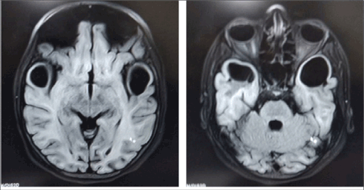
Figure 2. T1 weighted MRI brain; well defined symmetrical subcortical cysts in anterior temporal lobe which are hypointense on T1WI
A 18 month old boy born from consanguineous marriage in a muslim family, presented to our department for MRI brain with history of delayed development milestones, especially motor and social. He was born out of normal delivery with uneventful birth history. No history of abnormal body movements and prolonged fever. Abnormally large head is noticed by parents 5 months after birth, which was normal at birth as per parents. On examination, the child has macrocephaly with a HC of 54 cm, (>95th percentile). Rest of physical and systemic examination was normal. MRI brain revealed extensive diffuse homogenous dysmyelination involving the subcortical and deep white matter of the cerebral hemispheres with sparing of basal ganglia and frontal white matter region, consistent with MLC (Figures 3 and 4).
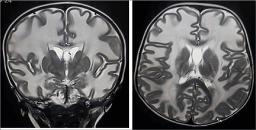
Figure 3. T2 weighted coronal and axial MR shows bilateral symmetrical white matter hyperintensity suggestive of dysmyelination
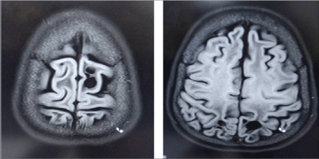
Figure 4. T2 FLAIR axial MR shows subcortical cysts involving high left frontal and parietal regions
A 12 months old boy, born of non- consanguineous marriage in a Muslim community from a Kashmir, presented with delayed development of milestones with one episode of abnormal body movements. He was not able to sit or stand himself. Birth history was uneventful. There is progressive increase in head circumference after birth and measures 50 cm (>97th percentile) at presentation. Sensory system was normal, and no cerebellar signs are seen. MRI brain revealed extensive bilaterally symmetrical white matter changes, which are hypointense on T1 weighted images and hyperintense on T2/FLAIR images suggestive of extensive demyelination. Also, well defined symmetrical subcortical cysts are noted in anterior temporal lobe, consistent with diagnosis of MLC (Figures 5 and 6).
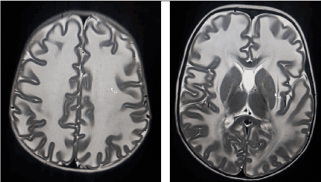
Figure 5. T2 WI showing extensive dysmyelination of bilateral subcortical white matter with sparing of basal ganglia and deep white matter
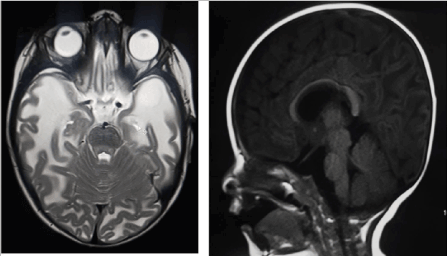
Figure 6. T2 WI MRI brain shows extensive dysmyelination with subcortical cyst formation in left anterior temporal lobe. Saggital FLAIR image shows enlarged, macrocephalic head
MLC is an uncommon disorder and has typical clinical presentation. Infantile onset macrocephaly is characteristic, but neurologic deterioration is often delayed. Macrocephaly was present in all the three cases and often observed/ developed during first year of their life. Neurological deteriorations manifest in form of delayed motor development which later on progress to involve pyramidal and cerebellar regions and leads to gait ataxia, hypotonia, and frequent fall while walking. With further progression of disease leads to spastic tetra paresis and cognitive decline. Developmental delay seen in two of three early presenting cases and cognitive decline with progressed motor symptoms seen in 17-year-old case diagnosed as case of MLC.
The diagnosis of MLC can be made with confidence in patients with typical clinical findings and characteristic abnormalities on cranial MR. Slow deterioration of motor functions with cerebellar ataxia and mild spasticity usually starts in early childhood.
Some patients have extrapyramidal movement abnormalities with the dystonia and athetosis, usually as a late finding. Mental decline occurs later and is much milder than motor decline. Most patients have epileptic seizures. In typical cases, the MR findings are often diagnostic of MLC. MR shows 'swollen white matter' and diffuse supratentorial symmetrical white matter changes in cerebral hemispheres with relative sparing of central white matter structures like the corpus callosum, internal capsule, and brain stem. Subcortical cysts are almost always present in the anterior temporal region and are also frequently noted in frontoparietal region. Grey matter is usually spared. Gradually the white matter swelling decreases and cerebral atrophy may ensue. The subcortical cysts may increase in size and number. In India, majority of the patients belong to the Agarwal community [4]. Our patient did not belong to this community. Indian patients with megalencephaly and MR changes that show extensive white matter changes with temporal cysts should raise suspicion for MLC [4]. This disease has been assigned to the gene, MLC1, and is localized on chromosome 22qtel [3]. Moderate decrease in NAA/ Cr and Choline/Cr ratios have been reported in patients with MLC on MR spectroscopy [6].
The differential diagnosis of MLC includes Canavan's disease, Alexander disease, infantile-onsetGM2 and GM1 gangliosidosis. These conditions have relentlessly progressive infantile onset leukoencephalopathy that is frequently fatal within first decade of life, however MLC has remarkably slow course of deterioration in neurologic function. MLC must also be included in differential diagnosis of macrocephaly with early onset leukoencephalopathy [7].
- Van Der Knaap MS, Barth PG, Stroink H, van Nieuwenhuizen O, Arts WF, et al. (1995) Leukoencephalopathy with swelling and a discrepantly mild clinical course in eight children. Ann Neurol 37: 324-334. [Crossref]
- Koussa S, Roukoz H, Rizk T, Megarbane A (2005) Megalencephalic leucoencephalopathy with subcortical cysts: a study of a Lebanese family and a review of the literature. Rev Neurol (Paris) 161: 183-191. [Crossref]
- Leegwater PA, Boor PK, Yuan BQ, van der Steen J, Visser A, et al. (2002) Identification of novel mutations in MLC1 responsible for megalencephalic leukoencephalopathy with subcortical cysts. Hum Genet 110: 279-283. [Crossref]
- Singhal BS, Gursahani RD, Udani VP, Biniwale AA (1996) Megalencephalic leukodystrophy in an Asian Indian ethnic group. Pediatr Neurol 14: 291-296. [Crossref]
- Ben-Zeev B, Gross V, Kushnir T, Shalev R, Hoffman C, et al. (2001) Vacuolating megalencephalic leukoencephalopathy in 12 Israeli patients. J Child Neurol 16: 93-99. [Crossref]
- Brockmann K, Finsterbusch J, Terwey B, Frahm J, Hanefeld F (2003) Megalencephalic leucoencephalopathy with subcortical cysts in an adult: quantitative proton MR spectroscopy and diffusion tensor MRI. Neuroradiology 45: 137-142. [Crossref]
- Krishnan KH, Pauline CL, Kumaresan G, Mallika TK (2005) Megalencephalic leukoencephalopathy with subcortical cysts. Indian Pediatr 42: 60-63.