A 76-year-old man with end-stage renal disease on intermittent hemodialysis was brought to the emergency department with a one-week history of fatigue and exertional dyspnea. On initial assessment, his blood pressure was 134/90 mmHg, heart rate was 76 bpm and oxygen saturation was 78% on ambient air. Other findings included an elevated jugular venous pressure, distant heart sounds and absent air entry throughout the left lung field. Chest X-ray (Figure 1A) revealed nearly complete opacification of the left hemithorax with contralateral tracheal deviation. Shortly thereafter, he became hemodynamically unstable. Immediate bedside ultrasound demonstrated a large pericardial effusion with evidence of tamponade, including diastolic collapse of the right atrium and right ventricle. Urgent pericardiocentesis was successfully performed to stabilize the patient. A subsequent CT chest (Figure 1B) demonstrated a massive pericardial effusion causing complete collapse of the left lung. In total, 4.3L of fluid were drained, resulting in re-expansion of the left lung and hemodynamic stabilization. Twenty-four hours later, a transthoracic echocardiogram demonstrated a residual posterior effusion with no evidence of cardiac tamponade. Repeat chest X-ray (Figure 2) demonstrated improved aeration of both lungs with residual pericardial effusion and atelectasis. Tests for malignancy, infection and tuberculosis were negative. Despite regular hemodialysis, the effusion reaccumulated one month later, requiring a repeat pericardiocentesis.
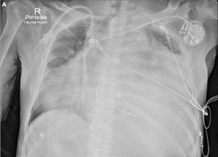
Figure 1A. Portable chest X-ray demonstrates nearly complete left hemithorax opacification with opacity extending into the right hemithorax. A haemodialysis line and dual-lead pacemaker are also present
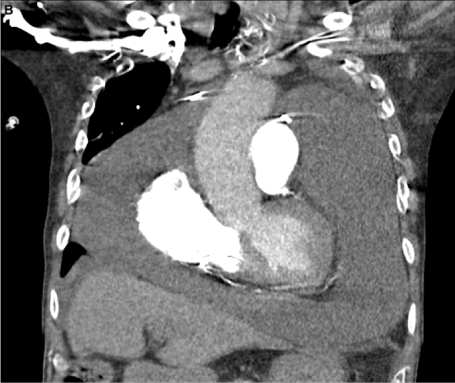
Figure 1B. Computed tomography of the chest demonstrates a massive pericardial effusion causing complete left lung collapse
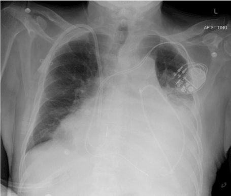
Figure 2. Chest X-ray performed after pericardial drain placement demonstrates improved aeration of the lungs bilaterally with residual pericardial effusion and atelectasis
Hemithorax opacification, often referred to as “hemithorax whiteout”, has a limited differential diagnosis. When the tracheal deviation is contralateral to the “whiteout”, the differential diagnosis includes pleural effusion, pulmonary mass, diaphragmatic hernia, and, as demonstrated in this case, massive pericardial effusion. As the parietal pericardium is relatively inelastic, tamponade can occur with as little as 100-200mL of fluid; however, this case demonstrates that when an effusion accumulates chronically, the pericardial space can accommodate several litres of fluid, which can lead to compression of not only the cardiac chambers, but also other adjacent structures.
Common causes of chronic pericardial effusions include pericarditis, tuberculous and viral infections, malignancy, connective tissue disease and uremia, but up to 50% are idiopathic.1 While the clinical presentation is often non-specific, up to one third of patients with chronic pericardial effusions develop cardiac tamponade [1,2]. Treatment should target the underlying etiology, but pericardiocentesis is indicated for cardiac tamponade, moderate-to-large symptomatic effusions and when suspicion for a neoplastic, purulent or tuberculous effusion exists [1]. If the effusion recurs, repeat pericardiocentesis or definitive surgical intervention with a pericardial window or pericardiectomy can be performed [3].
The authors have received no financial support for the research, authorship or publication of this article.
All authors report no conflict of interest
- Adler Y, Charron P, Imazio M, Badano L, Barón-Esquivias G, et al. (2015) ESC Guidelines for the diagnosis and management of pericardial diseases: The Task Force for the Diagnosis and Management of Pericardial Diseases of the European Society of Cardiology (ESC) Endorsed by: The European Association for Cardio-Thoracic Surgery (EACTS). Eur Heart J 36: 2921-2964. [Crossref]
- Sagristà-Sauleda J, Angel J, Permanyer-Miralda G, Soler-Soler J (1999) Long-Term Follow-up of Idiopathic Chronic Pericardial Effusion. N Engl J Med 341: 2054-2059. [Crossref]
- Imazio M, Adler Y (2013) Management of pericardial effusion. Eur Heart J 34: 1186-1197. [Crossref]