Madelung’s disease, also known as benign symmetric lipomatosis or Launois-Bensaude syndrome was first described by Sir Benjamin Brodie in 1846 from a series of patients he had encountered characterizing adipose tumors deposited preferentially on the neck [1]. It is a disorder of fat metabolism with deposition of multiple, symmetrical and painless non-encapsulated lipomas. Depending on the type of Madelung’s Disease, the adipose tissue deposition can be seen in the neck, upper extremities, trunk and hip area. About 90% of the cases described in the literature have been associated with chronic alcoholism and tobacco use and in a population of European/Mediterranean descent, reporting up to 1 in 25,000 cases in Italy to have the condition [2,3]. Rare cases have also been reported in Indians and one case in an African American male [4,5]. We present a case of a 48-year-old Hispanic male of Mexican ancestry with no significant history of alcoholism who presented to our emergency department with chief complaint of progressive proximal muscle weakness in upper and lower extremities resulting in an inability to walk or care for daily activities.
Madelung’s disease was first described in 1846 by Sir Benjamin Brodie as adipose tumors, in his book “Lectures illustrative of various subjects in pathology and surgery” [1]. Madelung later reported 33 cases in 1888 and Launois and Bensaude published 65 cases in 1898 [6,7]. It is a disorder of fat metabolism with abnormal deposition of multiple, non-encapsulated, painless lipomas which can be seen symmetrically in neck, (buffalo neck), upper extremities, back and chest (pseudo athletic appearance), and hip area. The location of the lipomas is what classifies the condition into 3 types. The exact pathophysiology for this condition is still considered unknown, but articles have suggested a defect in adrenergic-stimulated lipid mobilization and an increased LPL-dependent FFA incorporation to be responsible the abnormal accumulation of fat [8]. This abnormal growth of lipomas leads to anatomical disfigurement and can compromise adjacent structures such as trachea, lymphatics and vasculature, causing further complications which require surgical removal [9,10]. In addition, this condition has been seen associated with obstructive sleep apnea (OSA), gynecomastia as well as reports that have associated this disease with myopathy and lower extremity muscle weakness [4]. It is important to differentiate patients suffering from this condition from obesity due to the potential differences in treatment regimens available [9]. In regards to a genetic component, Madelung’s Disease has been reported to show some mitochondrial inheritance pattern although case reports in non-alcoholics have also suggested an autosomal recessive pattern of inheritance [11,12]. The Long-term outcome is not favorable for patients suffering from this condition, with many patients becoming bedbound as debility progresses [4]. The vast majority of cases in the literature have associated Madelung’s disease in patients of European descent and with a history of chronic alcoholism [13]. To our knowledge, up to this day there have been no reports on patients of Hispanic/Mexican descent
We present a 48-year-old Mexican-American male who arrived to our emergency department secondary to an acute worsening of chronic and progressive generalized muscle weakness. His past medical history was significant for multiple lipomas located in the back, posterior and anterior neck, deltoid area and chest which had been present for several years and had been progressively increasing in size. The proximal muscle weakness was noted 6 years prior to admission at which patient attributed his weakness and some back pain to a previous back injury. He then began having multiple falls which increased in frequency throughout the subsequent years, described as his legs “giving out” on him. Over the past year he began to need the use of a walker for ambulation, but could still manage to carry out his daily activities. He also described that for the past 6 years; he would have episodes of not being able to get out of bed for about 3-5 days due to weakness, but would gradually resolve and was able to tolerate activity as time passed. Over the course of the week prior to ED admission, patient began to notice that his lower extremities were progressively becoming weaker, followed by the proximal muscles of his upper extremities. At that moment patient was unable to stand from sitting position nor have his upper extremities elevated above shoulder level. With this weakness he also complained of occasional numbness and tingling in his upper and lower extremities and occasional joint pain. Patient and family did not describe him as a former heavy alcohol drinker. He reported drinking about 1 beer every month and quit several years before presentation of symptoms. He denied current or former tobacco or illicit drug use and used to work as an air conditioning service technician but for the past 5 years had been disabled due to his current disease state. Patient was partially dependent on activities of daily living (ADLs) for the past 9 days before ED admission, dependent in activities of independent daily living (AIDLs). His family history was significant for a father with DM Type II and mother and brother with similar history of multiple large lipomas with similar distribution to patient, which had been surgically removed
On physical exam patient had several non-tender large lipomas in his chest, back, posterior and anterior neck, and bilateral deltoid region (Figures 1A and 1B). On neurologic exam while patient in supine position, he had adequate strength in the arms and showed no muscle fatiguing with repeated deltoid testing. Biceps reflexes were absent and diminished in brachioradialis and triceps bilaterally. He did not cooperate adequately in the legs, and Hoover sign seemed to be present bilaterally. Ankle reflex was absent and patellar reflexes diminished in both lower extremities. Babinski and Hoffman signs were absent bilaterally. Rest of physical exam was unrevealing. CBC and BMP were non-significant, LFTs did reveal ALT of 127 and AST of 111, CK total of 565 with a CRP of 4.070, LDH of 705 and Aldolase of 16.3, TFTs were within normal limits, Lipid panel was significant for HDL of 21 and Triglyceride levels of 277. Patients Vitamin D 25-H was 12.3 and a Uric Acid of 10.2. Serum lactate was 68.4 mg/dL, serum pyruvate 0.3 mg/dL with and elevated lactate to pyruvate ratio of 228:1. Other rheumatologic and coagulation studies were negative. MRI or the lumbar spine showed evidence of degenerative disease of the spine with moderate bilateral neural foraminal narrowing at L5-S1. Subsequent imaging studies included MRI of the thoracic spine with no findings, as well as MRI of the cervical spine, which showed moderate bilateral neural foraminal narrowing at C5-C6. All of the MRIs of the spinal cord did show non-encapsulated lipomas within each of the regions examined (Figure 1B). CT of the head was benign and ultrasound of the neck confirmed the existence of a large lipoma on the anterior neck. Biopsy of the lipoma revealed benign fat. A muscle biopsy could not be performed in hospital due to lack of staff available to do the procedure.
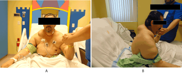
Figure 1. On physical exam patient had several non-tender large lipomas in his chest, back, posterior and anterior neck, and bilateral deltoid region.
Neurology and Rheumatology services were consulted. Neurology considered an atypical Guillain-Barre Syndrome as a differential diagnosis and ruled out other neurologic conditions after physical exam and lab findings. Rheumatology service ruled out a rheumatologic condition with a negative rheumatologic panel. Although patient did have a lipoma biopsy done, muscle biopsy for testing of the myoclonus epilepsy and ragged red fibers (MERRF) gene was unable to be done due to lack of availability of service. Patient was clinically diagnosed with Madelung’s Disease Type I due to the accumulation of fat around the neck/nape of the neck, shoulders, upper arms, and upper back. His initial muscle weakness did improve slightly, although he remained with the need for assistance in basic needs. Patient was discharged for a surgery follow up to discuss the possibility of surgical removal of lipomas as well as to try to obtain a muscle biopsy as outpatient. He was also referred for physical and occupational therapy. Due to patient being unfunded, social worker was contacted.
Initial thought process for the patients generalized weakness and noted lower extremity muscle wasting was neurological, such as myasthenia gravis, Guillain barre syndrome, cerebrovascular accidents, disease of the spinal cord, myopathies and muscular dystrophies, all of which were ruled out based on consultants, labs and imaging. The possibility of there being a relationship between the increase adiposity in the upper body with the neurological manifestations was not considered. Upon discussion with the patient's primary care physician and admitting physician, they had categorized the patient as being obese with no relevance given to the symmetrical distribution of lipomas of the upper extremities, neck and torso.
There is no definitive treatment for Madelung’s Disease. There are proposed recommendations such as alcohol abstinence in alcoholics, lymphatic decongestive therapy (LDT) and surgery [9]. Other considerations include B2 adrenergic agonists, fibrates, growth hormone, lifestyle changes and local SAT injections, although none of them offer definitive treatment. There has been a consideration of a plant-based diet which has shown some improvement, although no studies have been done on this regard. Surgical removal of lipomas is considered the best treatment option [2]. For this patient we did consider surgical consult.
Madelung’s disease is a rare disorder of fat metabolism and accumulation of painless, symmetrical non-encapsulated lipomas in the upper body. This condition is very rare and is seen mostly in patients of European/Mediterranean descent with an estimated incidence rate of 1 in 25,000 in the Italian population and affects males up to 30 times more frequently than females [3,13-15] Majority of cases reported have been on patients of Mediterranean descent and alcoholic males. The exact pathophysiology of this condition is still unknown, but in vitro studies have evidenced a defective tissue lipolysis and excess adipose triglyceride accumulation is due to reduced fatty acid release, an abnormal lipogenesis induced by catecholamine, and deposition of brown adipose tissue [8]. Three types of this condition have been described in the literature [9,16]. Type 1 presents with tumor like lipomas in the head/neck and back with a characteristic dorsocervical pad (buffalo hump). Complications seen with Type 1 are tracheal and esophageal compression and the risk for superior vena cava syndrome [17] which can be present in 15-20% of patients and one case reported a patient presenting with severe respiratory dyspnea which required the use of a tracheostomy due to the compression of the fatty tissue on the laryngotracheal region. Additionally, patients with Type I and Type II should be evaluated for sleep apnea due to the distribution of lipomas [8]. Type II presents with fat accumulation in the shoulder girdle, upper arms, thorax, back, abdomen and upper buttocks. Type III can be seen mainly in women and is considered the rarest type which presents with fat accumulation on the hip and thigh areas [9]. Several associated conditions have been seen in patients presenting with Madelung's disease, although not pathognomonic, are metabolic disturbances such as glucose intolerance, increased secretion of insulin, hyperuricemia, renal tubular acidosis, alterations in liver enzymes which can be attributed to alcoholism and abnormal function of thyroid, adrenal and pituitary glands. In our patient, he did have hyperuricemia and elevations in liver enzymes, which in his case was not attributed to underlying alcoholism. Reports have also associated polyneuropathy associated with Madelung's disease, which can eventually be the principal cause of severe disability [12]. One case report mentioned a patient whose muscle weakness progressed over time 7 years after onset of symptoms and eventually became bedridden and dependent on others for activities of daily living [4]. Reports have also associated sudden death secondary to a slowly progressive axonal sensory and autonomic peripheral neuropathies after the development of Madelung's disease, which leads to impairment of autonomic function [8]. Possible causes of myopathy include alcoholic myopathy and mitochondrial myopathy, the latter supported by a high lactate to pyruvate ratio, as described in one case report [4]. Madelung's disease has been suggested to be of mitochondrial inheritance when seen in familial cases. The patient presented in this case report did also suggested mitochondrial inheritance, with his mother and siblings with features of Madelung's. One study found multiple deletions of mitochondrial DNA and mutation of the myoclonus epilepsy and ragged red fibers (MERRF) tRNA (Lys) A>G (83344) mutation [18,19]. The diagnosis is clinical, although imaging, specialists and biopsy should be warranted to exclude other conditions and the treatment for Madelung's Disease is mainly surgical [9]. The overall recurrence rate following surgery is approximately 63%, open surgery results in a recurrence rate of 51% compared to a recurrence rate of 95% with liposuction [14]. Non-surgical approaches alcohol abstinence in those with history of alcoholism, lymphatic drainage therapy and an animal free diet as well as other treatment modalities considered are B2-Adrenergic agonists, Fibrates and Growth Hormone therapy.
- Lectures illustrative of various subjects in pathology and surgery (1846) London, Longman.
- Adamo C, Vescio G, Battaglia M, Gallelli G, Musella S (2001) Madelung's disease: case report and discussion of treatment options. Ann Plast Surg 46: 43-45. [Crossref]
- Enzi G, Angelini C, Negrin P, Armani M, Pierobon S, et al. (1985) Sensory, motor, and autonomic neuropathy in patients with multiple symmetric lipomatosis. Medicine (Baltimore) 64: 388-393.
- Suresh Chandran CJ, Godge YR, Oak PJ, Ravat SH (2009) Madelung's disease with myopathy. Ann Indian Acad Neurol 12: 131-132. [Crossref]
- Sarhill N, Kumar A, Cook L, Tahir A, Barakat K (2006) Madelung’s Disease in an African-American Patient. Hospital Physician pp: 35-38.
- Madelung O (1888) Uber den Fetthals. Langenbecks Archiv Klin Chirurg 37: 106.
- Lanois P, FFB (1898) L’ade´nolipomatose symmetrique. Bull Soc me´d Hop Paris 1: 289.
- Enzi G, Favaretto L, Martini S, Fellin R, Baritussio A, et al. (1983) Metabolic abnormalities in multiple symmetric lipomatosis: elevated lipoprotein lipase activity in adipose tissue with hyperalphalipoproteinemia. J Lipid Res 24: 566-574
- Herbst KL (2012) Rare adipose disorders (RADs) masquerading as obesity. Acta Pharmacol Sin 33: 155-172. [Crossref]
- Laure B, Sury F, Tayeb T, Corre P, Goga D (2011) Launois-Bensaude syndrome involving the orbits. J Craniomaxillofac Surg 39: 21-23. [Crossref]
- Berkoviv SF, Andermann F, Shoubridge EA, Carpenter S, Robitaiile Y, et al. (1991) Mithocondrial dysfunction in multiple symmetrical lipomatosis. Ann Neurol 29: 566-569.
- Chalk CH, Mills KR, Jacobs JM, Donaghy M (1990) Familial multiple symmetric lipomatosis with peripheral neuropathy. Neurology 40: 1246-1250.
- Enzi G, Biondetti PR, Fiore D, Mazzoleni F (1982) Computed tomography of deep fat masses in multiple symmetrical lipomatosis. Radiology 144: 121-124.
- Adamo C, Vescio G, Battaglia M, Gallelli G, Musella S (2001) Madelung's disease: case report and discussion of treatment options. Ann Plast Surg 46: 43-45. [Crossref].
- Ross M, Goodman MM (1992) Multiple symmetric lipomatosis (Launois-Bensaude syndrome). Int J Dermatol 31: 80-82.
- Harsch IA, Michaeli P, Hahn EG, Ficker JH, Konturek PC (2003) Launois-Bensaude syndrome in a female with type 2 diabetes. Med Sci Monit 9: CS5-8. [Crossref].
- Borriello M, Lucidi A, Carbone A, Iannone V, Ferrandina G (2012) Malignant transformation of Madelung's disease in a patient with a coincidental diagnosis of breast cancer: a case report. Diagn Pathol 7: 116.
- Chong PS, Vucic S, Hedley-Whyte ET, Dreyer M, Cros D (2003) Multiple symmetric lipomatosis (Madelung's disease) caused by the MERRF (A8344G) mutation: a report of two cases and review of the literature. J Clin Neuromuscul Dis 5: 1-7.
- Perera U, Kennedy BA, Hegele RA (2018) Multiple Symmetric Lipomatosis (Madelung Disease) in a Large Canadian Family with the Mitochondrial MTTK c.8344A>G Variant. J Investig Med High Impact Case Rep 6: 2324709618802867.