Objective: The purpose of our study was to ascertain the outcome of patients with iatrogenic anterior skull base injuries during functional endoscopic sinus surgery (FESS) and to identify factors regarding the patient, cerebrospinal fluid (CSF) leaks, and a treatment that may influence the results of the injury.
Study Design and Setting: A retrospective analysis review of the patients in an otorhinolaryngology and neurosurgery tertiary referral center.
Methods: 398 patients with medically refractory chronic rhinosinusitis who underwent FESS during a 2-year period were reviewed and analyzed. Additionally, we present two rare cases of iatrogenic skull base injuries during routine FESS with intracranial complications.
Results: Complications occur mostly because of the close proximity of crucial anatomical structures such as the sinuses to the anterior cranial fossa, internal carotid arteries and orbit. The overall major complication rate was 2.8%; and the intracranial complication rate was 1.3%. The risk of an injury is related to the history of a previous surgery, the extent and severity of a disease, and anatomical variation. Intraoperative penetration of the skull base with laceration of the dura and the associated CSF leaks are not often immediately detected and thus cause life-threatening situations for patients.
Conclusions: Although the improvement of FESS is notorious, concerning risks of complications still occur while carrying out FESS. The awareness of the location of skull base and orbit, together with early identification of anatomic landmarks during FESS, plays a pivotal role when it comes to avoiding complications. An exhaustive preoperative evaluation of the patient and a closer follow-up can help to further diminish negative outcomes.
anterior skull base injury, iatrogenic lesion, sinonasal surgery complication, functional endoscopic sinus surgery, brain pathology
Chronic rhinosinusitis (CRS) is a highly prevalent disorder, affecting approximately 11% of the patients in Europe and 12% in the USA [1,2]. Its impact on the patients’ quality of life [1] that often, unfortunately, does not completely improve with medical therapies. In these situations, functional endoscopic sinus surgery (FESS) is the mainstay of surgical treatment for this condition [1,3]. Nevertheless, endoscopic sinus surgery is also the leading cause of litigation in otolaryngology [4,5]. Despite the recent technical and instrumentation improvements in FESS, even in the best hands, disastrous complications during endoscopic surgery are still possible. Preoperative assessment of the paranasal sinuses and their relationship with the anterior skull base is critical for preventing major complications such as skull base penetration, cerebrospinal fluid (CSF) fistulas, brain injury or even a lethal outcome.
There are certain anatomic variations that may predispose the endoscopic surgeon to an inadvertent injury of the anterior skull base [6]. The olfactory fossa, for instance, is a depression in anterior cranial cavity whose floor is formed by the cribriform plate of ethmoid roof. The lateral lamella of the cribriform plate is also the thinnest area of the anterior skull base. Keros classified the profundity of the cribriform plate in three categories [7]. Its depth is less than 3 mm in Keros type I, 4-7 mm in Keros type II and more than 7 mm in Keros type III. Recent radiological analysis of the lateral lamella dimensions has revealed that Keros type II was the most common type identified (74.6%). Prevalence of the dangerous Keros type III was the rarest type (7.9%) [8]. The bigger the depth of the cribriform plate, the greater chance there is for an inadvertent intracranial penetration with resultant CSF leak. According to a questionnaire-based study to practicing otolaryngologists, about 25% of otolaryngologists have experienced an intraoperative CSF leak during endoscopic sinus surgery within a five-year period [9].
Dural lesions with CSF leak are the most frequent manifestations of iatrogenic skull base penetration. The incidence of iatrogenic CSF leaks is more common than accidental traumatic CSF leaks, making up 41-56% of CSF leaks in several series from large tertiary care centers [10,11]. Usually, the prognosis of patients with isolated CSF leaks after FESS is good when treated promptly [12]. If unrecognized or untreated, CSF leaks can lead to serious complications such as development of meningitis, which can endanger the patient’s vitality [13]. Much more rarely, brain injury and major hemorrhage from laceration of neuro-vascular structures, including the internal carotid artery, have been described in patients treated with FESS [5,14-16]. A delayed discovery of these potentially devastating complications postoperatively makes the localization of a via falsa more difficult. Also, the management of serious intracranial complications of FESS leading to death can be challenging because of the complexity and severity of injuries. Thus, surgical management is best optimized using a multidisciplinary team consisting of neurosurgeons and otolaryngologists [17,18]. However, despite an increased complication rate, the rarity of life-threatening intracranial injury during endoscopic sinus surgery makes the development of an evidence-based management strategy difficult. The aim of the present study is to investigate the occurrence of intracranial complications after perforating injuries of the anterior skull base secondary to FESS in patients referred for neurosurgical care and treatment. It also serves to underline the potential impact of life-threatening brain injury during endoscopic sinus surgery and to identify factors regarding the patient, CSF fistula, and treatment that may influence the results of the injury.
During a 2-year period, 398 patients with medically refractory chronic rhinosinusitis were treated by the departments of otolaryngology, neurosurgery, or anaesthesiology at Paracelsus Medical University (PMU) Nuremberg. Study patients were identified from an anonymized observational database called “SAB-Datenbak Nürnberg” using the International Statistical Classification of Diseases, 10th Revision-based search (ICD-10 of WHO). Data generated or analysed particularly for this study are all included in this published article.
The research was approved by the Institutional Reviewboard at PMU (IRB-2021-003). All patient information used in this study is based on a completely anonymous or pseudonymous and retrospective data. Informed consent of the patients or their relatives was obtained during the initial hospital stay or during the telephone interview in the follow up. The study was performed in accordance with the ethical standards laid down in 1964 declaration of Helsinki and its later amendments.
Inclusion criteria were all patients between 18 and 75 years of age who underwent a routine functional endoscopic sinus surgery (FESS) for medically refractory chronic rhinosinusitis (CRS). Excluded from the study were those <18 or >75 years old patients with underlying immunodeficiencies, cystic fibrosis or neoplasia.
All patient data with iatrogenic anterior skull base injuries after FESS and cerebral lesions referred to the Department of Neurosurgery were retrospectively collected. The demographic data, imaging studies, operative reports, hospital notes, and follow-up records were reviewed. Five patients with intracranial complications were identified during the study period. Ten patients had undergone an outpatient surgery. All patients underwent a thin-layer computed tomography (CT) to detect the side where the instrument has penetrated the skull base and to outline the cerebral lesions. Each CT was reviewed by the authors, investigating the presence of anatomic variations according to the classification proposed by Keros [7]. Clinical findings included anosmia/hyposmia, headache, hypogeusia, nasal obstruction, postnasal drip and ronchopathy.
Statistical analysis was conducted with IBM SPSS Version 22 for Windows. Descriptive statistics including means and standard deviations were used to summarize the main characteristics of the patient cohort. For metric variables, the Mann-Whitney-U test was performed. For categorical variables, we used the chi-square test and the Fisher’s exact test was calculated for categories with frequencies within the categories smaller than five. Statistical significance was set at p < 0.05.
Three hundred and ninety-eight patients were included (Table 1). Ages ranged from 18 to 75 years, with a mean age of 46.2 ± 14.1 years; there were 152 (38.2%) females and 246 (61.8%) males. Ten of the 398 patients were managed as outpatients. The other 388 patients spent at least one postoperative night in hospital. The average hospital stay was 6 days.
Table 1. Preoperative characteristics of the total cohort of patients with FESS. Frequency (%) unless otherwise specified. COPD: Chronic Obstructive Pulmonary Disease; CRSsNP: Chronic Rhinosinusitis Sine Nasal Polyposis; CRSwNP: Chronic Rhinosinusitis with Nasal Polyposis; GERD: Gastroesophageal Reflux Disease; IGS: Image-Guided Surgery; OF: Olfactory Fossa; OSA: Obstructive Sleep Apnea; SD: Standard Deviation.
Age, mean (SD) |
46.2 (14.1) years |
Sex |
|
Female |
38.2 |
Male |
61.8 |
IGS |
3.8 |
Outpatient |
2.5 |
FESS Indication |
|
CRSsNP |
19.3 |
CRSwNP |
80.7 |
Keros type |
|
I (0–3 mm) |
9.1 |
II (4–7 mm) |
69.6 |
III (8–16 mm) |
21.3 |
Asymmetry
OF depth, mean (SD) |
28.7
|
left (mm) |
6.05 (1.98) |
right (mm) |
5.81 (1.90) |
Concurrent symptoms |
|
Anosmia/Hyposmia |
24.6 |
Headache |
34.2 |
Hypogeusia |
1.8 |
Nasal obstruction |
90.2 |
Postnasal drip |
35.4 |
Rhonchopathy |
5.0 |
Comorbidities |
|
Asthma |
8.0 |
COPD |
2.3 |
GERD |
0.8 |
OSA |
1.5 |
Type-2 diabetes |
2.0 |
In the overall population, indications for FESS compromised chronic rhinosinusitis with polyps (CRSwNP) in 80.7% and chronic rhinosinusitis without nasal polyps (CRSsNP) in 19.3%. Also, 15.1% of patients had revision surgery. The most common symptom at presentation was nasal obstruction (found in 90.2% of patients); 35.4% of patients had postnasal drip, 34.2% had headaches, and 24.6% suffered from hyposmia or anosmia. In addition, 5% of them complained of rhonchopathy, and 1.8% of patients presented with complaints of hypogeusia.
A total of 11 major complications from FESS were identified (Table 2), representing a major complication rate of 2.76%. Among these major complications, five intracranial complications (complication rate of 1.25%) and six orbital complications (complication rate of 1.5%) were identified. In this dataset, two patients had iatrogenic perforations of the anterior skull base (1%) noticed due to cerebralspinal fluid leak (1%), or excessive bleeding during the procedure. Two more perforations were detected after the procedure, one being detected early after the surgery and the second first two weeks after the surgery.
Table 2. Results of Statistical Analysis of Complications of FESS. Fisher’s exact test used. CSF, cerebrospinal fluid; ICH, intracerebral hemorrhage.
Total complications |
22 (5.5) |
18 (4.7) |
4 (26.7) p = 0.006 |
14 (3.6) |
7 (70) p < 0.001 |
Major complications |
11 (2.8) |
9 (2.3) |
2 (13.3) n. s. |
7 (1.8) |
4 (40) p < 0.001 |
Intracranial: |
5 (1.3) |
4 (1.0) |
1 (6.7) |
3 (0.8) |
2 (20) |
Skull base perforation
CSF leak |
4 (1.0)
4 (1.0) |
4 (1.0)
4 (1.0) |
n. s.
0 (0) |
2 (0.5)
2 (0.5) |
p = 0.006
2 (20) |
ICH |
3 (0.8) |
3 (0.8) |
n. s. |
1 (0.3) |
p = 0.003 |
Meningitis |
1 (0.3) |
0 (0) |
0 (0) |
1 (0.3) |
2 (20) |
Death |
1 (0.3) |
1 (0.3) |
n. s.
0 (0) n. s.
1 (6.7) p = 0.038
0 (0) n. s. |
0 (0) |
p = 0.003
2 (20) p = 0.002
0 (0) n. s.
1 (10) p = 0.025 |
Orbital |
6 (1.5) |
5 (1.3) |
1 (6.7) n. s. |
4 (1) |
2 (20) p = 0.008 |
Minor complications |
11 (2.8) |
9 (2.3) |
2 (13.3) n. s. |
8 (2.1) |
3 (30) p = 0.002 |
Synechiae
Hemorrhage |
1 (0.3)
10 (2.5) |
0 (0)
9 (2.3) |
1 (6.7) p = 0.038
1 (6.7) n. s. |
0 (0)
8 (2.1) |
1 (10) p = 0.025
2 (20) p = 0.023 |
According to the Keros classification [7], 9.1% of the patients were classified as type I, 69.6% of the patients as type II, and 21.3% as type III. In addition, 28.7% of the patients had asymmetry between the two olfactory fossae, with the left side averaging 6.05 mm and the right 5.81 mm.
CT imaging studies showed a perforating injury of the cribriform plate in three patients, followed by the frontal sinus in one patient. Three of the four patients with perforation of the anterior skull base evidenced frontal lobe lesions with hematoma (0.8%). The perforation canal in the frontal lobe reached the lateral ventricle in one patient and was associated with subarachnoid and intraventricular hemorrhage. Also, in another patient CT revealed a cerebral infarction of the anterio-medial flow due to injury of the artery. One patient with subarachnoid and intraventricular hemorrhage developed acute hydrocephalus, and an external ventricular drain was placed. Further, a rapidly increasing intracranial pressure (ICP) resulted in widespread brain infarction in another patient. Despite pharmacologic and surgical measurements to control ICP, the patient had a catastrophic course resulting in early brain death (0.3%). The other two patients recovered and had no neurological deficits. However, no routine neuropsychologic testing was performed.
A subgroup of 15 patients who underwent FESS with Image-guided techniques showed no significant difference regarding orbital (6.7% vs. 1.3%) or intracranial complications respectively (6.7% vs. 1.04%). One patient who underwent FESS with assistance of a navigation system developed meningitis (6.7% vs. 0%, P = 0.038). No complications such as iatrogenic skull base perforation, laceration of the dura, nerve lesion, or intracerebral bleeding occurred during or after surgery with navigation system.
Case 1
A 49-year-old man arrived at the clinic with a history of frontal headaches and clear rhinorrhea. The patient underwent functional endoscopic sinus surgery (FESS) for chronic sinusitis two weeks ago. He developed severe headaches after the procedure without other neurological abnormalities. A computed tomography (CT) scan revealed a perforation injury through the right-sided ethmoidal roof with intracerebral hemorrhage (ICH) on the fronto-basal right side with ventricular collapse and perifocal edema (Figure 1). Upon presentation, he was awake, alert and appeared to be neurologically intact. Routine blood screening did not reveal any clinically significant abnormalities. However, the patient was admitted to the intensive care unit (ICU) due to pronounced morphologic findings.
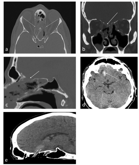
Figure 1. Computed tomography (CT) scan of a patient with a cerebrospinal fluid (CSF) leak following sinus surgery reveals lateral wall fractures of ethmoidal cells and fracture of the lamina cribrosa (bone window), white arrows in axial plane (a), coronal plane (b) and sagittal plane (c). Frontobasal hemorrhagic cerebral contusion (soft tissue window), white arrow in axial plane (d). Trajectory of iatrogenic frontal cerebral hemorrage (white arrows) associated with fracture of cribriform plate (e).
He underwent a CT angiography to exclude vascular lacerations or aneurysm without appreciable results. A CT scan of the paranasal sinuses revealed a bony defect of the cribriform plate and lateral wall fractures of ethmoidal cells (Figure 1). A β2-transferrin testing confirmed a cerebrospinal fluid (CSF) leak. The patient was taken into surgery for endoscopic endonasal repair of the skull base defect using a middle turbinate mucoperiosteal flap. An external ventricular drain was inserted. The patient’s clinical condition improved, and the drain was removed five days later. He was discharged 12 days postoperatively. One month after surgery, the examination demonstrated no neurological deficit or rhinorrhea. A cerebral CT scan showed no evidence of intracerebral hemorrhage.
Case 2
A 46-year-old male with deviated nasal septum, turbinate hyperplasia, obstructive sleep apnea as well as chronic rhinosinusitis was admitted for FESS. The operation was initially considered as successful. The operative note reported no complications. However, the patient did not recover from general anesthesia following a new-onset generalized seizure. Thereafter a cranial computed tomography (CCT) was obtained, which showed a posterior left fronto-basal intraparenchymal hemorrhage (Figure 2). The patient was taken to the ICU. A follow-up non-contrast CT scan of the head did not reveal appreciable changes. A further investigation with CT angiography did not show evidence of aneurysm, vascular malformation, or pseudoaneurysm. In addition, a direct coronal CT scan showed a left cribriform fracture (Figure 2). Intracranial pressure (ICP) was measured using an intraparenchymal sensor that showed an ICP of >40 mmHg during the tenth postoperative day. CCT was emergently repeated and revealed an increasing ICH with increased midline shift. The patient was taken to the operating room for emergent decompressive hemicraniectomy. After a temporary relief of brain compression, the patient developed dilated pupils and further imaging demonstrated ischemia in the antero-medial flow area and edema formation, resulting in lethal brainstem compression and caudal herniation of the cerebellum through the foramen magnum. Due to unfavorable prognosis, further therapy was abandoned, and the patient deceased a few days later.
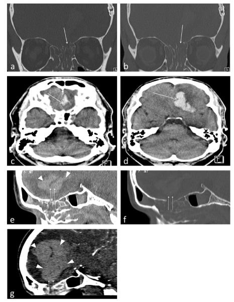
Figure 2. Fracture of lamina cribrosa (bone window), white arrows in coronal plane (a,b). Frontobasal hemorrhagic cerebral contusion (soft tissue window), white arrows in axial plane (c,d). Trajectory of fracture of cribrotic plate (soft tissue window (e), bone window (f), arrows), sagittal image from a computed tomographic angiogram (CTA) of the brain at the level of the skull base injury, demonstrating frontal cerebral hemorrhage (arrowheads) (g).
At autopsy, there was evidence of subarachnoid hemorrhage. An examination of the base of the skull showed an approximately 15 by 8 mm perforation of the left cribriform plate. Above the bone perforation of the lamina cribrosa, a disruption of the dura was noticed (Figure 3a). The examination of the sinonasal cavity showed surgical and traumatic changes of the ethmoid sinuses with hemorrhage (Figure 3b and 3c). The left frontal lobe was lacerated, with associated disruption of the left olfactory nerve (Figure 3c). The left anterior cerebral artery showed a laceration of the vessel wall with adjoining parenchymal and subarachnoid hemorrhage. Lack of preexisting vascular pathology like aneurysm or vascular malformation excluded concurrent etiologies for the hemorrhage. Subsequent sectioning of the fresh brain revealed extensive parenchymal destruction and hemorrhage immediately underlying the laceration within the left frontal lobe (Figure 3e and 3f). Also evident were global ischemic changes of the brain. The cause of death was certified as complications of sinus surgery, with a perforation of the skull base with hemorrhagic tissue destruction of the brain, while the manner of death was considered accidental.
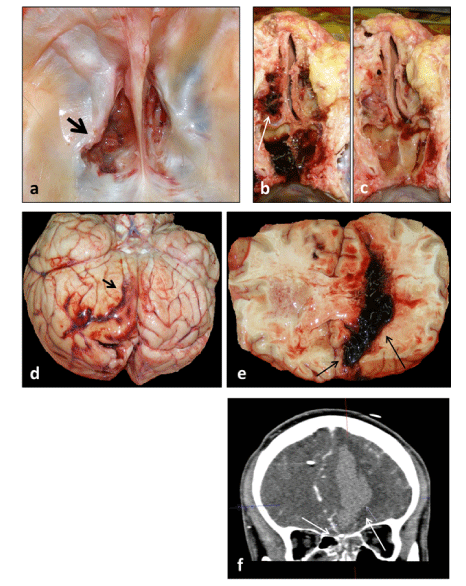
Figure 3. The skull base penetration after ESS causes an immediate increase in supratentorial pressure through hemorrhage in left hemisphere and edema, this may result in death because of impact of the cerebellum and medulla into the foramen magnum. The medial part of anterior fossa at autopsy, showing a left sided defect of the cribriform plate with laceration of dura mater (black arrow) (a). Superior aspect of the sinonasal cavity, with hemorrhage (b-c). Inferior aspect of fresh brain, with left frontal laceration of brain (white arrow) and thin subarachnoid hemorrhages are over the cerebral hemisphere (d). This coronal section of fresh brain shows an intracerebral hemorrhage with massive extension resulted from trauma (iatrogenic brain injury) (e-f).
Our study shows that even in modern practice routine, functional endoscopic sinus surgery may result in fatal and rarely in lethal intracranial complications with mechanical destruction of cerebral parenchyma. We examined the frequency of intracranial complications following endoscopic sinus surgery in a cohort of 398 patients. The risk of major complications according to ERS1 was at 2.8% respectively. In this cohort, patients who had more sinuses operated on, or who especially had a greater height of the lateral lamella of the cribriform plate were more likely to have an intracranial complication. Iatrogenic CSF leakage is the most common anterior skull base complication, even if its rate in the endoscopic approaches is about 1%.
There are several published studies assessing the highly variable incidence of complications from endoscopic sinus surgery. Institutional reports from the last decades indicated major complication rates up to 3% [19-22]. A case series of 997 patients, who underwent endoscopic sinus surgery, observed a direct relationship between the experience of the surgeon and the possibility of uncontrolled maneuvers with iatrogenic complications (a resident 12.67% vs. an experienced surgeon 2.4%) [23].
A successful sinus surgery highly depends on accurate identification of anatomic landmarks within a circumscribed surgical field. The anatomy of the paranasal sinuses shows a remarkable degree of individual variation. Bleeding from the sinonasal mucosa impairs endoscopic visualization and may lead to complications due to the close proximity to the skull base and vital neurovascular structures. Therefore, an understanding of the endoscopic view of sinonasal anatomy is essential. CT scans (thin slices, high resolution) are helpful to assess the extension of chronic rhinosinusitis and thus help in the classification of CRS [1]. Moreover, CT scans show the bony landmarks of all paranasal sinuses and their pneumatization or changes in previously operated patients [24]. The use of intraoperative navigation to integrate the knowledge of endoscopic and CT anatomy may help in identifying a difficult anatomical situation such as an asymmetry of the olfactory fossa between the left and right sides or a deep olfactory fossa and reduce the risks of endonasal surgery in complex cases, especially in revision surgeries [25]. In our cohort, we found an asymmetry between the two olfactory fossae in 28% of patients, with the right side averaging 5.81 mm and the left 6.05 mm. The incidence of a deep lateral lamella of the cribriform plate (Keros type III) was 21% compared to the incidence described by Keros in his initial studies (18%) [7]. This is consistent with previous reports describing that an increased depth of the olfactory fossa poses a risk factor for iatrogenic CSF leak [26]. Furthermore, the analysis of 15 patients undergoing image-guided surgery (IGS) and 383 patients undergoing endoscopic sinonasal surgery without image guidance showed no statistically significant difference in the incidence of major intraoperative or postoperative complications and the number of revision procedures. However, a higher incidence of CSF leak was noted in the group undergoing surgery without image guidance (1.04% vs. 0%). Total complications were significantly greater in the IGS group (4.7% vs. 26.7%, p = 0.006). Because the risk of intracranial complication is relatively rare, a very large population-based study may be necessary to show any positive benefits from the use of navigation systems.
CSF leakage may occur both intraoperatively and postoperatively. When patients report symptoms such as headache, hyposmia, and/or symptoms that are consistent with CSF rhinorrhea after sinus surgery, a diagnosis of CSF leakage should be considered, even if the procedure itself was seemingly uneventful (Case 1 – 2). In our study, 60% of CSF leaks were diagnosed postoperatively, with the majority occurring in primary surgery (1.2%) and in the outpatient surgery (20%). This suggests that skull base injury during endoscopic sinus surgery is a barely seen entitiy in high risk patients. Our data suggest that the laceration of the dura with CSF leak is often not recognized during primary surgery for CRS, leading to significant morbidity. This can usually be recognized at the time of surgery by a “washout” of clear fluid from the area of the injury, caused by dilution of the blood covering the surrounding tissues. Endoscopic surgeons must always assume that there is always a high risk of CSF leak, especially when it comes to any sudden increase in bleeding that occurs in the vicinity of the skull base.
In the current investigation, delays in the recognition of skull base penetration leading to CSF rhinorrhea and a frontal lobe injury after a routine functional endoscopic sinus surgery were associated with increased neurological complications and, specifically in outpatient surgery, significantly increased mortality (p = 0.025). Whereas, in some instances, brain lesions after endoscopic sinus surgery are asymptomatic and even unrecognized during the postoperative period [27] and discovered incidentally by a routine request for other reasons many years later [28]. Lesions of frontal lobe and neurovascular structures still represent a feared complication, resulting in death in one patient caused by an endoscope itself (Case 2). Also, vascular complications such as central retinal artery occlusion [29] and the development of a pseudoaneurysms [30,31] are described. Serious complications leading to death after endonasal surgery are intracerebral injuries and abundant bleeding after vascular injury [30,32].
The two cases presented here, with very different outcomes, clearly show the ever-present risk of a so-called major intracranial complication due to intracranial injury during FESS. The lesion was noticed intraoperatively only in two of the 398 operations (0.5%), thus first countermeasures could be taken to prevent a catastrophic outcome. For example, the risk of an ascending infection was reduced. Iatrogenic brain pathologies, after a routine endoscopic sinus surgery, might have a higher percentage than assumed because they may remain undetected with a considerable delay.27 Notably, such lesions might not be described in studies involving only experienced and high volume centers. Such data, possibly, may only be captured by the collection of large systematic case series concentrating on the specific problem scrutiny.
Moreover, we identified only a small-sized cohort of outpatients or patients who underwent FESS with the navigation system. Thus, making it difficult to draw definite conclusions about safety aspects of outpatient surgery. A very large population-based study may be necessary to show any positive benefits from the use of navigation systems.
Analysis of two years’ activity in the Paracelsus Medical University (PMU) Nuremberg showed that even in modern practice routine, FESS may result in severe or fatal intracranial complications with mechanical destruction of cerebral parenchyma. Careful review and understanding of the patient's symptoms, anatomy, and pathology are essential to derive a personalized approach unique to them. The awareness of the location of skull base and orbit, together with early identification of anatomic landmarks during sinonasal surgery, is paramount when it comes to avoiding complications. FESS is usually safe and effective, but life-threatening anterior skull base injuries can occur during surgery and affect the outcome of patients; therefore, this paper suggests close follow-up for routine endoscopic sinus surgery in order to help provide the best outcome for one’s patients.
Our thanks go to all participating departments.
EY conceived and designed the study. EY, JP and SS collected the data and EY, SS, CG and JP analyzed the data. EY wrote the manuscript. All authors contributed to the preparation of the final manuscript.
- Fokkens WJ, Lund VJ, Hopkins C, Hellings PW, Kern R, et al. (2020) European Position Paper on Rhinosinusitis and Nasal Polyps 2020. Rhinology 58: 1-464. [Crossref]
- Hirsch AG, Stewart WF, Sundaresan AS, Young AJ, Kennedy TL, et al. (2017) Nasal and sinus symptoms and chronic rhinosinusitis in a population-based sample. Allergy 72: 274-281. [Crossref]
- Rudmik L, Soler ZM, Hopkins C, Schlosser RJ, Peters A, et al. (2016) Defining appropriateness criteria for endoscopic sinus surgery during management of uncomplicated adult chronic rhinosinusitis: a RAND/UCLA appropriateness study. Int Forum Allergy Rhinol 6: 557-567. [Crossref]
- Tolisano AM, Justin GA, Ruhl DS, Cable BB (2016) Rhinology and medical malpractice: An update of the medicolegal landscape of the last ten years. Laryngoscope 126: 14-19. [Crossref]
- Stankiewicz JA, Hotaling J (2015) Medicolegal Issues in Endoscopic Sinus Surgery and Complications. Otolaryngol Clin North Am 48: 827-837. [Crossref]
- Meyers RM, Valvassori G (1988) Interpretation of anatomic variations of computed tomography scans of the sinuses: a surgeon's perspective. Laryngoscope 108: 422-425. [Crossref]
- Keros P (1962) On the practical value of differences in the level of the lamina cribrosa of the ethmoid. Z Laryngol Rhinol Otol 41: 809-813. [Crossref]
- Babu AC, Nair M, Kuriakose AM (2018) Olfactory fossa depth: CT analysis of 1200 patients. Indian J Radiol Imaging 28: 395-400. [Crossref]
- Platt MP, Shaye D, Parnes SM (2007) Management of unexpected cerebrospinal fluid fistulae during endoscopic sinus surgery. Am J Rhinol 21: 611-614. [Crossref]
- Tabaee A, Kassenoff TL, Kacker A, Anand VK (2005) The efficacy of computer assisted surgery in the endoscopic management of cerebrospinal fluid rhinorrhea. Otolaryngol Head Neck Surg 133: 936-943. [Crossref]
- Banks CA, Palmer JN, Chiu AG, O'Malley Jr BW, Woodworth BA, et al. Endoscopic closure of CSF rhinorrhea: 193 cases over 21 years. Otolaryngol Head Neck Surg 140: 826-833. [Crossref]
- Marshall AH, Jones NS, Robertson IJ (2001) CSF rhinorrhoea: the place of endoscopic sinus surgery. Br J Neurosurg 15: 8-12. [Crossref]
- Bernal-Sprekelsen M, Bleda-Vazquez C, Carrau RL (2000) Ascending meningitis secondary to traumatic cerebrospinal fluid leaks. Am J Rhinol 14: 257-259. [Crossref]
- Stankiewicz JA, Chow JM (2004) The low skull base: an invitation to disaster. Am J Rhinol 18: 35-40. [Crossref]
- Weidenbecher M, Huk WJ, Iro H (2005) Internal carotid artery injury during functional endoscopic sinus surgery and its management. Eur Arch Otorhinolaryngol 262: 640-645. [Crossref]
- Chin OY, Ghosh R, Fang CH, Baredes S, Liu JK, et al. (2016) Internal carotid artery injury in endoscopic endonasal surgery: A systematic review. Laryngoscope 126: 582-590. [Crossref]
- McLaughlin N, Carrau RL, Kelly DF, Prevedello DM, Kassam AB (2013) Teamwork in skull base surgery: An avenue for improvement in patient care. Surg Neurol Int 4: 36. [Crossref]
- Olofsson J (2010) Multidisciplinary team a prerequisite in endoscopic endonasal skull base surgery. Eur Arch Otorhinolaryngol 267: 647.
- Stankiewicz JA, Lal D, Connor M, Welch K (2011) Complications in endoscopic sinus surgery for chronic rhinosinusitis: a 25-year experience. Laryngoscope 121: 2684-2701. [Crossref]
- Weber R, Draf W, Keerl R, Schick B, Saha A (1997) Endonasal microendoscopic pansinusoperation in chronic sinusitis. II. Results and complications. Am J Otolaryngol 18: 247-253. [Crossref]
- Siedek V, Pilzweger E, Betz C, Berghaus A, Leunig A (2013) Complications in endonasal sinus surgery: a 5-year retrospective study of 2,596 patients. Eur Arch Otorhinolaryngol 270: 141-148. [Crossref]
- Oeken J, Torpel J (2008) The influence of navigation on endoscopic sinus surgery. HNO 56: 151-154, 156-157. [Crossref]
- Chou TW, Chen PS, Lin HC, Lee KS, Tsai HH, et al. (2016) Multiple analyses of factors related to complications in endoscopic sinus surgery. J Chin Med Assoc 79: 88-92. [Crossref]
- Bansberg SF, Harner SG, Forbes G (1987) Relationship of the optic nerve to the paranasal sinuses as shown by computed tomography. Otolaryngol Head Neck Surg 96: 331-335. [Crossref]
- Dalgorf DM, Sacks R, Wormald PJ, Naidoo Y, Panizza B, et al. (2013) Image-guided surgery influences perioperative morbidity from endoscopic sinus surgery: a systematic review and meta-analysis. Otolaryngol Head Neck Surg 149: 17-29. [Crossref]
- Heaton CM, Goldberg AN, Pletcher SD, Glastonbury CM (2012) Sinus anatomy associated with inadvertent cerebrospinal fluid leak during functional endoscopic sinus surgery. Laryngoscope 122: 1446-1449. [Crossref]
- Scharpf J, Dean R, Stultz T, Citardi MJ (2005) The magnetic resonance imaging profile of occult intracranial violations as a result of sinus surgery. Am J Otolaryngol 26: 411-414. [Crossref]
- Karaman E, Isildak H, Yilmaz M, Enver O, Albayram S (2011) Encephalomalacia in the frontal lobe: complication of the endoscopic sinus surgery. J Craniofac Surg 22: 2374-2375. [Crossref]
- Kurt MM, Akpolat C, Fethallah B (2019) A rare complication of endoscopic sinus surgery: Retinal artery occlusion. Eur J Ophthalmol 29: 9-11. [Crossref]
- Golinelli G, Toso A, Taranto F, Aluffi P, Pia F (2012) Delayed carotid pseudoaneurysm: a life-threatening complication after endoscopic sinus surgery. J Craniofac Surg 23: 1822-1824. [Crossref]
- Munich SA, Cress MC, Rangel-Castilla L, Krishna C, Siddiqui AH, et al. (2016) Importance of repeat angiography in the diagnosis of iatrogenic anterior cerebral artery territory pseudoaneurysm following endoscopic sinus surgery. J Neurointerv Surg 8: e20. [Crossref]
- Al-Afif S, Hermann EJ, Hatipoglu Majernik G, Nakamura M, Raab P, et al. (2017) Severe Cerebral Complications Secondary to Perforation Injury of the Anterior Skull Base During Sinonasal Surgery: An Underappreciated Problem? World Neurosurg 108: 783-790. [Crossref]
Editorial Information
Editor-in-Chief
Chin-Lung Kuo
Taoyuan Armed Forces General Hospital
Taiwan
Article Type
Research Article
Publication history
Received date: June 01, 2021
Accepted date: June 18, 2021
Published date: June 23, 2021
Copyright
©2021 Yakubov E. This is an open-access article distributed under the terms of the Creative Commons Attribution License, which permits unrestricted use, distribution, and reproduction in any medium, provided the original author and source are credited.
Citation
Yakubov E, Gaschler C, Schmid S, Eibl T, Hammer A, et al. (2021) Life-threatening anterior skull base injuries after endoscopic sinonasal surgery. Otorhinolaryngol Head Neck Surg 6: doi: 10.15761/OHNS.1000271