Introduction: Anaplastic thyroid carcinoma (ATC) is a rare but aggressive thyroid malignancy. It is the most primary thyroid tumor malignancy with distant metastasis to bone, lungs, and rarely to meningeal.
Leptomeningeal metastasis of ATC is not well documented in English literature.
Case report: We report the case of a 65 years old patient with Balance disorders and confusion as the revealing signs. Anamnesis revealed an intracranial hypertension syndrome with cerebellar ataxia evolving since four weeks. Physical examination found a firm and fixed thyroid nodule moving upon swallowing. Brain MRI found subarachnoid space lesions consistent with meningeal carcinomatosis and the Chest CT revealed military opacities compatible with pulmonary metastasis. Thyroid ultrasound showed a nodule in the left lobe with microcalcifications rated as T-RADS 4C, and the nodular biopsy revealed an undifferentiated thyroid carcinoma metastasis. The patient died three weeks later.
Discussion: Several factors have been reported to worsen it including old age, poor general condition, rapid growth of a pre-existing goiter, tumor size, local extension and distant spread.
Conclusion: Although ATC metastasis may occur in all sites, meningeal localization is rare and may be explained by its hematological spread.
anaplastic thyroid carcinoma- Leptomeningeal metastasis- poor prognostic factor
Abbreviations
ATC: Anaplastic thyroid carcinoma
Anaplastic thyroid carcinomas (ATC) are undifferentiated tumors of the thyroid follicular epithelium and an extremely aggressive neoplasm that accounts for 1-3% of all thyroid cancers with a dismal median survival of four to five months .It’s the most primary thyroid tumor malignancy with distant metastasis to bone, lungs, and rarely to the brain and meningeal . Metastasis of ATC to dura is not well documented in English literature.
We report a case of a locally insidious ATC revealed by an unusual meningeal carcinomatosis.
This report describes a case of leptomeningeal metastases as the initial manifestation of ATC in a 65-year old woman with a history of type 2 diabetes mellitus controlled under treatment who presented confusion as the revealing sign. Anamnesis revealed an intracranial hypertension syndrome evolving since four weeks with headache, vomiting, spatiotemporal confusion and paroxysmal vertigo with photophobia. The patient report also dyspnea, nocturnal cough and unexplainable weight loss of 12 kg. At the physical examination, the patient was normotensive, polypneique without a focal deficit or vestibular signs. The pulmonary examination found bilateral rales. The neck examination found a moderate stiffness with a painless fixed anterior neck mass. Endoscopic laryngeal examination revealed a decreased mobility of the right vocal cord and arytenoid. The neck ultrasound scan showed a nodule in the left lobe with microcalcifications classified T- RADS 4C, this nodule was hypo functional in technetium thyroid scintigraphy concluding to “cold nodule”. The FNAC showed many atypical spindle cells strongly suspect with a hyperchromatic nucleus and abundant cytoplasm. The biopsy confirmed the diagnosis of anaplasic carcinoma.
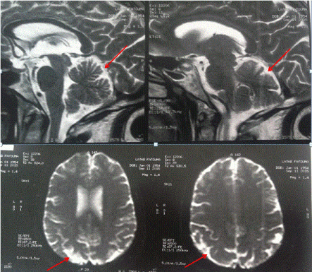
The brain MRI found a diffuse meningeal carcinomatosis and the chest CT scan found a bilateral diffuse miliary opacities.
Because of the general weakness of the patient, she couldn’t tolerate any palliative treatment, she died three weeks later.
Leptomeningeal metastasis tumors are a weighty cause of mortality in cancer patients. the primary tumor is most commonly seen in the lung, breast, colon and kidney and very rarely in the thyroid , and exceptionally of its undifferentiated follicular epithelium [1,2].
An insidiously local ATC is unusual, this tumor usually appear as a rapidly enlarging neck mass, often associated with dysphonia, dyspnea, dysphagia, and neck stiffness. Half patients have distant metastases at diagnosis, the lungs are the most common site involved [3]. However leptomeningeal metastasis is an uncommon site it may be explained by its hematological spread [3] and according to our knowledge it has been never reported before.
Most of the patients with ATC do not live longer than one year after the diagnosis establishment due to local and/or regional tumor progression leading to airways and esophageal obstructions [4].
Patients with ATC are older than those with differentiated thyroid carcinoma, mean age is 65 years at diagnosis, and fewer than 10% are younger than 50 years, 70% of tumors occur in women [5] .The presence of distant metastases in extracranial sites like lung and bone is a risk factor for the development of leptomeningeal and brain metastases [2].
The metastasis of thyroid carcinoma depend on the subtype. However, for each of the histologic subtypes, metastasis to the leptomeningeal and brain is decidedly unusual, reportedly occurring in approximately 1% of all cases of thyroid carcinoma [2]. Although the overall incidence of leptomeningeal and brain metastases is low, a study published by Gandhi and al. reported that 18% of patients with distant metastases from papillary thyroid carcinoma developed leptomeningeal and brain metastases during their disease course, and that the brain was a common second location for distant spread [6].

Figure 1. Leptomeningeal contrast enhancement of the right cerebellar hemispheric compatible with a secondary location.
Survival period of patients with leptomeningeal metastasis of ATC is very short after diagnosis. However, other several factors have been reported to worsen it including old age, poor general condition, rapid growth of a pre-existing goiter, tumor size, local extension and distant spread. These factors attest of tumor aggressiveness, hence the difficulty to insure a complete surgical resection [5]. In some patients in weak condition, searching for metastasis is not systematic because of the decision to deliver palliative treatment [5] .In respectable tumors, the decision for adjuvant therapy using chemotherapy or radiotherapy was taken on a case-by-case basis after discussion in a multidisciplinary staff including surgeons, cancerologists and/or radiotherapists. Cytotoxic drugs such as doxorubicin, cisplatin and paclitaxel are the agents generally used in ATC metastasis. However they aren’t capable of altering the lethality of this malignancy, which suggests the need for additional therapeutic innovations [7].
In advanced disease, the role of surgery is less well established and the cytotoxic drugs show a poor effectiveness because the failure to penetrate the blood brain barrier. The roles of radioiodine therapy, external beam radiation therapy and gamma knife radiosurgery are still unclear [3].
authors suggest that the most successful management of ATC with leptomenigeal metastasis include a local control with a combination of tracheal stenting together with radiotherapy, and by controlling the initial sites of metastases with chemotherapy [7].
Therapeutic researches are trying to explore ATC and develop the targeted therapies. To date, the best treatment of this deadly disease remains the prevention which involves the appropriate management of ATC in elderly patients.
- Ayaz T, Sahin SB, Sahin OZ, Akdogan R, Gucer R (2015) Anaplastic thyroid carcinoma presenting with gastric metastasis: a case report. Hippokratia 19: 85-87.
- Solomon B, Rischin D, Lyons B (2000) Leptomeningeal metastases from anaplastic thyroid carcinoma. Aust NZ J Med 30
- Melike Pekmezci, Arie Perry (2013) Neuropathology of brain metastases. Surg Neurol Int 4(Suppl 4): S245–S255. [Crossref]
- 4. Sugitani I, Kasai N, Fujimoto Y, Yanagisawa A (2001) Prognostic factors and therapeutic strategy for anaplastic carcinoma of the thyroid. World J Surg 25: 617–622. [Crossref]
- O’Neill JP, Shaha AR (2013) Anaplastic thyroid cancer . Oral Oncol 49:702–706. [Crossref]
- Grandhi (2015) Follicular carcinoma of thyroid presenting as brain metastasis. Romanian Neurosurgery.
- Rochea B, Larroumets G (2010) Epidemiology, clinical presentation, treatment and prognosis of a regional series of 26 anaplastic thyroid carcinomas. Annales d’Endocrinologie 71: 38–45.

