Background: Coronary artery anomalies are a cause of sudden cardiac death. The most debatable are anomalous origination of a coronary artery from an opposite sinus of Valsalva (ACAOS).
Cases: We present 2 cases of patients with newly discovered anomalous origination of the left coronary artery from the right sinus of Valsalva (L-ACAOS). Both patients underwent cardiac computed tomography for evaluation of coronary anatomy along with other forms of functional testing. Patients are treated medically without recurrence of symptoms despite the high-risk nature of the anomalies.
Summary: After review of the literature, we have found that the risk of sudden cardiac death in patients with congenital coronary anomalies, even among variants considered the highest risk, may be overestimated. In addition, the exact prevalence of coronary anomalies in the general population is currently underestimated. The true prevalence of congenital coronary anomalies and overall risk of sudden cardiac death in this population are not well known. Surgical intervention remains the mainstay of therapy in certain patients though recent investigations into the pathophysiology of these abnormalities have shown that the risk of surgery may outweigh the minimal reduction in risk of sudden cardiac death.
Coronary artery anomalies (CAA) are usually encountered incidentally during coronary angiography, they remain an important cause of debilitating cardiac symptoms and in some instances sudden cardiac death.
Coronary artery anomalies occur in 0.3-0.9% of the population without structural heart defects and 3-6% of those with congenital heart defects [1,2]. They are encountered in 1% of those undergoing cardiac catheterization [3]. The largest angiographic series, comprising 126595 coronary angiograms [4], detected 1686 coronary anomalies, with a prevalence of 1.3%; there were 22 (0.01%) cases with anomalous left main coronary artery (LMCA) arising from the right coronary sinus (RCS) and 136 (0.08%) with anomalous right coronary artery (RCA) arising from the left coronary sinus (LCS). Most people with underlying CAA will never have an angiogram, as they are generally asymptomatic. There is evidence of familial clustering of CAA, but no gene defects have yet been identified [5].
In a retrospective review of the Department of Defense data from 1977 to 2001, nontraumatic sudden death occurred in 126 military recruits out of over 6million cases reviewed. Of these patients, the cause of death was identified as a coronary artery anomaly in 39 patients with 21 of those having an anomalous coronary artery origin. All 21 of these patients were found to have an anomalous origination of the left coronary artery from the right sinus of Valsalva (L-ACAOS) with an interarterial course between the pulmonary artery and aorta [6,7].
Anomalous origin of the left coronary artery from the right sinus of Valsalva may be further separated into four separate subtypes:
(1) The left main coronary artery tracks anteriorly over the right ventricular outflow tract.
(2) The left main coronary artery takes an intramyocardial course before resurfacing at the proximal portion of the interventricular groove.
(3) The left main coronary artery courses between the aorta and the pulmonary artery.
(4) The left main coronary artery passes posteriorly around the aortic root [8,9] (Figure 1).
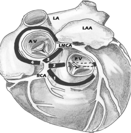
Figure 1. Diagrammatic representation of the four possible pathways for the anomalous left main coronary artery (LMCA) arising from the right coronary cusp. 1, anterior to the right ventricular outflow tract (anterior course); 2, trans-septal or intraseptal course; 3, interarterial course; 4, retro aortic course Of these, the first anomalous configuration is classically considered the most dangerous placing patients at the highest risk of sudden cardiac death.
In this paper, we present two examples of the four subtypes of anomalous origin of the left coronary artery from the right sinus of Valsalva.
Case 1:
A 44-year-old female patient nonsmoker with hypothyroidism, hypertension and dyslipidemia was admitted for atypical chest pain of several month duration. She didn’t report any history of syncope. ECG was normal. Cardiac biomarkers were negative, TTE was normal. The patient underwent coronary CT angiogram for assessment of coronary anatomy. There was no significant coronary artery disease, but the CT shows a left main coronary artery originating from the right sinus of Valsalva with subsequent intraseptal course (Figure 2). Invasive coronary angiography (Figure 3) was completed with normal fractional flow reserve (FFR) measurements in the proximal left anterior descending. The patient is currently doing well without further symptoms on optimal medical therapy alone.
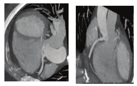
Figure 2. Cardiac CT showing anomalous left main originating from the right sinus of Valsalva and subsequent intraseptal course.
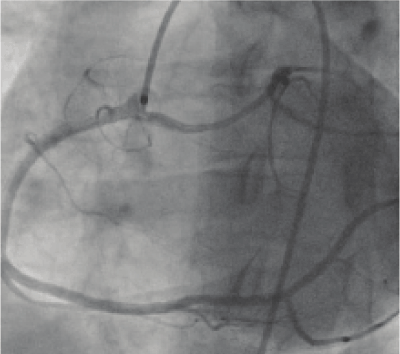
Figure 3. Left anterior oblique view of left main coronary artery with an anomalous origin off the right sinus of Valsalva with intraseptal course.
Case 2:
A 75-year-old female patient nonsmoker with hypertension was admitted for atypical chest pain. She has no history of near syncope or syncope. ECG was normal, cardiac enzymes were negative. TTE was normal. She underwent a stress echocardiography with no electrocardiographic changes and no angina but evidence of mid anteroseptal wall hypokinesis at peak exercise. The patient underwent invasive coronary angiography (Figure 4) showing no significant coronary artery disease but a left main coronary artery with an anomalous takeoff from the right coronary cusp. Cardiac CT showed the left main to have an anomalous takeoff from the right coronary cusp with a course between the great vessels (Figures 5). The patient is currently doing well without further symptoms on optimal medical therapy alone.
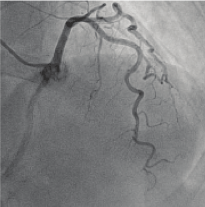
Figure 4. Right anterior oblique-cranial view of left main coronary artery with an anomalous origin off the right sinus of Valsalva with interarterial course.
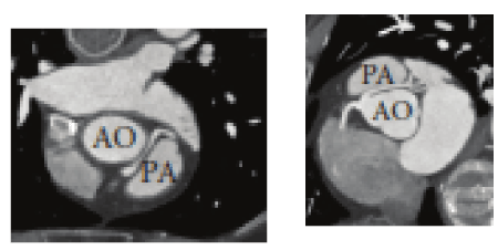
Figure 5. Cardiac CT images of anomalous left main coronary artery off the right sinus of Valsalva with a course between the pulmonary artery (PA) and aorta (AO).
The overall incidence of the abnormal aortic origin of the coronary arteries is estimated to be approximately 0.64% of births [10]. The most common anomaly is the origin of the left circumflex artery from the right sinus of Valsalva, followed by the origin of a single coronary artery from the left sinus of Valsalva, origin of both the right and the left coronary arteries from the right sinus of Valsalva, and the LAD originating from the right sinus of Valsalva. The prevalence of the RCA arising from the left sinus of Valsalva is approximately 0.17% while the rarest anomaly is the left coronary artery arising from the right sinus of Valsalva and its prevalence is around 0.047% [11]. There are four major variations in the course taken by the LMCA after its origin from the right sinus of Valsalva, in relation to the aorta and the pulmonary trunk, on its way to the left ventricle. The LMCA can have a septal course beginning at the right ventricular infundibulum; it can have an anterior course, a retro-aortic course, or an interarterial course between the aorta and the main pulmonary artery [12,13]. The clinical presentation in AAOCA is commonly anginal chest pain or syncope that occurs mostly with exercise or other strenuous activities and may unfortunately be sudden cardiac death as first presentation [14]. Coronary artery anomalies represent a life-threatening form of congenital cardiac pathology. The underlying cause of sudden cardiac death in patients with congenital coronary abnormalities is the source of some debate with multiple competing theories. Death may result from contortion of the vessel’s slit-like, tangential origin during exercise leading to ischemia and resultant arrhythmia. Another theory purports that compression of the anomalous coronary occurs with dilation of the aorta and pulmonary artery during exercise. Itis also possible that frequent episodes of myocardial ischemia lead to myocardial fibrosis and potential nidus for a deadly arrhythmia [7,15].
The severity of ischemia is related to degree of coronary intussusception, level of intramural hypoplasia, and lateral compression of the vessel by expansion of the aorta as discovered by Anjelini and associates when evaluating three patients with L-ACAOS by intravascular ultrasound both at rest and after pharmacologic challenge with saline/atropine/dobutamine [6,9,15]. The true cause of death is likely an aggregate form of the currently proposed theories.
In addition to contention over the true mechanism of death related to coronary artery anomalies, the true prevalence of disease within the general population is unknown.
Congenital coronary anomalies are likely underrecognized as completing an anatomic assessment in a very large portion of the population would seem unfeasible.
The angiographic study completed by Yamanaka and Hobbs found the overall incidence of coronary artery anomalies in a population of more than 120,000 patients to be 1.3%. In this same study, “benign” coronary anomalies were the most frequently discovered with an incidence of 1.07%. L-ACAOS was found in only 22 patients comprising a fairly small portion of abnormalities considered potentially serious [11]. If these same statistics were theoretically applied to the 6.3 million military recruits reviewed by Eckart et al., the overall number of the expected coronary artery anomalies would be 81,900. In the same study, the expected number of military recruits with L-ACAOS would be 1,071. In total, the review found only 21 deaths attributable to an anomalous coronary artery origin, all of which were the L-ACAOS variant. This suggests that the prevalence of congenital coronary anomalies in the general population is much higher than previously predicted. Also, it is very likely that the overall risk of sudden cardiac death in patients with congenital coronary anomalies is much less than previously reported [7,15]. In fact, this has been suggested by a group from the New York University School of Medicine in reviews published in 2005 as well as in 2012 [15,16].
Both patients presented in this paper were found to have a different anatomic configuration of the L-ACAOS variant. Cases included one with an intramural course and the other with an interarterial course.
Both patients have remained symptom free since the initial presentation. No one underwent surgical correction of their unique coronary anomaly as the risk of surgery outweighed the potential benefit of correction based on completed functional studies.
The patient with an interarterial course, considered to be the most insidious variant, developed symptoms in the eighth decade of life potentially due to prolonged hypertension and associated changes in aortic morphology.
In conclusion, the left coronary artery arising from the right sinus of Valsalva is not a frequently encountered congenital coronary artery anomaly. Congenital coronary artery anomalies remain as a cause of sudden cardiac death though it appears that the risk is lower than previously thought. The true prevalence of coronary artery anomalies in the general population is unknown. In addition, the absolute risk of sudden cardiac death is difficult to predict and hinges upon multiple variables. Treatment of coronary artery anomalies is controversial and dependent on the discovered anatomy. Surgery is the mainstay of treatment though beta blockers and calcium channel blockers have been utilized to lessen ischemic symptoms. We presented two patients with two different variants of left coronary artery from the right sinus of Valsalva. Patients are currently being treated medically and continue to remain symptom free. As imaging with cardiac computed tomography continues, maintenance of a congenital coronary anomaly registry would help to broaden our medical knowledge and improve diagnostic and therapeutic modalities for future generations.
- Liberthson RR, Dinsmore RE, Fallon JT (1979) Aberrant coronary artery origin from the aorta. Report of 18 patients, review of literature and delineation of natural history and management. Circulation 59: 748-754. [Crossref]
- Roberts WC (1986) Major anomalies of coronary arterial origin seen in adulthood. Am Heart J 111: 941-963. [Crossref]v
- Taylor AJ, Rogan KM, Virmani R (1992) Sudden cardiac death associated with isolated congenital coronary artery anomalies. J Am Coll Cardiol 20: 640-647. [Crossref]
- Yamanaka O, Hobbs RE (1990) Coronary artery anomalies in 126,595 patients undergoing coronary arteriography. Cathet Cardiovasc Diagn 21: 28-40. [crossref]
- Brothers JA, Stephens P, Gaynor JW (2008) Anomalous aortic origin of a coronary artery with an interarterial course: should family screening be routine? J Am Coll Cardiol 51: 2062-2064. [Crossref]
- Angelini P, Walmsley RP, Libreros A, Ott DA (2006) Symptomatic anomalous origination of the left coronary artery from the opposite sinus of valsalva: clinical presentations, diagnosis, and surgical repair. Tex Heart Inst J 33: 171-179. [Crossref]
- Eckart RE, Scoville SL, Campbell CL (2004) Sudden death in young adults: a 25-year review of autopsies in military recruits. Ann Intern Med 141: 829-834. [Crossref]
- Hauser M (2005) Congenital anomalies of the coronary arteries. Heart 91: 1240-1245. [Crossref]
- Angelini P (2009) Anomalous origin of the left coronary artery from the opposite sinus of valsalva: typical and atypical features. Tex Heart Inst J 36: 313-315. [Crossref]
- Kimbiris D, Iskandrian AS, Segal BL, Bemis CE (1978) Anomalous aortic origin of coronary arteries. Circulation 58: 606-615.
- Yamanaka O and Hobbs RE (1990) Coronary artery anomalies in 126,595 patients undergoing coronary arteriography. Cathet Cardiovasc Diagn 21: 28–40. [Crossref]
- Verma PK and Singh Bisht D (2015) Anomalous origin of left main coronary artery from right sinus of Valsalva: a case report. Angiology 3: 4.
- Marler AT, Malik JA, Slim AM (2013) Anomalous left main coronary artery: case series of different courses and literature review. Case Rep Vasc Med 2013: 380952.
- Basso C, Maron BJ, Corrado D, Thiene G (2000) Clinical profile of congenital coronary artery anomalies with origin from the wrong aortic sinus leading to sudden death in young competitive athletes. J Am Coll Cardiol 35: 1493-1501. [Crossref]
- Pe˜nalver JM, Mosca RS, Weitz D, Phoon CK (2012) Anomalous aortic origin of coronary arteries from the opposite sinus: a critical appraisal of risk. BMC Cardiovasc Disord 12: 83, 2012. [Crossref]
- Mirchandani S and Phoon CKL (2005) Management of anomalous coronary arteries from the contralateral sinus. Int J Cardiol 102: 383-389. [Crossref]