Abstract
Objective: The aim of the work was to report a rare case of isolated unilateral papillo-retinal coloboma discovered late in the course of a decrease of visual acuity and strabismus.
Observation: This is a 7-year-old female child with a history of strabismus in the right eye, brought in by his parents for consultation for a decrease of visual acuity that cannot be improved by optical correction. The examination on admission revealed a reduced visual acuity to light perception in the right eye and 10/10 in the left eye. The anterior segment was normal in both eyes. The fundus examination revealed, in the right eye, a well-defined whitish excavation within the enlarged optic disc in favor of a coloboma of the papilla, including the optic papilla in its lower part and overflowing into the retina, in favor of a papillo-retinal coloboma.
Discussion: Papillo-retinal coloboma is a rare congenital anomaly. It is easy to diagnose clinically and most often it manifests itself as strabismus and as a decrease of visual acuity. It can cause refractive errors with deep amblyopia demonstrated in this clinical case. This malformation can be isolated or most often associated with other malformation anomalies.
Key words
papillo-retinal coloboma, child, organic amblyopia, strabismus
Introduction
Coloboma of the papilla is a congenital papillary excavation resulting from a defect in the closure of the embryonic fissure that occurs at the 7th week of intrauterine life [1,2]. The resulting tissue absence can vary in size and overflow into the chorioretinitis [3]. Coloboma of the papilla is classically located in the lower part of the fetal fissure [1,3]. This malformation can be isolated or most often associated with other malformation anomalies [2,3]. They can lead to refractive errors with deep amblyopia [3]. The aim of this work was to report a rare case of isolated unilateral papillo-retinal coloboma in front of a late discovery decrease of visual acuity.
Observation
This is a 7-year-old female child, with no general or particular ophthalmological history, who was brought in by his parents for a consultation for a decrease of visual acuity in the right eye in an ophthalmology medical center. The onset of symptoms is believed to date back to the age of three by the discovery of strabismus in the right eye in which the diagnosis of a congenital malformation was established. However, faced with the persistence of the deviation of the eye and the appearance of a decrease of visual acuity, the father of the child brought her back in consultation for better care. Visual acuity was limited to simple light perception in the right eye and 10/10 in the left eye. The anterior segment was normal in both eyes. On examination of the fundus of the right eye, a whitish, well-defined excavation within an enlarged optic disc suggesting a coloboma of the papilla was revealed. The lower part of the neuro-retinal ring of the optic disc was missing, while its upper part was relatively spared. This papillary coloboma affects the retina in its inferomedial part. The retinal vessels ran over the bare sclera. An irregular pigmented border of varying size was observed at the junction with the normal retina. Macular ectopia was also noted. It was a type 2 papillo-retinal coloboma including the papilla but does not exceed it in its upper part according to the Ida Mann classification completed by that of Gopal V where the papilla is in the coloboma and is then colobomatous (Figure 1). Myopia and astigmatism [(165-5D) - 17D] were noted with the automatic refractometer. Optical coherence tomography (OCT) revealed a decrease in the mean macular thickness to 214 μm (Figure 1). B-mode eye ultrasound was not performed. These examinations were completed by an orbito- cerebral computed tomography, an electrocardiogram, a cardiac ultrasound and a renal ultrasound which were not also performed. However, the clinical examination of the other systems was unremarkable. An ophthalmologic surveillance was instituted to look for possible complications.
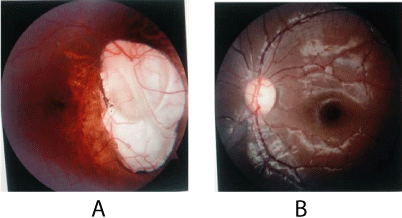
Figure 1. Retinophotography of Right eye (A) and left eye (B). (A): papillary coloboma (green arrow) made of a whitish, well-defined excavation within an enlarged optic disc reaching the retina in its inferomedial part (blue arrow). An irregular pigmented border (arrow in red) is observed at the junction with the normal retina in favor of a type 2 papillo-retinal coloboma including the papilla but which does not exceed it in its upper part according to the Ida Mann classification completed by that of Gopal V where the papilla is in the coloboma and is colobomatous, (B): on the left the fundus is normal
Discussion
The conditions for diagnosing papillary coloboma vary. It can manifest itself as early nystagmus for large chorioretinal or papillary colobomas or be accidentally discovered for smaller ones. In addition, the diagnosis can be established in front of an early strabismus secondary to a unilateral organic amblyopia, for asymmetric or unilateral forms [3] as was the case of this girl who was accompanied by her parents in ophthalmological consultation for a convergent right strabismus. These signs of visual impairment could be explained by the fact that in general the young child does not quickly realize the decrease of visual acuity, especially when it is monocular. Visual acuity depends on the size of the coloboma, the extent of macular involvement, and the associated eyeball anomalies such as nystagmus [3] seen in this clinical case. Macular atrophy with an average thickness to 234 μm associated with macular ectopia and nystagmus in this study, could justify the severe decrease of visual acuity in this girl which was reduced to simple light perception. The diagnosis of coloboma is clinical and most often easy. The appearance of papillary coloboma is that of a whitish excavation, well demarcated within an enlarged optic disc. When the papillary coloboma reaches the retina in its inferomedial part, it is papillo-retinal coloboma exposing the sclera on which the retinal vessels run. At the junction with the normal retina is a pigmented border which is sometimes irregular and of variable width as noted in this clinical case. Thus, it is a type 2 papillo-retinal coloboma including the papilla but which did not exceed it in its upper part according to the Ida Mann classification completed by that of Gopal V where the papilla is in the coloboma and is colobomatous [3]. Histologically, according to the authors [4], there is no choroidal tissue, pigment epithelium or normal retina within the coloboma. Instead, there is a tissue corresponding to an extension of the retina called the intercalated membrane (IM). The benefit of the OCT would be to show the transition between the normal retina and the IM. This limit can be gradual or abrupt. This area could not be highlighted at the OCT in this work, due to the poor acquisition during the performance of this examination. The histological differentiation and the anatomical instability of this area could explain the high frequency of rhegmatogenic retinal detachment associated with papillo-retinal colobomas. Detachment most often occurs from an area of weakness on this junction line. OCT is therefore essential imaging in the monitoring of complications such as rhegmatogenic retinal detachment. The angle between the normal retina and the fundus of the coloboma is also well measured by B-mode eye ultrasound. This could not be performed due to the failure of the probe. Chorioretinal colobomas are characterized by their extreme anatomo-clinical variability. It is important to document coloboma with photographs, OCT and to classify it. The most commonly used classification is that of Ida Mann [3] in seven stages completed by that of Gopal [3], the interest of which is prognostic. In this case, it is a Type 2 from Ida Mann and a Type V from Gopal. Several syndromes involving a coloboma are described. In the first place we have the CHARGE syndrome characterized by a large clinical polymorphism associating coloboma, cardiac anomalies, choanal atresia, growth retardation and delay in development, genital anomalies, anomalies of the ears [3]. In this child, no anomaly was clinically detected.
Differential diagnoses include scars from toxoplasmosis and other infectious agents for chorioretinal colobomas. Bindweed flower papillae and papillo-renal syndrome papillae where the excavation in both cases is centered, would represent the differential diagnosis of papillary colobomas [3]. Colobomatous dimple, double papillae would also be differential diagnoses of papilla colobomas. All these congenital anomalies of the optic papilla must be recognized because they are a source of organic amblyopia with sometimes a functional part requiring early rehabilitation as shown in this clinical case where a strong ametropia was noted. They can also be complicated by detachment of the retina, choroidal neo-vessels hence regular monitoring. They also require a precise initial assessment in search of malformations and / or pathologies which may, be life-threatening. The management of papillo-retinal coloboma would be based on measures to prevent rhegmatogenic retinal detachment. Sports at risk of eye trauma should be prohibited or it should be advisable to practice these sports with plastic-framed glasses in order to limit impacts as much as possible. Essalime [5] reported a case of retinal detachment on chorioretinal coloboma. The early diagnosis of retinal detachment is based on the education of parents to detect the slightest visual anomalies and then of the child to practice self-monitoring of the sight [3], hence the interest of this monitoring instituted in this clinical case [6].
Conclusion
Papillo-retinal coloboma is a rare birth defect of the papilla. It is easy to diagnose clinically and most often manifests itself by early strabismus and decrease of visual acuity. It can cause refractive errors with deep amblyopia.
References
- Brémond-Gignac D, Milazzo S (2011) Les anomalies congénitales du nerf optique de l’enfant. Les Cahiers d'Ophtalmologie 146: 33-34.
- Denis D, Hugo J, Beylerian M, Ramtohul P, Aziz A, et al. (2019) Les anomalies congénitales de la papille. J Fr Ophtalmol 42: 778-789.
- LebranChu P (2017) Pathologie du nerf optique, 533-555, «in» Ophtalmologie pédiatrique, Denis D, Bui Quoc E, Aziz-Alessi A, Paris, Elsevier Masson, 539-539, 2017.
- Diallo S, Bakayoko S, Coulibaly B, Sidibe MK, Guirou N (2018) Colobome chorioretinien bilateral : à propos d’un cas. Pan Af Med J 30: 261.
- Denis D, Girard N, Levy-Mozziconacci A, Berbis J, Matonti F (2013) Colobome oculaire et résultats de l’IRM cérébrale: résultats préliminaires. J Fr Ophtalmol 36: 210-220.
- Essalime K, Mazzouz H, Lahbil D, El Kettanil, Lamaril H, et al. (2008) Décollement de rétine sur colobome choriorétinien : à propos d’un cas. J Fr Ophtalmol 31 Supplément 1: 172.