Abstract
Gossypiboma is an uncommon surgical iatrogenic complication due to the retention of textile material inside natural cavities, especially in abdomen. Most patients are symptomatic, and clinical history and physical examination combined with computed tomography are essential to establish the diagnosis. In response to the presence of a foreign body, the host can develop two different types of inflammatory response: aseptic and septic reaction, determining the most common signs and symptoms. Prevention is the key point to control the complication and is based on a systematic and systematized conference of such materials at the end of an operation. The purpose of this case report is to show the typical case of aseptic reaction, it most frequent clinical course and alert the surgeons about the severity including legacy aspects.
Key words
Gossypiboma, textiloma, post-operative complications
Introduction
Gossypiboma is an operative iatrogenic complication arising from the retention of textile material into natural cavities of the body, particularly in the abdomen. However, the term does not apply to foreign bodies of other nature [1,2].
Textilomas, as these can also be referred to, were first described in 1884 by Wilson, an American surgeon who published 30 cases occurring in the U.S.A and Europe and emphasized the severity of the complication [3]. The incidence of gossypibomas (0.1%/laparotomy) may be underestimated due to neglect of notification in surgery reports, considering the possibility of legal implications under the current judicialization of Medicine [4,5]. In the U.S.A, 1.500 new cases are estimated to occur per year (1 event/300 or 1,500 operations) [4,5]. Gynecological and digestive tract interventions are the main surgeries connected to gossypibomas [6]. According to Dakubo et al., 36% of them occur after gastrointestinal surgeries, 27% after caesarean sections, 18% after hysterectomies, and 13.5% after oncologic surgeries [4].
Most patients are symptomatic, and clinical history, physical examination and imaging methods are essential for diagnosis. The complaints are related to the type of inflammatory reaction developed by the host in response to the presence of a foreign body. Thus, the disease can manifest as abdominal tumors isolated by a fibrous capsule, intestinal obstruction, multiple intestinal fistulae or peritonitis [5].
The purpose of this case report is to alert surgeons about the severity of gossypibomas; review the pathophysiological and diagnosis aspects and highlight the recommended preventive measures.
Case report
A 51-year-old female patient, smoker, suffering from systemic hypertension and diabetes mellitus type 2, sought medical care due to a bulge in the periumbilical region, noticed approximately two months before, after a blunt abdominal trauma caused by falling from her own height. During the preceding months, she had recurrent episodes of postprandial cramping and distension with spontaneous resolution. She also mentioned a cesarean operation 17 years before. The abdomen showed a painless, irreducible periumbilical tumor, fixed to deep plans, without local guarding and tenderness or inflammatory signs on physical examination.
2021 Copyright OAT. All rights reserv
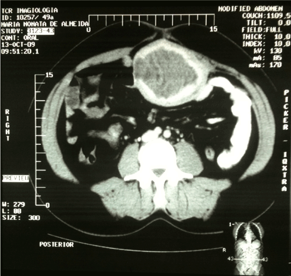
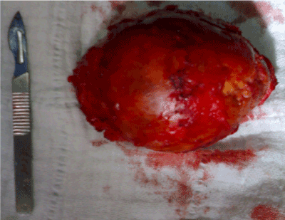
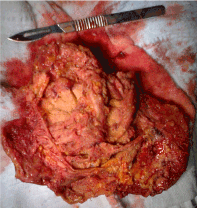
A computed tomography of the abdomen (Figure- 1) showed an expansive lesion with heterogeneous density, well-defined borders, measuring 8.8 x 7.3 cm, with heterogeneous enhancement after intravenous contrast infusion located in close contact with the anterior abdominal wall at the umbilicus level. The patient underwent an exploratory laparotomy, which operative findings confirmed a large well-defined tumor with clear cleavage plane in relation to the wall and abdominal viscera. An important inflammatory process and adhesions were observed between the tumor and intestinal loops. The patient underwent total monoblock tumor excision including the peritoneal-aponeurotic segment (Figures- 2 and 3). Five days after the surgery, intestinal obstruction was diagnosed, and the patient underwent laparotomy for adhesiolysis. The anatomopathological examination of the surgical specimen diagnosed the presence of granulation tissue compatible with an inflammatory reaction to a foreign body involving textile material. The patient was discharged ten days after surgery in a good health, without any other complication and has been on outpatient.

Figure 1. Abdomen CT showing well-limited expansive heterogeneous tumor with a fibrous capsule in close contact with the anterior abdominal wall.

Figure 2. Macroscopic aspect of the surgical specimen showing a solid inflammatory tumor with a well-limited round-shaped dense and fibrotic capsule.

Figure 3. Macroscopic aspect of an open surgical specimen, showing gaps and vacuoles, fulfilled by purulent sterile secretion and the presence of a partially degraded textile foreign body.
Dicussion
The most important manuscript about gossypiboma was published by Crossen in 1940. He studied 307 cases, defined the main features of the disease and proposed ways to prevent its occurrence [6]. According to him, the presence of textile material in serous cavities of the human body can trigger two distinct types of inflammatory reaction: aseptic or pyogenic. The aseptic or fibrinous type of reaction stems from a significant fibroblastic process with adhesions between the sterile textile material and the abdominal viscera without eroding its wall. In most cases, the aseptic reaction gossypiboma remains asymptomatic for a long time, surrounded by a dense inflammatory capsule, which gives it a solid-cystic tumor aspect, as we can see in this case report [7]. The differential diagnosis with others intra-abdominal tumors is crucial to enable proper treatment. However, textile material is microscopically observed and surrounded by granulomatous and fibrotic tissue with dense lymphocytic infiltration and presence of giant cells [5,8,9] (Figures- 1, 2 and 3).
The paper documents the asseptic pathophysiology of the disease and stresses the clinical elements that enable the suspected diagnosis. These include a history of gynecological operation, surgical scars and a palpable abdominal tumor without local guarding and tenderness. The tomography represents the gold standard approach to diagnose the gossypiboma [1]. The typical finding is a spongiform appearance with small air bubbles inside, giving it a dark image contrasting with white parts surrounding them. Another feature is the presence of a high-density thin capsule around a non-homogeneous low-density content enhanced after injection of a contrast medium [5].
Surgical morbidity ranges from 18 - 62%, and infection is the most common complication. Mortality rate ranges from 11 - 35%, in accordance to the size of the sample analyzed and foreign body stay time. In addition, patients under high surgical risk, with aseptic gossypibomas near to vital structures and few clinical symptoms, could be followed up clinically [5,10,11]. The most important way to manage the gossypibomas is its prevention, what relies on simple operative measures within the reach of all surgeons. The re-interventions for treatment of this surgical complication are always difficult. Therefore, it is recommended its withdrawal early in the postoperative course, when observing only a foreign body lock without integration with the surrounding tissues.
The use of radiopaque marked textile material; systematic and systematized abdominal cavity review; conference of the compresses used before peritoneal synthesis; not use of small compresses or gauze; not use of compresses to help abdominal wall synthesis are simple and fundamentals strategies that have to be assimilated by all surgical team. In case of doubt, a X-ray before closing the wall in the operate room should be done [11]. At the same time, the Surgeon needs to be alert for potential factors that predispose to complication which include: obesity, emergency operations, intraoperative hemorrhage, long surgery time, surgical team shifting, and operations at difficult access sites [5,7].
Therefore, although such an error is of human nature, gossypiboma is a severe underreported disease arising from operative negligence by the surgical team with potential legal implications. It can be avoided by simple coordinated intra-operative measures, for which surgeon's leadership and appropriate team training are of utmost importance.
Acknowledgment
The authors would like to thank MD. Sandra Marcia Carvalho Ribeiro Costa for her considerable help in the histological assessment of the removed specimens.
Conflict of interest disclosure statement
The authors have no conflicts of interest ties or financial funding source's to disclose.
References
- Alayo E, Attar B, Benjamin GO (2010) A case of recurrent abdominal pain due to a gossypiboma with spontaneous resolution. Clinical Gastroenterology and Hepatology 8: 13-14.
- Aminian A (2008) Gossypiboma: A case-report. Cases Journal 1: 201-203.
- Mefire AC, Tchounzou R, Guifo ML, Fokou M, Pagbe JJ, et al. (2009) Retained sponge after abdominal surgery: experience of a third world country. Panafrican Medical Journal 2:10.
- Dakubo J, Clegg-Lamprey JN, Hodasi WM, Obaka HE, Toboh H, et al. (2009) An intra abdominal gossypiboma. Ghana Medical Journal 43: 43-45.
- Iglesias AC, Salomão RM (2007) Gossipiboma abdominal – Análise de 15 casos. Revista do Colégio Brasileiro de Cirurgiões 34: 106-113.
- Debnath D, Buxton JK, Koruth NM (2005) Two years of wait and 7000 miles of journey: the tale of a gossypiboma. Int Surg 90: 130-133. [Crossref]
- Sharma D, Pratap A, Tandon A, Shukla RC, Shukla VK (2008) Unconsidered cause of bowel obstruction – Gossypiboma. Can J Surg 51: 34-35. [Crossref]
- Kaiser CW, Friedman S, Spurling KP, Slowick T, Kaiser HA (1996) The retained surgical sponge. Ann Surg 224: 79-84. [Crossref]
- Akbulut S, Sevenic MM, Basak F, Aksoy S, Cakabay B (2009) Transmural migration of a compress to the stomach after splenectomy- A case report. Cases Journal 2:75-79.
- Gencosmanoglu R, Inceoglu R (2003) An unusual cause of small bowel obstruction: gossypiboma--case report. BMC Surg 3: 6. [Crossref]
- Lauwers PR, Van Hee RH (2000) Intraperitoneal gossypibomas: the need to count sponges. World J Surg 24: 521-527. [Crossref]


