The influence of the treatment of the surface of IPS Empress II (emax) glass-ceramic dental material with different silanization protocols on the surface behavior during the cementation with tooth dentine was evaluated. IPS Empress II (emax) veneers were treated with different silanization protocols used in dentistry nowadays. For the cementation with tooth dentine were used two different cements and one flow composite resin generally used for tooth resin restorations. After the cementation the interfaces were observed with a scanning electron microscope (SEM). The observation subject was the perfection or not of the cementation along the connection of the cement with IPS Empress II (e-max) and tooth dentine and how the silane treatment of the ceramic surface reacted to the final cementation appearance.
dental materials, IPS Empress II (e max), silane agents, silanization protocols, resin cements, cementation, interface
The evolution of dental-technology is closely bound up with the progress in ceramics. Thus, several high quality dental ceramics and glass-ceramics are currently available, such as Reflex, IPS Empress I, IPS Empress II, Vitablocks, In Ceram, etc. These materials display outstanding features, in terms of their chemical, mechanical, and aesthetic properties, resulting from their chemical composition, inspired processing, and fine microstructure.
It is a well-known fact in dental practice that the surface treatment of both the ceramic part and the dentine matter of the tooth strongly influence the cementation [1]. More specifically, a literature survey shows that etching with hydrofluoric acid (HF) markedly increases the bond strength. Actually, this etching method is considered to be more effective than other methods, such as sand-blast, or laser, for example [2,3]. Besides that, silane treatment applied on the ceramic surface has a very good effect as to increasing the surface roughness and consequently, on bond strength with the natural dentine of the tooth [4]. However, the dentists should precisely follow the protocols suggested by the manufacturers of each type of silane.
Therefore, the aim of this study is to shed light on the influence of the treatment of the surface of IPS Empress II (e-max) glass-ceramic dental material with one silane agent at different time of silanization protocols and how this affects the cementation process with two different resin cements and one flow composite resin, with the tooth dentine.
Natural, fresh extracted, third molars were prepared with dental high-speed handpiece under water spray for IPS Empress II (e-max) veneers. An impression copy was taken, and a cast was made of the prepared surfaces. Cores of IPS Empress II (e-max) were pressed and ceramic veneers were constracted individual for each tooth that was prepared. For acid etching, HF aqueous solution 10% was used for the ceramic surfaces and H3(PO4)2 aqueous solution 35% for the teeth.
As far as the silanization process is concerned, the following silane agent was used; Ceramic Primer Plus (by Kuraray, Tokio).
For the cementation process the resin cements that they were used are; Panavia F (by Kuraray), Panavia V5 (by Kuraray, Tokio). As a third cementation agent HFO Enamel Plus (by Micerium, Italy) flow composite resin was used with the additional use of dentin primer and an adhesive system known as FL primer/adhesive (by Kerr, Germany)
Nine IPS Empress II (e-max) veneers were manufactured by the dental lab that were etched with HF solution for 30s and washed with distilled water for 10 s and finally air dried. After the etching, the etched surfaces of the glass-ceramic samples were subjected to silanization treatment (6 samples) and no silane treatment (3 samples). For the silane agent two protocols were followed (1) silane treatment for 20s and then being air dried and (2) silane treatment for 24 hours and then being air dried.
The veneers were permanetaly cemented and light-cured with dental LED lamb (900-1100 W) for 40s each one separately with finger pressure for stabilization on the tooth. Using a dental high speed handpiece under a water spray the restored teeth were dinided into two pieces so it could be able to observe the cemented interface under a scanning electron microscope (SEM).
The interface samples that we created for scanning electron microscope (SEM) and finally observed followed the below silanization and cementation protocols:
- No silane treatment on IPS Empress II (e-max) with ED Primer II for tooth dentine and cementation with Panavia F 2.0 (Figure 1)
- Silanization with Ceramic Primer Plus for 20 s on IPS Empress II (e-max) with ED Primer II for tooth dentine and cementation with Panavia F 2.0 (Figure 2)
- Silanization with Ceramic Primer Plus for 24 hours on IPS Empress II (e-max) with ED Primer II for tooth dentine and cementation with Panavia F 2.0 (Figure 3)
- No silane treatment on IPS Empress II (e-max) with Tooth Primer for dentine and cementation with Panavia V5 (Figure 4)
- Silanization with Ceramic Primer Plus for 20 s on IPS Empress II (e-max) with Tooth Primer on dentine and cementation with Panavia V5 (Figure 5)
- Silanization with Ceramic Primer Plus for 24 hours on IPS Empress II (e-max) with Tooth Primer for dentine and cementation with Panavia V5 (Figure 6)
- No silane treatment on IPS Empress II (e-max) with FL primer/adhesive system for dentin and cementation with HFO Enamel Plus flow composite resin (Figure 7)
- Silanization with Ceramic Primer Plus for 20 s on IPS Empress II (e-max) with FL primer/adhesive system for dentine and cementation with HFO Enamel Plus flow composite resin (Figure 8)
- Silanization with Ceramic Primer Plus for 24 hours on IPS Empress II (e-max) with FL primer/adhesine system for dentine and cementation with HFO Enamel Plus flow composite resin (Figure 9)
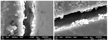
Figure 1. No silane treatment on IPS Empress II (e-max) with ED Primer II for tooth dentine and cementation with Panavia F 2.0
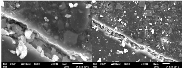
Figure 2. Silanization with Ceramic Primer Plus for 20 s on IPS Empress II (e-max) with ED Primer II for tooth dentine and cementation with Panavia F 2.0
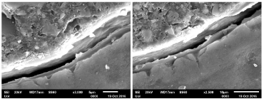
Figure 3. Silanization with Ceramic Primer Plus for 24 hours on IPS Empress II (e-max) with ED Primer II for tooth dentine and cementation with Panavia F 2.0
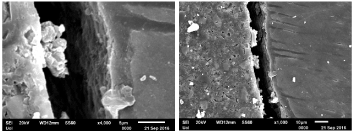
Figure 4. No silane treatment on IPS Empress II (e-max) with Tooth Primer for dentine and cementation with Panavia V5

Figure 5. Silanization with Ceramic Primer Plus for 20 s on IPS Empress II (e-max) with Tooth Primer on dentine and cementation with Panavia V5
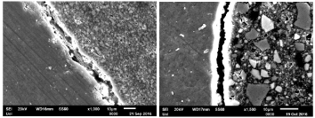
Figure 6. Silanization with Ceramic Primer Plus for 24 hours on IPS Empress II (e-max) with Tooth Primer for dentine and cementation with Panavia V5
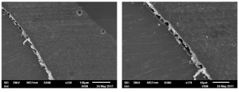
Figure 7. No silane treatment on IPS Empress II (e-max) with FL primer/ adhesive system for dentin and cementation with HFO Enamel Plus flow composite resin
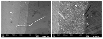
Figure 8. Silanization with Ceramic Primer Plus for 20 s on IPS Empress II ( e-max) with FL primer/ adhesive system for dentine and cementation with HFO Enamel Plus flow composite resin
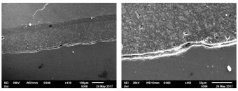
Figure 9. Silanization with Ceramic Primer Plus for 24 hours on IPS Empress II (e-max) with FL primer/ adhesine system for dentine and cementation with HFO Enamel Plus flow composite resin
After the observation with scanning electron microscope (SEM) the pictures that where taken following the above protocols are:
After the observation with scanning electron microscope (SEM) it was revieled that the cementation of IPS Empress II (e-max) has some serious issues when we talk about the contact with dentine matter of the tooth. Specifically, with the cementation protocols that we have followed some of the interfaces resulted bad homogeneity as cemented parts of a restored tooth
At these samples we didn’t succeed an absolute cementation between the glass-ceramic and tooth dentine despites these protocols are in common use as a daily routine at any dental practice. Further investigation with the EDS-spectrum for the elemental analysis of the cemented interface of the IPS Empress II (e-max) and the dentine matter of the tooth showed us that at the gap area of the cement and the dentine, there was not no elemental existence. (Figures 10-12) That describes a lack of the cementation process to connect absolutely the dentine matter of the tooth with the cement surface. So, the quality of the cementation at the above protocols is not the absolute that we expected to be.
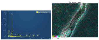
Figure 10. The EDS-spectrum for the elemental analysis of the cemented interface of the IPS Empress II (e-max)
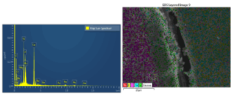
Figure 11. The dentine matter of the tooth showed us that at the gap area of the cement
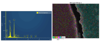
Figure 12. The dentine, there was not no elemental existence
Nevertheless, at the same time we found out the best quality at the cemented interface by following the exact protocol that the manufactures propose for these kinds of cementation (Figure 5). We have managed to achive the highest level of homogeneity among the IPS Empress II (e-max) surface, the cement and the dentine matter of the tooth without revealing any gaps. The EDS-spectrum analysis showed the further absence of elemental gaps (Figure 13). This protocol is: Silanization with Ceramic Primer Plus for 20s on IPS Empress II (e-max) surface with Tooth Primer on dentine and cementation with Panavia V5 (kuraray, Tokio).
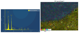
Figure 13. The EDS-spectrum analysis showed the further absence of elemental gaps
A different cementation protocol achieved a high cementation quality (Figure 8) nevertheless it has to be under further research procedure since it doesn’t follow the daily dental practice routine.
As far as the main aim of this study is concerned the properties of using a silane agent is nevertheless an advance for the cementation procedure between IPS Empress II (e-max) and the tooth dentine [5].
As it is concerned in a dental-practice daily routine, the quality that it can be achieved from the cementation process between a glass-ceramic system and tooth dentine is main factor for a strong and long-term stability for our restorations [6,7]. The results from the observation of the cemented interfaces of IPS Empress II (e-max) and tooth dentine recorded a chemical reaction among the substances that participate to the cementation process with no question about the importance about the silane treatment. More investigation is needed about the bond-strength in each case of silane treatment and cementation protocol, so that we would be able to end with the most secure and stable way to cement IPS Empress II (e-max) with tooth dentine.
- Elsaka SE (2014) Bond strength of novel CAD/CAM restorative materials to self –adhesive resin cement: the effect of surface treatments. J Adhes Dent 16: 531-540. [Crossref]
- Della Bona A, Donassollo TA, Demarco FF, Barrett AA, Mecholsky Jr JJ, et al. (2007) Characterization and surface treatment effects on topography of a glass-intiltrated alumina/zirconia-reinforced ceramic. Dent Mater 23: 769-775. [Crossref]
- Chaiyabutr Y, Mc Gowan S, Phillips KM, Kois JC, Giordano RA, et al. (2008) The effect of hydrofluoric acid surface treatment and bond strength of a zirconia veneering ceramic. J Prosthet Dent 100: 194-202. [Crossref]
- Queiros JR, Benetti P, Ozcan M, De Oliveira LF, Della Bona A, et al. (2012) Surface characterization of feldspathic ceramic, using ATR FT-IR and ellipsometry after various silanization protocols. Dent Mater 28: 189-196. [Crossref]
- Pekkan G, Hekimoglou C (2009) Evaluation of shear and tensile bond strength between dentin and ceramics using dual-polymerizing resin. J Prosthod Dent 102: 242-251. [Crossref]
- Shimada Y, Yamagushi S, Tagami J (2002) Micro-shear bond strength of dual-cured resin cement to glass ceramics. Dental Materials 18: 380-388
- Guarda GB, Goncalves LS, Correr AB, Moraes RR, Sinhoreti MAC, et al. (2010) Luting glass ceramic restorations using a self-adhesive resin cement under different dentin conditions. J Appl Ora Sci 18: 244-248. [Crossref]