Clinical Image
A three month old boy was referred by his pediatrician at the age of 3 months for intermittent inspiratory stridor since birth which was more in supine position. There was a history of preterm delivery at 35 weeks of gestation and child was discharged a week later. There is no history of any feeding difficulty or history of apnea or cyanosis. On examination, the child had mild inspiratory stridor with no distress. Child had no retrognathia or craniofacial dysmorphism. Flexible fiberoptic nasopharyngolaryngoscopy revealed omega-shaped epiglottis, foreshortened aryepiglottic folds, prolapsing arytenoids during inspiration with inability to visualise vocal cords (Figure 1). A clinical diagnosis of classical laryngomalacia was made. The parents were reassured about the condition and advised to be in close follow up.
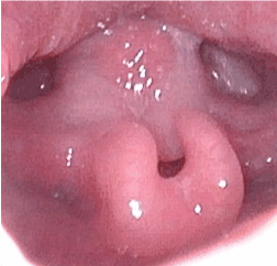
Figure 1. Flexible fiberoptic nasopharyngolaryngoscopy revealing omega-shaped epiglottis, foreshortened aryepiglottic folds, prolapsing arytenoids with inability to visualize vocal cords.
Laryngomalacia, the most common cause of stridor in newborns, affects 45-75% of all infants with congenital stridor [1]. It presents with mild inspiratory stridor that can worsen with feeding, crying, supine positioning, and agitation. The symptoms begin at birth or within the first few weeks of life and is usually diagnosed within the first 4 months of life [2]. Chronic hypoxia from airway obstruction can lead to pulmonary hypertension if not recognized early and managed. Although other conditions like tracheomalacia, asthma, bronchiolitis, and reactive airway disease may mimic layngomalcia, the diagnosis of laryngomalacia is suspected by the typical clinical history and it should be confirmed by flexible laryngoscopy in an awake infant. It can be associated with gastroesophageal reflux disease (GERD), neurologic disease, congenital heart disease, part of a syndrome or genetic disorder [3]. Those with mild disease can be managed expectantly. The most common indications for surgery are stridor with respiratory compromise and feeding difficulties with failure to thrive. Although laser supraglottoplasty is the mainstay surgical treatment, tracheotomy is reserved in children who continue to have life-threatening airway obstruction and who fail to improve after supraglottoplasty [1,2].
Consent
Informed written consent has been obtained from the parents for publication of the image.
References
- Richter GT, Thompson DM (2008) The surgical management of laryngomalacia. Otolaryngol Clin North Am 41: 837-864. [Crossref]
- Landry AM, Thompson DM (2012) Laryngomalacia: disease presentation, spectrum, and management. Int J Pediatr: 753526.
- Dickson JM, Richter GT, Meinzen-Derr J, Rutter MJ, Thompson DM (2009) Secondary airway lesions in infants with laryngomalacia. Ann Otol Rhinol Laryngol 118: 37-43. [Crossref]