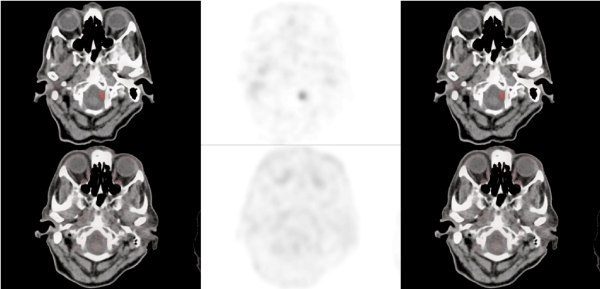
A female patient who was diagnosed as pancreatic neuroendocrine tumor and the primary tumor was surgically removed. The Ga-68 DOTATATE PET/CT imaging which was performed for tumor staging revealed significant uptake in the brain steam. Additional FDG PET/CT showed no increased uptake at the site of the lesion. MR could not exclude the diagnosis of metastasis. However follow up imaging showed no change in the tumor size thus diagnosed as meningioma as a Ga-68 DOTATATE accumulating lesion.
meningioma, Ga-68, DOTATAE, FDG, PET.
Neuroendocrine tumors usually accumulate Ga-68 DOTATATE PET/CT in a significant amount depending on the several factors related to the receptor status of the tumor. In recent years Ga-68 DOTATATE imaging is the important part of staging algorithm in neuroendocrine tumors. As a whole body imaging modality unexpected sites of metastasis as well as frequent sites could be demonstrated [1]. In a previous case series including 4210 patients; 6 patients were found to have brain metastasis [1].
Several benign lesions of the brain also demonstrate significant amount of Ga-68 DOTATATE due to the presence of somatostatine receptors. In a previous analysis including optic pathway tumors Ga-68 DOTATATE PET/CT imaging identified 8 of the 10 meningiomas successfully and influenced therapy decision in various tumors [2]. Meningiomas are benign tumors which is common in adult women and usually do not require treatment [3]. There was previous report of cases about incidental detection of meningiomas in the literature [4]. This is the first report of a case presented as incidentally detected menengioma by Ga-68 DOTATATE imaging.
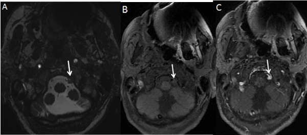
62 years old female patient presented with primary pancreas carcinoma was operated. Due to the pathology of neuroendocrine tumor primary staging with Ga-68 DOTATATE and F-18 FDG PET/CT was performed. Ga-68 DOTATATE images showed atypical brain steam uptake as a focus (Figure 1a). F-18 FDG PET/CT did not show increased activity in the lesion (Figure 1b). Further MR imaging showed the lesion but could not verify or exclude metastasis of neuroendocrine tumor at first imaging. However, second follow up imaging showed no change in tumor size thus diagnosis of benign tumor with high probability of meningioma was decided (Figure 2).

Figure 1. Ga-68 DOTATATE CT, PET and fusion PET/CT respectively images of the patients in axial projection demonstrates the increased activity in in left lateral brain steam region (A: upper line) however F-18 FDG PET/CT images do not show FDG uptake at the lesion site (B)

Figure 2. Axial Fiesta (A) and T1-weighted (B) images at foramen magnum level show a lesion (arrow) adjacent to the left hypoglossal canal and vertebral artery. Axial contrast-enhanced T1-weighted image (C) demonstrates homogeneously enhancing lesion (arrow) with dural tail
Meningiomas usually accumulate Ga-68 DOTATATE due to presense of SSTR 2 and affinity of the agent to the receptor. However, there are exceptional cases that do not show increased activity on Ga-68 DOTATATE [5]. Previous reports demonstrated higher sensitivity for the detection of meningiomas with Ga-68 DOTATATE PET/CT compared to MR especially in special cases in the problematic localizations [5]. Furthermore Y-90 DOTATOC treatment has been implicated in this group of patients with successful results [6]. Meningiomas are usually benign tumors and do not require treatment. However if the patient is symptomatic or radiological progression occurs there are treatment options. These ‘nonbenign menegioma’s might be evaluated by means of Ga-68 DOTATATE PET/CT as a pretreatment imaging method [7]. This imaging modality has prognostic value in the preoperative imaging of this neoplasm which can predict risk of recurrence according to a previous study [7]. Another study compared the value of intra arterial versus intravenous methods of Ga-68 DOTA imaging in patients with inoperable meningioma and achieved significantly increased accumulation [8].
A single previous case report including incidental detection of meningioma in the medulla spinalis by Ga-68 DOTATATE in a patient with neuroendocrine tumor has been presented [9]. However this is the report of first case with a single focus of meningioma mimicking metastasis of a known neuroendocrine tumor. It is important to be aware of potential pitfalls in Ga-68 DOTATATE imaging in order to provide true management of the patients with neuroendocrine tumors.
- Carreras C,Kulkarni HR,Baum RP (2013) Rare metastases detected by (68)Ga-somatostatin receptor PET/CT in patients with neuroendocrine tumors. Recent Results Cancer Res 194:379-384.
- Klingenstein A,Haug AR, Miller C, Hintschich C (2015) Ga-68-DOTA-TATE PET/CT for discrimination of tumors of the optic pathway. Orbit 34(1):16-22.
- WilhelmH (2013) Meningioma of the anterior visual pathways: Epidemiology and clinical symptoms. Ophthalmologe110:403-407
- VernooijMW,IkramMA,TangheHL (2007)Incidental findings on brain MRI in the general population. N Engl J Med357:1821-1828.
- Afshar-Oromieh A,Giesel FL,Linhart HG, Haberkorn U, Haufe S, Combs SE, et al. (2012) Detection of cranial meningiomas: Comparison of ⁶⁸Ga-DOTATOC PET/CT and contrast-enhanced MRI. Eur J Nucl Med Mol Imaging 39(9):1409-1415.
- Bartolomei M,Bodei L,De Cicco C, Grana CM, Cremonesi M, Botteri E, et al. (2009) Peptide receptor radionuclide therapy with (90)Y-DOTATOC in recurrent meningioma. Eur J Nucl Med Mol Imaging 36(9):1407-1416.
- Pelak MJ, d'Amico A (2019) The prognostic value of pretreatment Gallium-68DOTATATEpositron emission tomography/computed tomography in ırradiated non-benignmeningioma. Indian J Nucl Med 34(4):278-283.
- Verburg FA, Wiessmann M, Neuloh G, Mottaghy FM, Brockmann MA (2019) Intraindividual comparison of selective intraarterial versus systemic intravenous 68Ga-DOTATATEPET/CT in patients with inoperablemeningioma. Nuklearmedizin 58(1):23-27.
- Klinaki I,Al-Nahhas A,Soneji N,Win Z (2013) 68Ga DOTATATE PET/CT uptake in spinal lesions and MRI correlation on a patient with neuroendocrine tumor: potential pitfalls. Clin Nucl Med 38(12):e449-53.
Editorial Information
Editor-in-Chief
Article Type
Image Article
Publication history
Received: November 26, 2019
Accepted: December 09, 2019
Published: December 12, 2019
Copyright
©2019 Koç ZP. This is an open-access article distributed under the terms of the Creative Commons Attribution License, which permits unrestricted use, distribution, and reproduction in any medium, provided the original author and source are credited.
Citation
Koç ZP, Özcan PP, Sezer E, Özgür A (2019) Incidental detection of brain steam meningioma by Ga-68 DOTATATE PET/CT in a patient with neuroendocrine tumor. Glob Imaging Insights 4: DOI: 10.15761/GII.1000188
Corresponding author
Zehra Pınar Koç, MD, Prof
Mersin University Nuclear Medicine Department, Mersin/Turkey
E-mail : bhuvaneswari.bibleraaj@uhsm.nhs.uk


