Gout is frequently associated with nephrolithiasis. Uric acid nephrolithiasis in gout correlates with the degree of hyperuricemia and hyperuricosuria. A decrease in serum and tissue uric acid burden may result in improvement in uric acid nephrolithiasis in patients with gout. We present a case of severe chronic tophaceous gout with recurrent uric acid nephrolithiasis who had a remarkable improvement in his gout, as well as the resolution of uric acid renal calculi after treatment with pegloticase. Pegloticase has been approved for the treatment of chronic gout refractory to conventional therapy, however, its effects on uric acid nephrolithiasis have not been evaluated. Our case is first such reported case, and further studies evaluating the efficacy of pegloticase in improving uric acid calculi are needed to confirm our findings.
pegloticase, nephrolithiasis, gout
A 68-year-old Hmong man with a past history of chronic erosive tophaceous gout presented to the rheumatology clinic to establish care. His past medical history was significant for hypertension, chronic kidney disease, diabetes mellitus – type 2, and multiple episodes of nephrolithiasis, confirmed to be uric acid calculi on previous analyses. He reported multiple acute attacks of gout involving several joints at a time every month, needing frequent courses of corticosteroids. He had been on Febuxostat 80 mg daily and colchicine 0.6 mg daily for the past 11 months for management of his gout. He had previously been treated with allopurinol up to 800 mg daily without significant improvement in his urate burden. On exam, he had multiple soft tophaceous deposits on the elbows, wrists, hands, and feet bilaterally with active synovitis with erythema, warmth, and tenderness of left knee, left ankle and bilateral first metatarsophalangeal joints. Synovial fluid analysis from the left knee revealed intracellular and extracellular monosodium urate crystals with 15,000 polymorphonuclear cells per high power field. Laboratory workup revealed serum uric acid of 9.2 mg/dL and creatinine 1.5 mg/dL. C-reactive protein was 67 mg/L. He was started on prednisone 40 mg daily for the management of acute polyarticular gout attack, with which, his gout attack resolved after 10 days. He was then started on pegloticase 8mg/kg infusions every 2 weeks in addition to prednisone 5 mg daily and colchicine 0.6 mg daily and his febuxostat was discontinued.
3 months later, a remarkable improvement was noted in his tophaceous burden with the resolution of several tophi and reduction in the size of others. Serum uric acid was maintained at less than 0.2 mg/dL. He had 3 more gout attacks over the course of 6 months after starting pegloticase, all of which were successfully treated with oral corticosteroid courses. 12 months after initiating pegloticase, all of his tophi had resolved, and he remained acute attack free. Notably, the patient also reported no episodes of nephrolithiasis after initiating pegloticase. A computed tomography (CT) scan and ultrasound 3 months before initiating pegloticase had revealed several renal calculi bilaterally (Figure 1), while repeat CT scan and ultrasound 8 months after initiating pegloticase revealed resolution of all previous renal calculi with no new calculi (Figure 2).
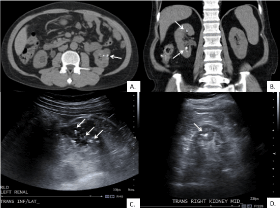
Figure 1. Images 3 months before initiating Pegloticase. (A) Axial CT scan with multiple renal calculi in the left kidney. (B) Coronal CT scan with multiple renal calculi in the right kidney. (C) Transverse ultrasound with multiple renal calculi in the left kidney. (D) Transverse ultrasound with multiple renal calculi in the right kidney. (Uric acid calculi marked by arrows)
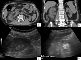
Figure 2. Images 8 months after initiating Pegloticase. No renal calculi seen at the same levels as Figure 1 on (A) Axial CT scan, (B) Coronal CT scan, (C) Transverse ultrasound of left kidney, and (D) Transverse ultrasound of right kidney
Gout is associated with nephrolithiasis in up to 25% of the cases, about 50% of which are asymptomatic [1]. The likelihood of uric acid nephrolithiasis correlates with the degree of hyperuricemia and hyperuricosuria. Hyperuricemia results from either overproduction or more commonly underexcretion of uric acid, which is the end product of purine metabolism. During the evolution, the humans lost the urate oxidase gene, a consequence of two independent mutations [2]. In mammals containing the urate oxidases, uric acid is converted to water-soluble allantoin, thus accounting for plasma concentrations of uric acid 5-10 times higher in humans compared to them. Pegloticase is recombinant PEGylated Uricase which converts uric acid to allantoin, thus rapidly resulting in a decrease in serum uric acid levels.
Pegloticase is approved for the treatment of gout in patients with chronic gout, refractory to conventional urate-lowering therapy [3]. The use of pegloticase leads to a rapid decline in serum uric acid levels within 24 hours [4]. As in our case, a complete resolution of tophi has been reported in more than 40% of patients within 13 weeks of therapy with pegloticase [5]. So far, there is no available data on improvement in uric acid nephrolithiasis in gout associated with pegloticase, and ours is the first such case. Rapid and persistent reduction in the serum and tissue uric acid burden due to pegloticase is likely responsible for the improvement in uric acid calculi. The cost of pegloticase therapy can be a prohibitory factor in its use even in patients where it may be indicated, and the cost-effectiveness of pegloticase in gout needs to be established. Further studies evaluating the efficacy of pegloticase in reducing uric acid calculi burden in patients with gout are needed to confirm the findings from our case.
- Shimizu T, Hori H (2009) The prevalence of nephrolithiasis in patients with primary gout: a cross-sectional study using helical computed tomography. J Rheumatol 36: 1958-1962.
- Oda M, Satta Y, Takenaka O, Takahata N (2002) Loss of urate oxidase activity in hominoids and its evolutionary implications. Mol Biol Evol 19: 640-653.
- http://www.accessdata.fda.gov/drugsatfdadocs/label/2010/125293s0000lbl.pdf
- Efficacy and safety of intravenous (IV) pegloticase (PGL) in treatment failure gout (TFG): results from GOUT 1 and GOUT 2 [abstract]. Ann Rheum Dis 68: S318.
- Baraf HS, Becker MA, Edwards NL, Gutierrez-Urena SR, Sundy JS, et al. (2008) Tophus response to pegloticase (PGL) therapy: Pooled results from GOUT1 and GOUT2, PGL phase 3 randomized, double blind, placebo-controlled trials. Arthritis Rheum 58: S176-S176.