Case report
Tricuspid pathology is usually associated with valvular disease of the left side of the heart. Its isolated presentation has a lower incidence. In any case its occurrence is related to a worsening of the patient's functional grade and a high mortality rate if it is not repaired or replaced [1-3].
Intervention of the tricuspid valve is one of the current challenges in the field of cardiac surgery. Its results are diverse depending on the series, related in part to its complex anatomy and, to a greater extent, due to its lower incidence and experience and to the change in its approach through the therapeutic strategy in recent years [1,4].
Compared to repair by annuloplasty, replacement of the tricuspid valve with implantation of mechanical or biological prosthesis leads to a higher mortality rate, although it associates with a lower incidence of residual tricuspid insufficiency. Any of both techniques may be chosen in patients with this type of disease [1,3,5]. Reintervention in this group of patients means a higher incidence of postoperative right ventricular insufficiency and a high morbidity and mortality rate (37% during hospitalization) [2,6,7].
Percutaneous techniques have become a reference in the treatment of aortic valve disease in patients with high perioperative risk, reducing their morbidity and mortality [7,8]. Currently, the possibility of performing percutaneous tricuspid valve replacement on already implanted valve prosthesis opens a new door in this group of patients, and could become the technique of choice in the coming years.
Our case was a 59-year-old woman with a personal history of hypertension, mild renal impairment (glomerular filtration rate of 50 ml/min), and long-standing right heart decompensation with diagnosis of isolated severe tricuspid insufficiency which was operated a year ago with the implant of a tricuspid valve (Carpentier 29).
After surgery patient presented new symptoms of right heart decompensation, with the diagnosis of tricuspid prosthetic dysfunction with isolated severe regurgitation associated with moderate-severe right ventricular dilation and preserved left ventricular function (EFLV 65%,), without any other echocardiographic finding. At operating room patients were monitored with 5-lead electrocardiogram, radial artery, pulseoximetry, capnography, urine bladder temperature and hypnosis (BIS XP®, Aspect Medical Systems, Newton, MA). Cardiac function was assessed with 3D echochardiography.
Intervention was performed under general anesthesia. During this the selected access for the placement of the tricuspid prosthesis was the femoral venous, central access scheduled was the right internal jugular. It had a dual objective, first to maintain a central access for the possible administration of amines, and second as an access option for the placement of the valve if technically the angulation of the different catheters in the right cavities from the femoral vein made impossible this technique. During the intervention, a 29mm Edwards-Sapiens 3 valve was implanted, requiring pacing to induce a transvalvular flow cessation during valve deployment and anchoring through stimulation of the interventricular septum.
2021 Copyright OAT. All rights reserv
There were no incidents with the selected access, nor with the placement and anchoring of the valve in its position. Echocardiogram allowed to evaluate the correct position of the valve and the ceasing of transvalvular gradients after implantation. At hemodynamic level there was an episode of transient hypotension during the introduction of the device containing the valve into the right cavities prior to its deployment, due to the decrease in venous return, which was resolved with the administration of 250 ml Ringer Lactate and 10 mg ephedrine. At the end of the procedure patient was transferred to the intensive care unit, where she was extubated at 6 hours and discharged at 48 hours without any incident. In the echocardiographic control at the end of the procedure it was emphasized the absence of significant transvalvular gradients, periprosthetic leakage or insufficiency.
Tricuspid valve disease has increased its importance in recent years, due to its high morbidity and mortality when manifested in isolation or associated with left valvular disease [9,10]. The latest studies on patients with tricuspid insufficiency secondary to left valve dysfunction, in whom surgical treatment of the tricuspid valve was not performed, show that in 60% of the cases there is no regression of the disease, which has made much more frequent the surgery on this valve affected as primary or secondary [1,4]. The perioperative mortality of this disease is high and is associated in a significant way with the patient's previous pathology, the cardiological status at the moment of the intervention, and the time of extracorporeal circulation and ischemia [2,6,11].
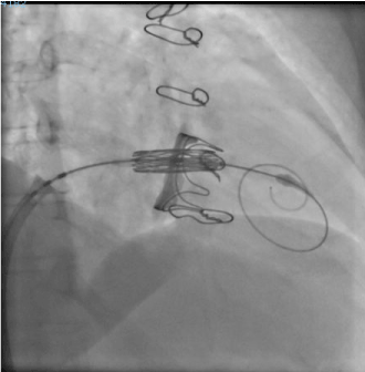
Figure 1. In the image appears the tricuspid valve expanded after radiographic control.
Patients requiring further surgical reoperation have a high morbidity and mortality with a poor postoperative prognosis in the short and medium term [5,12]. This is the reason why the possibility of performing this intervention percutaneously could open the doors to a greater survival in this group of patients. The main limitation of the technique is the need for a prior prosthetic valve or ring to be able to effectively deploy the new valve, being impossible on the native valve.
Regarding the perioperative management, several considerations are necessary. First the need to determine vascular access. If it is decided by the femoral, as was our case, the channeling of the right internal jugular as central access, allowed us to have this way as percutaneous access of rescue for the technique. The need to perform pacing for the cessation of transvalvular flow at the time of deployment and anchoring of the valve can be done through one of the guides used during the technique, supporting it on the interventricular septum and stimulating with a pacemaker generator or pharmacological treatment with adenosine, being more predictable in duration and intensity the first of them, which was the one realized in our patient.
Finally, the introduction of the valve-carrying device of significant size through the upper vena cava reduces venous return and produces transient hypotension that may require the delivery of inotropic and/or vasoactive support and/or fluid therapy support. Patients with indication for surgical reoperation following substitution surgery or tricuspid annuloplasty due to severe valvular dysfunction after previous intervention have a high risk and perioperative mortality. An alternative could be the percutaneous treatment of valvular dysfunction, being this one a lower complexity and risk procedure for this type of patients [8,13,14].
References
- Shah PM (2010) Tricuspid and pulmonary valve disease evaluation and management. Rev Esp Cardiol 63: 1349-1365. [Crossref]
- Rodríguez-Capitán J, Gómez-Doblas JJ, Fernández-López L, López Salguero R, Ruiz M, Leruite I, et al. (2013) Cirugía de la regurgitación tricuspídea grave: resultados a corto y largo plazo. Rev Esp Cardiol 66: 629-635.
- González-Santos JM, Arnáiz-García ME (2013) La corrección de la insuficiencia tricuspídea: una cuestión por resolver. Rev Esp Cardiol 66: 609-612.
- Irwin RB, Luckie M, Khattar RS (2010) Tricuspid regurgitation: contemporary management of a neglected valvular lesion. Postgrad Med J 86: 648-655. [Crossref]
- Bernal JM, Morales D, Revuelta C, Llorca J, Gutiérrez-Morlote J, et al. (2005) Reoperations after tricuspid valve repair. J Thorac Cardiovasc Surg 130: 498-503. [Crossref]
- Vassileva CM, Shabosky J, Boley T, Markwell S, Hazelrigg S (2012) Tricuspid valve surgery: the past 10 years from the Nationwide Inpatient Sample (NIS) database. J Thorac Cardiovasc Surg 143: 1042-1049. [Crossref]
- Weich H (2012) Transcatheter tricuspid valve replacement. Interventional Cardiology 7: 59-62.
- Schofer J, Bijuklic K, Tiburtius C, Hansen L, Groothuis A, Hahn RT (2015) First in Human transcatheter tricuspid valve repair in a patient with severely regurgitant tricuspid valve. J Am Coll Cardiol 65: 1190-1195. [Crossref]
- Nath J, Foster E, Heidenreich PA (2004) Impact of tricuspid regurgitation on long-term survival. J Am Coll Cardiol 43: 405-409. [Crossref]
- Bruce CJ, Connolly HM (2009) Right-sided valve disease deserves a little more respect. Circulation 119: 2726-2734. [Crossref]
- Anyanwu AC, Chikwe J, Adams DH (2008) Tricuspid valve repair for treatment and prevention of secondary tricuspid regurgitation in patients undergoing mitral valve surgery. Curr Cardiol Rep 10: 110-117. [Crossref]
- Singh SK, Tang GH, Maganti MD, Armstrong S, Williams WG, et al. (2006) Midterm outcomes of tricuspid valve repair versus replacement for organic tricuspid disease. Ann Thorac Surg 82: 1735-1741. [Crossref]
- Sánchez-Recalde A, Moreno R, Gonzále A, Domínguez F, Leyra F, López-Sedón JL (2014) Direct percutaneous implantation of an Edward-SAPIEN valve in tricuspid position in a degenerated bioprosthesis in a patient with ebstein anomaly. Rev Esp Cardiol 67: 769-780. [Crossref]
- Hoendermis ES, Douglas YL, van den Heuvel AFM (2012) Percutaneous Edwards SAPIEN valve implantation in the tricuspid position: case report and review of literature. Eurointervention 8: 628-633. [Crossref]