cervical cancer, sentinel lymph node, lymphatic dissemination, robotic-assisted approach, indocyanine-green, near-infrared fluorescence
Lymph node involvement is one of the major prognostic factors for cervical cancer (CC); thus, it is crucial to correctly identify high-risk patients in order to perform an accurate pelvic lymphadenectomy and to plan eventual adjuvant therapies. The mapping of sentinel lymph node for CC has increased considerably in recent years [1-4]. This fact is due to the greater attention for reducing the co-morbidities related to the performance of lymphatic dissection in patients with suspicious of early-stage CC (especially, micro invasive disease, FIGO [International Federation of Gynecology and Obstetrics] stage 1A1-1A2), which tends to have a low risk of tumor lymphatic dissemination.
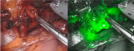
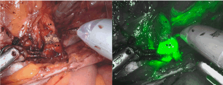
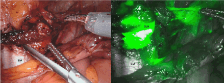
These images show the identification and extraction of sentinel lymph node (SNL) by using indocyanine-green (ICG) with near-infrared (NIR) fluorescence during a robot-assisted laparoscopic approach. The procedure was done with da Vinci Xi™ robot (Intuitive Surgical Inc., Sunnyvale, CA, USA). The patient was 38-year-old with the suspicious of micro invasive CC (FIGO stage 1A1). During the procedure, we also evaluated the feasibility of SLN identification and removal in a patient in which we supposed negativity for nodal tumor dissemination.
After the anesthesia and the robotic docking (time=23 min), a solution of 5 ml of ICG 1% was injected into the cervical stroma, superficially and deep into the tissue, at 3 and 9 o’clock positions by the use of a spinal needle (Figure 1).

Figure 1. Tracer injection at 3 and 9 o’clock positions in the cervix.
We accurately evaluated the abdominal and pelvic organs, before obtaining a peritoneal washing to send to cytological evaluation. A meticulous inspection of pararectal and paravesical spaces was done after the resection of each uterine round ligament and the subsequent dissection of the broad ligament. We evaluated the main lymph nodal regions of each side (obturator, external iliac, presacral) in order to detect the fluorescent SLNs (after 20 min from the tracer injection) via the special camera for NIR fluorescence (FireFly™, Intuitive Surgical Inc., Sunnyvale, CA, USA). This tool allows a rapid conversion from the normal robotic view to that which detects the fluorescent emission derived from ICG via the robotic camera. This is done by holding control pedal of the camera at the console and sliding the finger switch.
At the end of SLN dissection at both pelvic sides (time for the right side=12 min; time for the left side=15 min, total time of the SLN dissection=25 min, since the incision of each uterine round ligament), we sent the SLNs to definitive histological evaluation by following standardized ultra-staging criteria.
The figures 2 and 3 show the left side SLN in normal view (left) and by NIR fluorescent system (right). In upper part of the figure it is observed another lymph node (LN) of the same lymphatic chain with a clearly lower fluorescence. Both are located adjacent to the Extern Iliac Artery (EIA). In the figure 4, we can see the left SLN after its complete dissection (Figures 2-4).

Figure 2. The left side SLN in normal view (left) and by NIR fluorescent system (right).

Figure 3. The left side SLN in normal view (left) and by NIR fluorescent system (right).

Figure 4. The left SLN after its complete dissection.
After the end of this procedure, we performed a total hysterectomy with preservation of both ovaries by robot-assisted laparoscopic approach (time=97 min). The uterus was extracted via transvaginal route and was sent to definitive histological evaluation. The post-surgical period was regular (no major and minor complications) and the time of hospitalization was 3 days.
In the near future, our research group is going to confirm the diagnostic accuracy of the SLN biopsy by performing ING-based NIR fluorescence during robotic-assisted laparoscopic approach in patients with early-stage CC.
All procedures performed in studies involving human participants were in accordance with the ethical standards of the institutional and/or national research committee and with the 1964 Helsinki declaration and its later amendments or comparable ethical standards
- Kim JH, Kim DY, Suh DS, Kim JH, Kim YM, et al. (2018) The efficacy of sentinel lymph node mapping with indocyanine green in cervical cancer. World J Surg Oncol 16: 52. [Crossref]
- Salvo G, Ramirez PT, Levenback CF, Munsell MF, Euscher ED, et al. (2017) Sensitivity and negative predictive value for sentinel lymph node biopsy in women with early-stage cervical cancer. Gynecol Oncol 145: 96-101. [Crossref]
- Buda A, Bussi B, Di Martino G, Di Lorenzo P, Palazzi S, et al. (2016) Sentinel Lymph Node Mapping with Near-Infrared Fluorescent Imaging Using Indocyanine Green: A New Tool for Laparoscopic Platform in Patients with Endometrial and Cervical Cancer. J Minim Invasive Gynecol 23: 265-269. [Crossref]
- Van de Lande J, Torrenga B, Raijmakers PG, Hoekstra OS, van Baal MW, et al. (2007) Sentinel lymph node detection in early stage uterine cervix carcinoma: a systematic review. Gynecol Oncol S 106: 604-613. [Crossref]


