Introduction
Restrictive strabismus is usually caused by congenital cranial dysinnervation disorders1 or congenital orbital fibrosis2, in which extraocular muscle (EOM) can be reported fibrosis and cranial nerve also is maldevelopment with magnetic resonance imaging (MRI). While EOM enlargement also happened due to thyroid eye disease in adults or secondary to inflammatory myositis, neoplastic infiltrate3. However, congenital enlargement of EOM happened in young children, it is difficult to identify the etiology. We present 1 case with enlargement of lateral rectus muscle in young patients, which no confirmed pathology can be identified.
Key words
restrictive strabismus, enlargement, extraocular muscles, ocular motility
Case description
A 2-year-old boy presented in 2018 with right exotropia since birth. On examination he had noted right exotropia more than 140PD in primary gaze (Figure 1. Top left). The motility of the right eye was severely limited. Other ophthalmic examination was normal. Systemic examination was normal. Cycloplegic refraction was +5.50DS+1.75DC×85 in the right eye and +1.50DS+1.25DC×85 in the left eye.
The patient underwent MRI scans of brain and orbits, which revealed the left lateral rectus muscle belly enlarged diameter with the collapsed lateral orbital wall due to the compression from the muscle. The thickening belly located in the orbit apex. (Figure 1. Right). Thyroid function testing and inflammatory makers were normal. Considering the right exotropia existed from birth, and cannot version, which increased the risk of amplyopia. The refraction error also caused amplyopia. Procedure was decided to performed on the right eye.
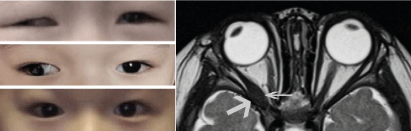
Figure 1. A 2-year-old male subject present with right exotropia since birth (Top left). MRI showed that left lateral rectus muscle belly had enlarged diameter with the corresponding area of the lateral orbital wall collapsed (Figure 1. Right). Six months postoperatively, the right exotropia was 40PD with 10 PD hypotropia in primary gaze (Figure 1. Middle left). Ocular alignment was very straight in primary gaze after 2nd procedure (Figure 1. Bottom left)
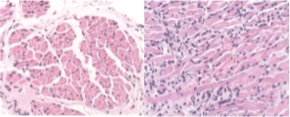
Figure 2. The biopsy of left lateral rectus muscle revealed dense and enlarged muscle fibrosis, and variability muscle size. No evidence showed tumor or inflammation from biopsy
Forced duction test at surgery confirmed a tight right lateral rectus. Right lateral canthus was cut for partial thickening lateral rectus muscle biopsy and release the adhesion between the lateral rectus and orbital wall. The right lateral rectus was hooked and relieved entirely. Then Forced duction test showed no limitation. A 1mm×2mm muscle biopsy was obtained from the left lateral rectus belly. The biopsy revealed dense and enlarged muscle fibrosis, and variability muscle size (Figure 2. Right). No evidence showed tumor or inflammation from biopsy. Right lateral rectus muscle biopsy was obtained. Six months postoperatively, the right exotropia was 40PD with 10 PD hypotropia in primary gaze (Figure 1. Middle left). Right eye had some adduction. Patching the left 4 hours each day with framed glassed. His vision acuity was 20/50 in the right and 20/30 in the left.
A second surgery performed at age 3. The right medial rectus was plicated 15mm. Orthotropia was obtained postoperatively in primary gaze (Figure 1. Bottom left). Abduction and adduction was limited after twice operations. Patching left eye was kept post-operation (Figure 2, 1-5).
Conclusion
Restrictive strabismus is usually caused by congenital cranial dysinnervation disorders1 or congenital orbital fibrosis 2, in which extraocular muscle (EOM) can be reported fibrosis and cranial nerve also is maldevelopment with magnetic resonance imaging (MRI). While EOM enlargement also happened due to thyroid eye disease in adults or secondary to inflammatory myositis, neoplastic infiltrate3. However, congenital enlargement of EOM happened in young children, it is difficult to identify the etiology. We present a case with enlargement of lateral rectus muscle in young child, which no confirmed pathology can be identified.
Congenital restricted strabismus can be usually caused by congenital cranial dysinnervation disorders (CCDD), congenital orbital fibrosis, which the EOM was maldevelopment and cannot contract well. The common diseases result in acquired restricted strabismus includes thyroid eye disease, EOM involvement secondary to the orbital fracture, heavy eye syndrome and etc. The differential diagnosis should be based on the detail history, ocular movement, lab test and imagings.
Idiopathic congenital enlargement of extraocular muscles was reported in young patients4. The patient in our reported had no systemic disease, and MRI scan revealed that lateral rectus belly was enlargement. Forced duction testing in surgery also confirmed the tight lateral rectus, which caused congenital restricted strabismus. The patient had tight lateral rectus, and the thicken belly of lateral rectus compressed the lateral orbital wall. We did biopsy and no tumor or inflammation tissue was detected.
The pathogenesis in the case was unclear. Graves disease seldom caused limited extraocular motility in children5. Surgery was performed according to the forced duction test. Detachment of the muscle or releasing the adhesion between the muscle and orbital wall could improve the ocular duction, regular recession-resection was not the first choice. The deviation angle or ocular duction may change with the first surgery. Patching is necessary for amplyopia treatment after improving the ocular motility.
The case is required further discussion on the etiology of enlargement, which only involved belly with sparing tendon and not all EOM are involved. Patient was still followed up to check ocular motility and treat amplyopia.
Declaration of conflicting interests
The author(s) declared no potential conflicts of interest with respect to the research, authorship, and/or publication of this article.
Funding
Rui Hao received support of National Natural Science Foundation of China (81800861) and General Project of Tianjin Health Science and Technology Fund (TJWJ2021MS041) for the research.
Informed consent
The patient gave the informed written consent to publish the case, the images.
References
- Gutowski NJ, Chilton JK (2015) The congenital cranial dysinnervation disorders. Arch Dis Child 100: 678-681. [Crossref]
- Kim N, Yang HK, Kim JH, Hwang JM (2018) Comparison of clinical and radiological findings between congenital orbital fibrosis and congenital fibrosis of the extraocular muscles. Curr Eye Res 43: 1471-1476. [Crossref]
- Metz HS (1999) Strabismus related to Graves ophthalmopathy. In:Rosenbaum AL, Santiago AP, eds. Clinical strabismus management: Principles and surgical techniques. London: W.B. Saunders Company 285-295.
- Sharma A, Foster RS, Suh DW, Smith D, Kraft SO, et al. (2016) Idiopathic enlargement of the extraocular muscles in young patients: A case series. Am J Ophthalmo 161: 206-213. [Crossref]
- Chan W, Wong GW, Fan DS, Cheng AC, Lam DS, et al. (2002) Ophthalmopathy in childhood Graves’ disease. Br J Ophthalmol 86: 740-742. [Crossref]