Take a look at the Recent articles

Heterotopic trigeminal pregnancy after in vitro fertilization and embryo transfer: A case report

Carlo V Bellieni

Neonatal Intensive Care Uniti, University Hospital, Siena, Italy

E-mail : bhuvaneswari.bibleraaj@uhsm.nhs.uk

Libera Troìa

Obstetrics and Gynecology, Department of Molecular and Developmental Medicine, University of Siena, Siena, Italy

Michela Torricelli

Obstetrics and Gynecology, Department of Molecular and Developmental Medicine, University of Siena, Siena, Italy

Matteo Benvenuti

Department of Legal Medicine, University of Siena, Italy

Giuseppe Buonocore

Neonatal Intensive Care Uniti, University Hospital, Siena, Italy

Filiberto Maria Severi

Landau Institute for Theoretical Physics, Russian Academy of Sciences, Kosygin str. 2, Moscow, Russia

DOI: 10.15761/GII.1000136

Article
Article Info
Author Info
Figures & Data

A caucasic 42-year-old woman, first pregnancy, with a diagnosis of female factor infertility caused by ovulation failure and azoospermia in the male partner, underwent an IVF procedure using sperm and ovocytes donors. The patient had 2 embryos transferred 26 days before she presented to the Obstetric Department with a chief symptom of abdominal pain.

Laboratory testing revealed a serum β-human chorionic gonadotropin level of 56779 mIU/mL.

A transvaginal pelvic sonographic examination demonstrated a intrauterine twin pregnancy without embryonic structures, a third gestational sac in the right uterine adnexa with an embryo measuring 4,3 mm length according to 5+5 weeks of amenorrhea with cardiac activity and the presence of free fluid in the pouch of Douglas.

A bilateral salpingectomy and uterine curettage was performed.
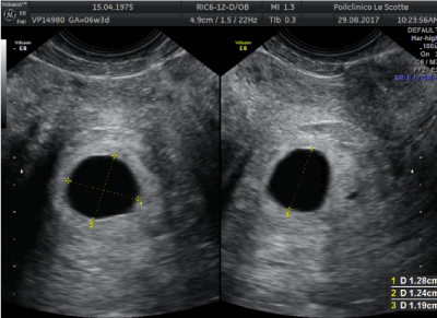
Figure 1. Transvaginal sagittal section showing the first empty intrauterine gestation sac.
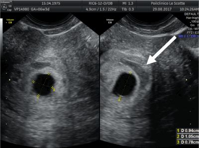
Figure 2. Second empty intrauterine gestation sac, located below the first and smaller dimensions. The arrow indicates an area of amniochorial detachment.
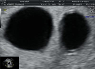
Figure 3. Ultrasonographic find of both intrauterine gestational sacs showing twin pregnancy.
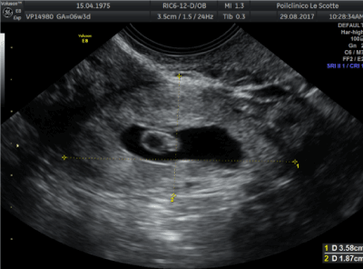
Figure 4. Ultrasound image showing an ectopic pregnancy in the ampullary region of the right fallopian tube with the presence of embryonic structures: gestational sac, yolk sac and fetal pole.
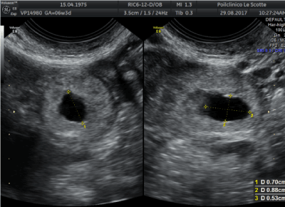
Figure 5. Ultrasonographiv find of the tubal sign, defined by the presence of a round hypoechoic adnexal collection with a well-defined hyperechoic rim. This represents a gestational sac (hypoechoic center) with surrounding trophoblast (hyperechoic rim).
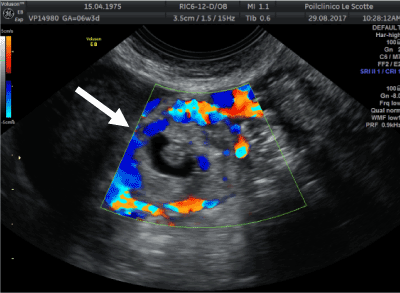
Figure 6. Tubal ectopic pregnancy by transvaginal ultrasound. The arrow indicates the ectopic gestation with circumferential Doppler flow, called the “Ring of Fire”.
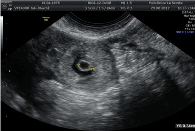
Figure 7. Ultrasound examination revealing the presence of yolk sac of tubal pregnancy.
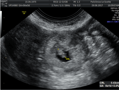
Figure 8. Ultrasound image showing the presence of an embryo with crown-rump length measurements of 4,3 mm, according to 5+5 weeks of amenorrhea.
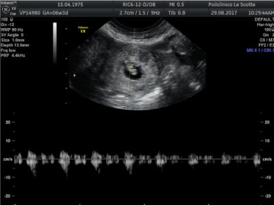
Figure 9. Fetal pole with visible fetal heartbeat detected at Power-Doppler.
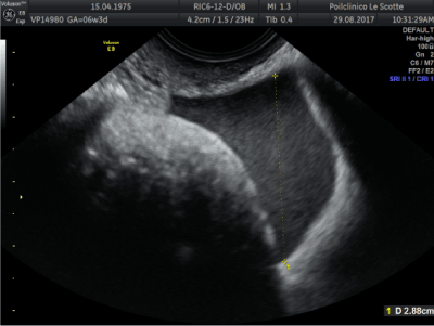
Figure 10. Ultrasonographic find of free fluid in the pouch of Douglas.

2021 Copyright OAT. All rights reserv

Article Type

Image Article

Publication history

Received date: September 02, 2017
Accepted date: September 18, 2017
Published date: September 21, 2017

Copyright

© 2017 Bellieni CV. This is an open-access article distributed under the terms of the Creative Commons Attribution License, which permits unrestricted use, distribution, and reproduction in any medium, provided the original author and source are credited.

Citation

Bellieni CV, Troìa L, Torricelli M, Benvenuti M, Buonocore G, Severi FM (2017) Heterotopic trigeminal pregnancy after in vitro fertilization and embryo transfer: A case report. Glob Imaging Insights 2: DOI: 10.15761/GII.1000136

Corresponding author

Filiberto Maria Severi, MD, PhD

Obstetrics & Gynecology, Department of Molecular and Developmental Medicine, University of Siena, Policlinico “Le Scotte” Viale Bracci, 53100 Siena, Italy

E-mail : bhuvaneswari.bibleraaj@uhsm.nhs.uk

Figure 1. Transvaginal sagittal section showing the first empty intrauterine gestation sac.

Figure 2. Second empty intrauterine gestation sac, located below the first and smaller dimensions. The arrow indicates an area of amniochorial detachment.

Figure 3. Ultrasonographic find of both intrauterine gestational sacs showing twin pregnancy.

Figure 4. Ultrasound image showing an ectopic pregnancy in the ampullary region of the right fallopian tube with the presence of embryonic structures: gestational sac, yolk sac and fetal pole.

Figure 5. Ultrasonographiv find of the tubal sign, defined by the presence of a round hypoechoic adnexal collection with a well-defined hyperechoic rim. This represents a gestational sac (hypoechoic center) with surrounding trophoblast (hyperechoic rim).

Figure 6. Tubal ectopic pregnancy by transvaginal ultrasound. The arrow indicates the ectopic gestation with circumferential Doppler flow, called the “Ring of Fire”.

Figure 7. Ultrasound examination revealing the presence of yolk sac of tubal pregnancy.

Figure 8. Ultrasound image showing the presence of an embryo with crown-rump length measurements of 4,3 mm, according to 5+5 weeks of amenorrhea.

Figure 9. Fetal pole with visible fetal heartbeat detected at Power-Doppler.

Figure 10. Ultrasonographic find of free fluid in the pouch of Douglas.