Background: Heterotopic Ossification (HO) is usually associated to musculoskeletal trauma or neurological injury.
Case Report: Our objective is to present a rare case of bilateral presence of HO in the hip joints in a patient with Guillain-Barre Syndrome (GBS) and compare it with other similar cases from the literature. Possible causative correlation of HO with different factors and effects on patients’ functionality are also discussed.
Clinical Rehabilitation Impactː HO is a rare complication of patients with GBS and should be suspected early when causative factors exist, as it plays a major role in their functional outcome
Heterotopic Ossification (HO) is usually associated to musculoskeletal trauma or neurological injury
Abbreviations
Heterotopic ossification (HO), Guillain Barre Syndrome (GBS)
Heterotopic Ossification (HO), also known as Myositis Ossificans (MO), is a metabolic disorder of calcium that results in new lamellar bone formation mostly in muscles and soft tissue near the joints, outside the normal skeletal system. It is often a complication of Central Nervous System (CNS) injury, Spinal Cord Injury (SCI), brain injury and other non-traumatic CNS diseases, although less frequently [1-4].
Guillain-Barre syndrome (GBS) is an inflammatory polyneuropathy, often precipitated by infections and presents by ascending progressive weakness that eventually leads to flaccid paralysis. There may also be sensory symptoms, pain and autonomic involvement [5]. 80% of the patients may achieve total or partial recovery, for 5% of them is fatal and 15% of the patients have neurologic and functional deficits.
HO is a rare complication of GBS [6], but when it’s presented within GBS it can have an important impact on the functional recovery of the patients. In this paper we present an unusual case of GBS complicated with HO in both hips and compare it with similar cases from the literature, aiming to evaluate causative factors of HO in GBS patients.
Patient information
A 67-year-old, otherwise healthy, male was admitted to our hospital with weakness in the lower extremities that later also affected the upper extremities and gradual onset. The patient reported a previous viral upper respiratory tract infection. His past medical, family and psychological history was not significant of serious neurological or musculoskeletal illnesses.
Clinical findings, Diagnostic assessment and Therapeutic intervention
Head CT was clear and cerebrospinal fluid exam was not compatible with CNS infection (0/mm3 cells and 29mg/dl protein). GBS was diagnosed and the patient started treatment with immunoglobulin (160g for 5 days). Even with the treatment, the patient gradually deteriorated, and it was necessary to be intubated. Two months following his admission to ICU, he was disconnected from the respirator and one week later, he was also decannulated. A day later he was transferred back to the neurology clinic, where he started daily kinesiotherapy and respiratory physiotherapy. He was finally transferred to our clinic (PRM). At that time the labs showed increased alkaline phosphatase (ALP) (368 IU/L) and normal serum calcium (9,3 mEq/L). Radiographic imaging of the pelvis showed no evidence of HO (Figure 1a) but due to local inflammation of the hip area and elevated ALP he was started on indomethacin 75 mg once daily for 6 weeks as well as etidronate 400mg twice daily for 3 months and once daily for another 3 months.
During his stay at our clinic, he continued with active assisted exercises of his hip with gradual sitting position, regular kinesiotherapy for the other joints of his extremities and swallow muscles exercises. Three months following admission, he regained muscle strength of his forearm and the Folley catheter was removed. In a new radiological assessment of pelvis and hips, HO was detected in the hip joints (most severe on the right) (Figure 1b) and was absent on both knees. The ALP remained increased almost throughout his stay in our clinic (range: 120-368 IU/L) and serum calcium remained within normal values.
On the fourth month after admission further improvement of his muscle strength in the upper and lower extremities and less in pelvis was observed. Serum ALP was decreased to normal. However, fixed flexion deformity (approximately 20 degrees) with difficulty in sitting and walking was evident.
Follow-up and Outcomes
Although patient gradually improved and was discharged, simple radiologic imaging was performed again, and HO was even more evident (Figure 1c). The CT of the pelvis showed bone lateral and posterior to the hip joints bilaterally. Total time of follow-up was 12 months. Bone scan after 12 months showed improvement although HO was still immature (increased radionuclide uptake in both hip joints). The patient was scheduled for surgical excision of HO when bone scan will not show increased uptake.
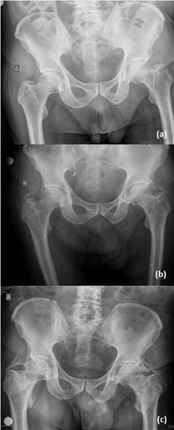
Figure 1. Simple radiologic imaging of the pelvis one month before HO diagnosis (a), on HO diagnosis (b), 4 months after diagnosis HO still present (c).
Discussion
A case of heterotopic ossification (HO) in a patient with Guillain-Barre Syndrome (GBS) is presented and the incidence, the influence on functionality and possible causative correlation between HO and GBS are also reported. In the literature, we found 13 cases of HO after GBS and including our case a total of 14 cases exist (Table 1). Zeilig, et al. [5] reported a 4% incidence of HO in patients with GBS. The incidence of HO in our PMR clinic is less than 3% (1 patient out of 40 GBS admitted patients over 11-year time period).
HO is a rare complication in the context of other neurologic disorders. It has been described more often following CNS trauma [2] and less frequently after other neurologic incidents. There has been a link between HO and peripheral nerve disorders, GBS included. Our patient was a male which happens to be the gender most commonly affected by HO after GBS (66% of cases). The mean age of the patients was 42 years old while our patient was 67 years old. The hip joint was more severely affected in our patient which is also in accordance with the international literature (7 out of 9 cases) [5-7].
Risk factors reported were mechanical ventilation [5,8,9], prolonged immobilization [7-9] and hypoxia [5,8]. Our patient was only mechanically ventilated. One of the patients also had fixed flexion deformity(15o) [9]. Also, our patient had fixed flexion deformity and the typical signs of inflammation [5,7,9].
Diagnostically simple radiologic imaging, 3-phase bone scan, MRI and SPECT/CT was used [5,7,9,10]. All these imaging techniques, except MRI, were performed in our patient.
Lab values reported were alkaline phosphatase, serum calcium and serum phosphate [5,7]. Of these, only alkaline phosphatase was mentioned to be increased, including our patient.
Treatment options consisted of medications, radiotherapy, surgery and rehabilitation therapy [5,7-9]. Our patient was treated with medications, rehabilitation therapy and will receive surgical treatment (excision of HO) after reevaluation.
Zeilig, et al. [5] and Kerdoncuff, et al. [6] reported the time of follow-up, which varied between 6 months and 15 years. In the former study, patients presented on admission to rehabilitation requiring assistance for ADL and mobility. During the long-term follow-up, patients presented with affected posture and gait cycle due to severe limitations in hip ROM. In the latter study, the patient had permanent disability which was not cause by the HO rather than the neurologic sequelae of GBS. Shawgi, et al. [10] also reported that their patient had improvement in the pain score despite the stiffness of the knees that was present throughout the follow-up. Kerdoncuff, et al. [6] also reported that their three patients had great functional handicap during the follow-up. Our patient has been followed for 12 months and has functional limitations in his postural and walking ability, but he did not complain of pain in the end of his follow-up.
Table 1. Cases of HO after GBS.
Study |
# of cases |
Ryu SR, et al. |
1 |
Zeilig G, et al. |
4 |
Vaishya R, et al. |
1 |
Shawgi M |
1 |
Kerdoncuff V, et al. |
3 |
Ohnmar H, et al. |
1 |
Bernard V |
1 |
Hung JCC, et al. |
1 |
PMR clinic |
1 |
A unique complication of HO in a patient with GBS and a systematic literature review is presented. Physicians (and therapists) should suspect HO in symptomatic (related to large joints) patients with GBS when risk factors exist.
The authors declare no conflict of interest.
No financial support/equipment/drugs have been received for this work
AP,DV,AT Conceived and/or designed the work that led to the submission, acquired data, and/or played an important role in interpreting the results.
AP, DV, AT Drafted or revised the manuscript.
AP, IG, AK Approved the final version
No
56th ISCoS Annual Scientific Meeting, Dublin, Ireland
No
- Sell S, Phillips O, Handel M (2004) No difference between two doses of diclofenac in prophylaxis of heterotopic ossifications after total hip arthroplasty. Acta Orthop Scand 75: 45-49. [Crossref]
- Van Kuijk AA, Geurts AC, van Kuppevelt HJ (2002) Neurogenic heterotopic ossification in spinal cord injury. Spinal Cord 40: 313-326. [Crossref]
- Garland DE (1991) A clinical perspective on common forms of acquired heterotopic ossification. Clin Orthop Relat Res 263: 13-29. [Crossref]
- Wharton GW (1975) Heterotopic ossification. Clin Orthop Relat Res 142-149. [Crossref]
- Zeilig G, Weingarden HP, Levy R, Peer I, Ohry A, et al. (2006) Heterotopic ossification in Guillain-Barre syndrome: incidence and effects on functional outcome with long-term follow-up. Arch Phys Med Rehabil 87: 92-95. [Crossref]
- Kerdoncuff V, Sauleau P, Petrilli S, Duruflé A, Ben Beroukh K, et al. (2002) Heterotopic ossification in Guillain-Barre syndrome. Ann Readapt Med Phys 45: 198-203. [Crossref]
- Ryu SR, Kim JH, Choi IS, Han JY, Lee SG (2008) Heterotopic ossification as an unusual complication after Guillain-Barre syndrome: a case report. Arch Phys Med Rehabil 89: 564-567. [Crossref]
- Ohnmar H, Roohi SA, Naicker AS (2010) Massive heterotopic ossification in Guillain-Barre syndrome: a rare case report. Clin Ter 161: 529-532. [Crossref]
- Vaishya R, Agarwal AK, Vijay V, Vaish A (2016) Heterotopic Ossification Circumferentia Articularis (HOCA) of Both Knee Joints After Guillain-Barre Syndrome. Cureus 8: e480. [Crossref]
- Shawgi M (2012) Heterotopic ossification of the hips in a patient with Guillain Barre syndrome demonstrated on SPECT/CT. Clin Nucl Med 37: e253-254. [Crossref]