arachnoid cyst, hemorrhagic arachnoid cyst, third cranial nerve palsy, transient ischemic attack (TIA), internal carotid artery compression
Herein, we report an uncommon case of unilateral, acute and complete third cranial nerve palsy (TNP), along with 2 episodes of transient hemiparesis, related to compression of the third nerve and of the ipsilateral internal carotid artery (ICA) by a hemorrhagic arachnoid cyst (AC) of the suprasellar cistern.
A previously healthy 54-year-old woman presented a right orbital pain of sudden onset followed by progressive ptosis and diplopia of the right eye.
Two weeks later occurred 2 episodes of transient weakness of the left side of 5 minutes of duration each. On admission, the neurological examination revealed a complete right TNP and a left hemiparesis. She had no history of head trauma, nor hypertension, vascular risk factors or anticoagulation therapy.
The non-contrast brain CT showed a slightly hyperdense rounded mass of the opto-chiasmatic cistern (Figure 1a). Brain MRI performed 3 days later showed a well-defined rounded mass of 10mm of diameter, bulging in the suprasellar cistern and adjacent to the right medial temporal lobe (TL). The lesion was localized along the course of the cisternal segment of the right third cranial nerve. The mass contained a fluid-blood level, well-identified on T1 and T2/FLAIR and T2 GRE sequences (Figures 1b-1f). The presence of an interface between the lesion and the temporal lobe was the only sign of the extra-axial origin of the lesion. There was no change of the surrounding parenchyma and no enhancement after contrast (Figure 1g). On DWI, the lesion did not exhibit diffusion restriction and this sequence revealed an ischemic lesion of the posterior limb of right internal capsule (Figure 1h).
No vascular anomaly was seen on magnetic resonance angiography (MRA) of the circle of willis or on digital cerebral angiography. The mass slightly pushed interiorly the ICA (Figure 1i-1j). Follow-up MRI at day 7 showed no changes.
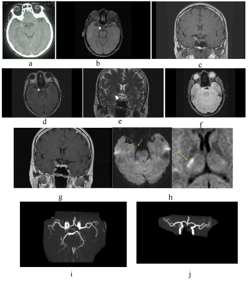
Figure 1. Non-enhanced CT (A), High-Resolution (HR) axial (B) and coronal (C) T1 MR images , FLAIR axial (D), HR coronal T2 (E) and post-contrast coronal T1 MR images (F) reveal a round-shaped well-delimited hemorrhagic nonenhancing mass within the right supraselllar cistern (10 x 10mm of diameter). Fluid-blood level is visualized within the cyst on T1, FLAIR/T2 images as well as on T2-GRE. The lesion has no restricted diffusion and the Diffusion-Weighted Images (DWI) show an ischemic lesion in the posterior limb of the right internal capsule (H). 3D TOF MR Angiography (MRA) of the circle of willis does not reveal any vascular malformation (I). MRA (vertebro-basilar system has been retrieved) shows a slight compression by the mass of the external wall of the internal carotid artery (ICA) (J).
Surgery was performed 2 months later. At surgery, as the third cranial nerve (TCN) was identified at the cavernous sinus entry and a little ruptured hematoma was visualized. A little quantity of fluid was aspired and sent for analysis.
Membranes around the area of the hematoma and resembling to arachnoid are collected. The third cranial nerve had a normal aspect but presented a cup-shaped segment at the site of the collapsed hematoma. No aneurysm or other vascular anomaly was observed.
Pathologic examination revealed slightly hemorrhagic CSF and fragments of arachnoid without concern for malignancy. The diagnosis of a hemorrhagic arachnoid cyst was established.
Immediately after surgery the TNP improved but the double vision persisted worse in vertical component.
Postoperatively, the patient demonstrated permanent TNP. At 3-years follow-up the patient had incomplete TNP and MRI showed no recurrence of the AC.
Common causes of isolated pupil-sparing TNP involve medical pathologies: diabetes mellitus and giant cell arteritis. Causes of isolated TNP involve compression pathologies: posterior circulation aneurysms, trauma, intracranial neoplasms.
The differential diagnosis of acute onset unilateral nonpupil sparing TNP must take in account in emergency, a vascular compressive lesion: direct vascular compression by the posterior cerebral artery (PCA) [1], by infundibular dilatation of the posterior communicating artery [2], or by an aberrant PCA [3] are well known. Few patients develop isolated TNP in the onset of ICA dissection.
A case of isolated TNP post-thrombectomy of an ICA terminus/PCA embolus for ICA dissection has also been reported [4]. However, the presence of a blood-fluid level within the lesion is not in favor of a vascular lesion.
Very rarely AC have been reported as causing cranial nerve palsy, especially TNP [5-9]. Furthermore, acute TNP caused by an AC of the suprasellar cistern is an extremely rare condition.
An AC is a benign, congenital fluid-filled CSF-containing structure whose wall is made of arachnoid structures. Arachnoid cysts account for approximately 1% of all intracranial mass lesions. The cyst can develop at any site in the subarachnoid space. The most common location of AC is the sylvian fissure and middle fossa. Other locations include the suprasellar region, the cerebello-pontine angle (CPA) and the interhemispheric fissure. The suprasellar region hosts approximately 9-11% of those lesions. They are usually asymptomatic and discovered incidentally on imaging. On CT as on MRI, AC present a CSF-like containing cyst. They do not enhance after contrast and do not present restriction diffusion.
They may become symptomatic in case of cyst rupture or of hemorrhage within the cyst after head trauma. In our patient, there was no recent history of head trauma.
Cranial nerve palsy related to AC have been reported in isolated cases) [5-9]. AC of the suprasellar cistern causing TNP have been reported in a few isolated cases [10,11] including a new entity of “de novo” AC [12].
Hemorrhagic AC of the suprasellar cistern causing acute onset of painful unilateral TNP, nonpupil sparing seems exceedingly rare. To the best of our knowledge only 1 case has been reported to date [10]. MRI, better than CT scan, show hemorrhage within a cystic mass. In our case, non-enhanced CT evidenced the presence of a round and dense structure in the right part of the suprasellar cistern. MRI visualized a blood-fluid level within a cyst-like extra-axial non-enhancing and well-delimited mass. T2-GRE sequence was very useful to identify blood products.
The differential diagnosis must take in account the suprasellar cystic lesions that may bleed: Rathke’s cleft cysts, neuroepithelial cyst [13] and more exceptionally parasellar dermoid tumor with intra-tumoral hemorrhage [14].
Finally, isolated TNP may be, in very rare occasions, the presenting symptom of bilateral chronic subdural hematomas [15,16].
This report highlights an unusual nonexpansile cause of acute and painful complete oculomotor palsy.
Although rare in the suprasellar region, hemorrhagic AC should be considered for patients presenting with isolated third cranial nerve palsy of acute and painful onset.
MRI is very useful in depicting in this localization an intracystic hemorrhage.
The authors have no conflict of interest and have not received any financial support.
- Jo YS, Kim SK, Kim DH, Kim JH, Na SJ (2015) Complete oculomotor nerve palsy caused by direct compression of the posterior cerebral artery. J Stroke Cerebrovasc Dis 24: e189-190. [Crossref]
- Fukushima Y, Imai H, Yoshino M, Kin T, Takasago M, et al. (2014) Ptosis as partial oculomotor nerve palsy due to compression by infundibular dilatation of posterior communicating artery, visualized with three-dimensional computer graphics: case report. Neurol Med Chir (Tokyo) 54: 214-218. [Crossref]
- Tan T, Tee JW, Wa2021 Copyright OAT. All rights reservecondary to aberrant posterior cerebral artery. BMJ Case Rep 2014. [Crossref]
- Kogan M, Natarajan SK, Kim N, Sawyer RN, Snyder KV, Siddiqui AH (2014) Third nerve palsy following carotid artery dissection and posterior cerebral artery thrombectomy: Case report and review of the literature. Surg Neurol Int 5: S497-500. [Crossref]
- Ohtsuka K, Hashimoto M, Nakamura Y (1998) Bilateral trochlear nerve palsy with arachnoid cyst of the quadrigeminal cistern. Am J Ophthalmol 125: 268-270. [Crossref]
- Pirotte B, Morelli D, Alessi G, Lubansu A, Verheulpen D, et al. (2005) Facial nerve palsy in posterior fossa arachnoid cysts: report of two cases. Childs Nerv Syst 21: 587-590. [Crossref]
- Hayden MG, Tornabene SV, Nguyen A, Thekdi A, Alksne JF (2007) Cerebellopontine angle cyst compressing the vagus nerve: case report. Neurosurgery 60: E1150. [Crossref]
- Hustler A, Joy H, Hodgkins P (2009) Isolated unilateral mydriasis with delayed oculomotor palsy secondary to intracranial arachnoid cyst. J AAPOS 13: 308-309. [Crossref]
- Jacob M, Gujar S, Trobe J, Gandhi D (2008) Spontaneous resolution of a Meckel's cave arachnoid cyst causing sixth cranial nerve palsy. J Neuroophthalmol 28: 186-191. [Crossref]
- Ide C, De Coene B, Gilliard C, Pollo C, Hoebeke M, et al. (1997) Hemorrhagic arachnoid cyst with third nerve paresis: CT and MR findings. AJNR Am J Neuroradiol 18: 1407-1410. [Crossref]
- Tempel ZJ, Johnson SA, Richard PS, Friedlander RM, Rothfus WE, Hamilton RL (2013) Parasellar arachnoid cyst presenting with a nonpupil sparing third nerve palsy mimicking a posterior communicating artery aneurysm in an adult. Surg Neurol Int 4: 87. [Crossref]
- Brewington D, Petrov D, Whitmore R, Liu G, Wolf R, et al. (2017) De novo intraneural arachnoid cyst presenting with complete third nerve palsy: Case report and literature review. World Neurosurg 98: 873. [Crossref]
- Park M, Lee SK, Choi J, Kim SH, Kim SH, et al. (2015) Differentiation between cystic pituitary adenomas and rathke cleft cysts: A diagnostic model using MRI. AJNR Am J Neuroradiol 36: 1866-1873. [Crossref]
- Mamata H, Matsumae M, Yanagimachi N, Matsuyama S, Takamiya Y, et al. (1998) Parasellar dermoid tumor with intra-tumoral hemorrhage. Eur Radiol 8: 1594-1597. [Crossref]
- Matsuda R, Hironaka Y, Kawai H, Su Park Y-S, Taoka T, Nakase H (2013) Unilateral oculomotor nerve palsy as an initial presentation of bilateral chronic subdural hematoma: Case report. Neurol Med Chir (Tokyo) 53: 616-619. [Crossref]
- Corrivetti F, Moschettoni L, Lunardi P (2016) Isolated oculomotor nerve palsy as presenting symptom of bilateral chronic subdural hematomas: Two consecutive case report and review of the literature. World Neurosurg 88: 686. [Crossref]