Abstract
Introduction: Deficit in the first metatarsophalangeal joint (MTPJ) extension range is suggested as one of the intrinsic risk factors for Plantar fasciitis. However, the relationship between MTPJ range and intensity of pain and dysfunction has not been explored. The purpose of this study was to determine if a difference exists in extension Range of Motion (ROM) of the first MTPJ between PF sufferers and healthy controls; and to explore its association with intensity of pain and dysfunction.
Methods: Thirty-six patients (22 female) with unilateral plantar fasciitis (19 on the dominant foot) and 21 healthy controls were recruited. Active and passive ROM of first MTPJ extension were assessed. In the healthy control, side-to-side difference on the joint range was compared using repeated measure analysis of variates with gender as a factor. Multivariate analysis of variates was used to compare MTPJ extension range between patients and healthy control matched with leg dominance, and with gender and body mass index as covariates. Partial correlation coefficient tests were conducted to assess relationships between MTPJ extension range and intensity of pain and dysfunction controlled with BMI, activity level and affected side.
Results: The active and passive MTPJ range was significantly greater in the dominant than non-dominant feet (all p < 0.05) in the healthy controls. Female patients had significantly larger passive MTPJ extension range (p = 0.034 and 0.006) for patients with the dominant and non-dominant feet being affected, respectively). Significant correlation was detected between the passive MTPJ extension range and intensity of pain (r = -0.54, p = 0.017) and pain score of foot functional index (r = -0.47, p = 0.042) in the female patients.
Conclusions: In female patients with plantar fasciitis, increased passive metatarsal phalangeal joint extension range is detected, and a larger extension range is associated with less pain.
Key words
first metatarsophalangeal joint, pain, plantar fasciitis, range of motion
Introduction
Plantar Fasciitis (PF) is one of the most common painful foot conditions treated by health care providers [1]. It is characterized by pain localized at the medial tubercle of the calcaneus which is exacerbated with weight bearing activities [2,3]. PF has high prevalence in both athletic and nonathletic populations [4]. The mean duration of symptoms ranged from 13.3-14.1months, with 5–10% of recalcitrant cases requiring surgery [5-7].
Mechanical origin has been thought to play an important role in developing PF [3,4]. It is suggested that plantar fascia provides truss support for the longitudinal arch without the function of muscles [3,8]. Environmental or intrinsic factors which lower the medial longitudinal arch induce tension in the fascia and can lead to the development of PF [3,4,9]. In this connection, extension of the first metatarsophalangeal joint (MTPJ) helps to elevate and stabilize the medial longitudinal arch throughout the stance phase of gait via “windlass effect” [10,11]. Restriction in extension of the first MTPJ may alter the mechanism to stabilize the medial longitudinal arch, and also reduce extensibility of the plantar fascia. Thereby, reduced extension of the first MTPJ is suggested as one of the intrinsic risk factors to develop plantar fasciitis [3,12]. However, Allen and Gross [8] found comparable passive ROMs of the first MTPJ in the affected and unaffected feet. Also, Howell [13] states that from his clinical experience patients with PF often demonstrate large extension range of the first MTPJ. He pointed out that majority of the previous studies, which reported the benefits of stretching exercises in treating patients with PF, did not measure the first MTPJ ROM. He further recommended strengthening instead of stretching for patients who demonstrated excessive extension ROM of the first MTPJ, as they may have a length associated muscle weakness [13].
In view of the contrasting findings regarding the extension ROM of the first MTPJ in patients with PF and its importance in guiding the selection of treatment strategies, the present study was designed to determine if there is a difference between patients with PF and health controls in terms of the extension range of the first MTPJ. We were also interested to know the relationship between the MTPJ extension range and intensity of pain and dysfunction in patients with PF. It was hypothesized that changes in the first MTPJ range would be observed in patients with plantar fasciitis when compared with control. Association between MTPJ extension range and pain intensity, dysfunction would exist in patients with PF.
Materials and methods
Participants
Thirty-six patients with plantar fasciitis were recruited from a local hospital. They had been diagnosed by experienced orthopaedic surgeons. Patients were considered eligible to participate if they fulfilled all of the following conditions: 1) age over 18; 2) only one foot affected; 3) symptoms having lasted more than 3 months; 4) intensity of pain equal or greater than 3 as self-assessed in past 7-day using a visual analogue scale (VAS); 5) no history of any systemic disease with manifestations similar to those of plantar fasciitis (including gout and seronegative arthritis). Patients with diseases that may affect lower limb vascularity, such as diabetes mellitus, peripheral vascular disease and foot trauma, were excluded from the study.
Twenty-one healthy subjects, with no history of heel pain in the previous 3 months and matched with age and gender, were invited to form the control group using convenience sampling from the community.
This study was approved by the human subject ethics sub-committees of the university and hospital and registered (Registration number ISRCTN49594569). Written consent was obtained from each subject after verbal explanation of the study.
Procedures
The study was conducted at the laboratory of a university. Each participant was invited to supply information on their age, gender, body weight, height and activity level. Each was assigned to a category of physical activity level (Sedentary/light or moderate/active) depending on the metabolic demands of the reported activities [14].
Active and passive extension ROM of 1st MTP joint
Range of joint motion was measured using a universal goniometer (Baseline, New York) [8,10]. The subject was supine lying with the knee in extension and was instructed to extend the first MTPJ as far as possible for the active joint range measurements [15]. The stationary arm was aligned with medial side of the long axis of the first metatarsal bone, and the movable arm was aligned with the lateral line of the proximal phalanx of the first toe. During the extension of the first MTP joint, the ankle joint was kept at 90° by a tailor-made brace (Figure 1). For passive joint motion, the examiner moved the tested joint until resistance was felt. Three trials were performed measuring the active and passive ranges of the first MTPJ of each foot. Averaged values were computed from the 3 trials [16,17]. Test-retest analyses on 17 subjects indicated good reliability with the ICC = 0.95.
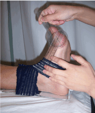
Figure 1. Measurement of extension ROM of the first MTP with the ankle fixed in a neutral position
Intensity of pain and dysfunction
Self-perceived pain rating using a visual analogue scale (VAS) [18] and dysfunction evaluation using the Chinese version of the foot functional index (FFI) [19] were conducted. The VAS in last 7-day was used to reflect the self-perceived intensity of pain. The FFI is a 23-item self-administered questionnaire. It comprises pain, disability and activity limitation subscales [20]. A higher score indicates greater dysfunction.
Statistical analysis
Descriptive statistics were used to summarize all the measured variables. Independent t-tests and Mann-Whitney U tests were performed to compare age, BMI and physical activity levels between the patient and control groups. Repeated analysis of variates was used to compare side-to-side differences in the control subjects’ first MTPJ extension range with gender as a factor. If side-to-side differences were detected, comparison were performed between patients with dominant foot being affected and dominant foot of the control; as well as patients with non-dominant foot being affected and non-dominant foot of the control. Univariate analysis of variances was used with group (Patients and control) and gender (male and female) as factors. Age, BMI and physical activity level would be regarded as a covariate if there was a significant group finding. Partial correlation coefficient tests were used to assess relationship between first MTPJ extension range, VAS, FFI and its sub-scales with gender and physical activity level as controlled factors. The level of significance was at p ≤ 0.05. The statistical analysis was performed with the SPSS v17 software packages.
Results
Patient characteristics
A total of 36 patients (mean age 44.3 years; range 25-57 years) satisfied the inclusion criteria and were recruited. Meanwhile 21 healthy subjects (mean age 45.1 years, range 33-58 years) participated as the controls.
Table 1 shows the characteristics of patients and the control group. The two groups had comparable age and gender distributions, but the patient group had a significantly greater average BMI (by 9.2 %, p = 0.014) than the controls. More than half of the patients (19 out of 36) had plantar fasciitis on their dominant side. Of the 36 patients, 36.1% had experienced plantar fasciitis for 12 months and beyond.
Table 1. Participant Characteristics*
|
|
Patient group
(n = 36) |
Control group
(n = 21) |
p value
|
|
Age, y |
44.31 ± 8.44
|
45.14 + 7.70
|
.71
|
Gender
|
Female
Male |
22
14 |
9
12 |
.19 |
|
BMI, kg/m2 |
25.38 ± 3.19 |
23.25 + 2.80 |
.01 |
Physical Activity |
Sedentary/light
Moderate/active
|
7
29
|
7
14
|
.34 |
Duration of symptoms |
3-6 months
> 6 and < 12 months
³ 12 months |
20
3
13 |
|
Affected foot |
(D/ND) |
19/17 |
|
|
BMI: body mass index; D: dominant; ND: non-dominant
*Values are presented as mean+ SD or numbers of subjects
Leg dominant effect in healthy controls
Among the healthy controls, significant side-to-side differences in range of motion of the first MTPJ was observed (p ≤ 0.05) (Table 2). The dominant side had significantly larger active (by 3.3° ± 3.1°) and passive (by 4.0°± 6.2°) MTPJ extension than the non-dominant side.
Table 2. Side-to-side comparisons of the first MTPJ extension range in the healthy controls.
|
Dominant foot
(n = 21) |
Non-dominant foot
(n = 21) |
side-to-side difference |
p value |
Active ROM Female |
68.70 ± 8.34 |
66.26 ± 7.47 |
3.33 ± 3.12 |
0.00 |
Male |
74.97 ± 6.71 |
70.97 ± 5.64 |
Passive ROM Female |
77.29 ± 4.84 |
73.70 ± 6.06 |
3.98 ± 6.19 |
0.01 |
Male |
87.41± 7.02 |
83.14 ± 7.24 |
ROM: range of motion; MTPJ: metatarsal phalangeal joint
Comparisons of patients with plantar fasciitis and healthy controls
Multivariate analysis of variance revealed that female patients with PF had larger passive first MTPJ extension in the dominant and non-dominant feet (all p < 0.05) (Table 3). There was no significant difference on the active ROM (p > 0.05). No significant difference between male patients and healthy control on the active and passive MTPJ extension range (all p > 0.05).
Table 3. Comparison between patients and healthy controls
|
Affected Foot |
1st MTPJ ROM |
Gender |
Patient |
Healthy control |
p value |
|
| |
|
Dominant |
Active ROM |
Female |
66.20 ± 15.40 |
68.70 ± 8.34 |
.689 |
|
Male |
71.04 ± 7.23 |
74.97 ± 6.71 |
.498 |
|
|
Passive ROM |
Female |
88.97 ± 12.73 |
77.29 ± 4.84 |
.034 |
|
Male |
79.30 ± 11.30 |
87.41 ± 7.02 |
.071 |
|
|
Non-Dominant |
Active ROM |
Female |
74.00 ± 11.27 |
66.26 ± 7.47 |
.095 |
|
Male |
75.67 ± 7.33 |
70.97 ± 5.64 |
.390 |
|
Passive ROM |
Female |
86.61 ± 10.64 |
73.70 ± 6.06 |
.006 |
|
| |
Male |
85.5 ± 36.79 |
83.14 ± 7.24 |
.478 |
|
ROM: range of motion; MTPJ: metatarsal phalangeal joint
Relationship between MTPJ range and intensity of pain and dysfunction
Table 4 shows that significant relationships were detected between passive first MTPJ extension range and intensity of pain (p = -0.54, p = 0.017, figure 2) and pain subscale of the FFI score (p = -0.47, p = 0.042) in female patients with PF. No relationships were detected in male patients with PF.
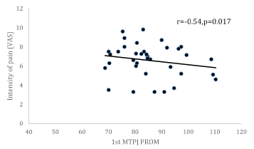
Figure 2. Measurement of extension ROM of the first MTP with the ankle fixed in a neutral position
Table 4. Relationship between passive first MTPJ extension range, pain and functional score
|
Gender |
|
1st MTPJ AROM |
1st MTPJ PROM |
Partial Correlation Coefficient (r) |
p value |
Partial Correlation Coefficient (r) |
p value |
|
Female |
VAS |
-.42 |
.070 |
-.54 |
.017 |
FFI_Pain |
-.39 |
.095 |
-.47 |
.042 |
FFI |
-.31 |
.204 |
-.32 |
.188 |
|
Male |
VAS |
.40 |
.221 |
.47 |
.140 |
FFI_Pain |
.38 |
.251 |
.46 |
.160 |
FFI |
.30 |
.367 |
.34 |
.311 |
MTPJ: metatarsal phalangeal joint; AROM: active range of motion; PROM: passive range of motion; VAS: visual analog scale; FFI_Pain: pain score of foot functional index; FFI: foot functional index
Discussion
This study was designed to delineate the first MTPJ ROM differences between patients with unilateral plantar fasciitis and healthy subjects and its association with pain and dysfunction. We detected an increased in passive extension range of the first MTPJ in female patients with plantar fasciitis compared with healthy controls. A greater joint range is associated with less intensity of pain in the female patients.
The result of this study showed dominant foot has more extension of the first MTPJ than non-dominant foot in healthy subjects, despite the difference are relatively small. We speculated that since the non-dominant side is the balance leg, subjects spend more time with the toes in extension on their dominant side in the push-off phase of gait, which made the dominant foot more susceptible to PF, e.g. in our patient group, we have more number of patients with dominant foot affected than non-dominant foot affected (19 vs. 17). We also observed smaller first MTP extension range in the female than male healthy subjects. In contrast, previous study has reported higher MTPJ ROM were found in female than male runners [21]. But they measured the MTPJ extension in a weight bearing position and did not stabilize the ankle joint. Different activity level and method to measure the MTPJ may all contribute to the different results between their study and our study. But both studies indicated differences in the first MTPJ ROM between males and females, which might also explain the risk profile difference between males and females. In our study, based on findings in healthy controls, comparison between healthy and patients with PF were conducted with gender, BMI, and leg dominance as controlled factors in later statistical analysis.
We detected greater first MTPJ extension in female subjects with PF than the healthy control. Creighton and Olson [10] reported decreased passive extension of the first MTPJ in runners with plantar fasciitis. The findings were based on small number of subjects (6 runners and 6 control). A later study from Allen and Gross [8] found comparable passive extension range of the first MTPJ in the affected and unaffected feet in patients with unilateral PF. But in that study, leg dominance was not being considered. In our study, we recruit more subjects and group comparison were conducted with control on leg dominance and gender. Our findings support the observation from Howell [13] that patients with PF have greater first MTPJ extension range. Moreover, we found MTPJ ROM is more significant in female patients. Previous studies have reported females have greater joint mobility of the foot and higher flaccidity foot ligaments than males [22] and analysis of foot radiographic images further proved the gender difference [23]. But due to the cross- sectional study design, we could not draw the conclusion regarding the cause and effect relationship between increased MTPJ ROM and development to PF.
In female patients, negative associations with extension range of first MTPJ and intensity of pain during walking and pain sub-scale of the FFI might suggest that a greater first MTPJ extension is associated with less intensity of pain; or less MTPJ extension is associated with greater intensity of pain. Based on our findings, there is a possibility that the female patients increased extension in their first MTPJ to minimize the stress on the plantar fascia. Why male subjects cannot have similar relationship might due to gender difference in joint laxity [22]. Our findings also support that stretching of plantar fascia can be used to ease the symptoms of patients with PF. On the other hand, due to the gender difference in MTPJ ROM, foot ligaments laxity as well as the muscle strength, for patients who demonstrate excessive MTPJ extension, there may be a length related weakness of the intrinsic foot muscles [24]. Therefore, strengthening exercises may need to be added to the treatment plan for this type of patients [25].
Increased BMI is reported frequently in patients with plantar fasciitis [26,27]. Theoretically, heavier individuals place more loading on the medial longitudinal arch, and this might increase the risk of developing plantar fasciitis [28,29]. Riddle et al. [27] have reported that an increase of BMI by more than 30 kg/m2 predicts a 5.6-fold increase in the risk of having plantar fasciitis. However, Rome et al. [17] reported no significant difference in average BMI for runners with and without heel pain. Butterworth et al. [30] have concluded that greater BMI is strongly associated with heel pain only in a non-athletic population. In this study, the majority of the patients were not athletes, yet we found significantly higher BMIs (average 25.38 kg/m2) in the patient group compared with the healthy controls (23.25 kg/m2). Note that in Asia people with a BMI of more than 25 kg/ m2 are regarded as obese [31]. In this study, about 55.56% of the patients belonged to the obese group and suffered from proximal heel pain associated with plantar fasciitis.
Clinical and research implications
Stretching [32,33] and strengthening foot and calf muscles [4,25] are found as an effective means to relieve the plantar heel pain. Whether the effect would be gender-specific needs further investigation. Findings from the present study indicates that female patients have greater passive extension first MTPJ range, strengthening exercises might be requested to stabilize the increased joint range and minimize stretch-related weakness of the intrinsic foot muscles.
In addition, weight control is one means to reduce loading and stress on the plantar fascia, but weight-bearing exercises might increase the impact on it. Indeed, any increase in physical activity has been identified as one of the risk factors for plantar fasciitis [26,27,34]. Non-weight bearing exercises with, for example, a stationary exercise bike, can reduce weight [35] without inducing mechanic loading on the fascia, and it could be an alternative for these individuals.
Limitatio
This was a cross-sectional observational study, so no cause and effect relationships could be established. Further longitudinal studies are warranted to confirm the cause and effect relationship between the first MTPJ ROM and plantar fasciitis.
Conclusions
Increase passive first MTPJ extension range was observed in female patients with plantar fasciitis. In the female patients, an increase in passive first MTPJ extension range is associated with less intensity of pain. Such findings suggest an adaptation in the first MTPJ range in female patients with PF. Therefore, strengthening exercises may need to be added to the treatment plan to minimize stretch-related weakness of the intrinsic foot muscles and stabilize the increased joint range.
Acknowledgment
The authors wish to thank the participants of this study.
Funding information
This study is supported by the university education funding from the Department of Rehabilitation Sciences of the Hong Kong Polytechnic University
Competing interest
The authors declare that they have no competing interests
References
- McPoil TG, Martin RL, Cornwall MW, Wukich DK, Irrgang JJ, et al. (2008) Heel pain--plantar fasciitis: clinical practice guidelines linked to the international classification of function, disability, and health from the orthopaedic section of the American Physical Therapy Association. J Orthop Sports Phys Ther 38: A1-1A18. [Crossref]
- Neufeld SK, Cerrato R (2008) Plantar fasciitis: evaluation and treatment. J Am Acad Orthop Surg 16: 338-346. [Crossref]
- Wearing SC, Smeathers JE, Urry SR, Hennig EM, Hills AP (2006) The pathomechanics of plantar fasciitis. Sports Med 36: 585-611. [Crossref]
- Martin RL, Davenport TE, Reischl SF, McPoil TG, Matheson JW, et al. (2014) Heel pain-plantar fasciitis: revision 2014. J Orthop Sports Phys Ther 44: A1-33. [Crossref]
- Klein SE, Dale AM, Hayes MH, Johnson JE, McCormick JJ, et al. (2012) Clinical presentation and self-reported patterns of pain and function in patients with plantar heel pain. Foot Ankle Int 33: 693-698. [Crossref]
- O'Malley MJ, Page A, Cook R (2000) Endoscopic plantar fasciotomy for chronic heel pain. Foot Ankle Int 21: 505-510. [Crossref]
- Yi TI, Lee GE, Seo IS, Huh WS, Yoon TH, et al. (2011) Clinical characteristics of the causes of plantar heel pain. Ann Rehabil Med 35: 507-513. [Crossref]
- Allen RH, Gross MT (2003) Toe flexors strength and passive extension range of motion of the first metatarsophalangeal joint in individuals with plantar fasciitis. J Orthop Sports Phys Ther 33: 468-478.
- McGonagle D, Marzo-Ortega H, O'Connor P, Gibbon W, Pease C, et al. (2002) The role of biomechanical factors and HLA-B27 in magnetic resonance imaging-determined bone changes in plantar fascia enthesopathy. Arthritis Rheum 46: 489-493. [Crossref]
- Creighton D, Olson VL (1987) Evaluation of range of motion of the first metatarsophalangeal joint in runners with plantar fasciitis. J Orthop Sports Phys Ther 8: 357-361. [Crossref]
- Mann RA, Hagy JL (1979) The function of the toes in walking, jogging and running. Clin Orthop Relat Re: 24-29. [Crossref]
- Martin JE, Hosch JC, Goforth WP, Murff RT, Lynch DM, et al. (2001) Mechanical treatment of plantar fasciitis. A prospective study. J Am Podiatr Med Assoc 91: 55-62. [Crossref]
- Howell D (2015) Letter to the Editor: 'Effect of stretching with and without muscle strengthening exercises for the foot and hip in patients with plantar fasciitis: A randomized controlled single-blind clinical trial'. Man Ther 23: e12 [Crosref]
- DuBose KD, Kirtland KA, Hooker SP, Fields RM (2004) Physical activity trends in South Carolina, 1994-2000. South Med J 97: 806-810. [Crossref]
- Pascual Huerta J, García JM, Matamoros EC, Matamoros JC, Martínez TD (2008) Relationship of body mass index, ankle dorsiflexion, and foot pronation on plantar fascia thickness in healthy, asymptomatic subjects. J Am Podiatr Med Assoc. 98: 379-385. [Crossref]
- Elveru RA, Rothstein JM, Lamb RL (1988) Goniometric reliability in a clinical setting. Subtalar and ankle joint measurements. Phys Ther 68: 672-677. [Crossref]
- Rome K, Howe T, Haslock I (2001) Risk factors associated with the development of plantar heel pain in athletes. The Foot 11: 119-125.
- Wewers ME, Lowe NK (1990) A critical review of visual analogue scales in the measurement of clinical phenomena. Res Nurs Health 13: 227-236. [Crossref]
- Wu SH, Liang HW, Hou WH (2008) Reliability and validity of the Taiwan Chinese version of the Foot Function Index. J Formos Med Assoc 107: 111-118. [Crossref]
- Budiman-Mak E, Conrad KJ, Roach KE (1991) The Foot Function Index: a measure of foot pain and disability. J Clin Epidemiol 44: 561-570. [Crossref]
- van der Worp MP, de Wijer A, Staal JB, Nijhuis- van der Sanden MW (2014) Reproducibility of and sex differences in common orthopaedic ankle and foot tests in runners. BMC Musculoskelet Disord 15: 171. [Crossref]
- Nagano K, Okuyama R, Taniguchi N, Yoshida T (2018) Gender difference in factors affecting the medial longitudinal arch height of the foot in healthy young adults. J Phys Ther Sci 30: 675-679. [ Crossref]
- Fukano M, Fukubayashi T (2012) Gender-based differences in the functional deformation of the foot longitudinal arch. Foot (Edinb) 22: 6-9. [Crossref]
- Shirley Sahrmann, Daniel C. Azevedo, Linda Van Dillen (2001) Diagnosis of movement impairment syndrome. Braz J Phys Ther 21: 391-399 [Crossref]
- Rathleff MS, Mølgaard CM, Fredberg U, Kaalund S, Andersen KB, et al. (2015) High-load strength training improves outcome in patients with plantar fasciitis: A randomized controlled trial with 12-month follow-up. Scand J Med Sci Sports 25: e292-300. [Crossref]
- Irving DB, Cook JL, Menz HB (2006) Factors associated with chronic plantar heel pain: a systematic review. J Sci Med Sport 9: 11-22. [Crossref]
- Riddle DL, Pulisic M, Pidcoe P, Johnson RE (2003) Risk factors for Plantar fasciitis: a matched case-control study. J Bone Joint Surg Am 85: 872-877. [Crossref]
- League AC (2008) Current concepts review: plantar fasciitis. Foot Ankle Int 29: 358-366. [Crossref]
- Cullen NP, Singh D (2006) Plantar fasciitis: a review. Br J Hosp Med (Lond) 67: 72-76. [Crossref]
- Butterworth PA, Landorf KB, Smith SE, Menz HB (2012) The association between body mass index and musculoskeletal foot disorders: a systematic review. Obes Rev 13: 630-642. [Crossref]
- Shiwaku K, Anuurad E, Enkhmaa B, Nogi A, Kitajima K, et al. (2004) Overweight Japanese with body mass indexes of 23.0-24.9 have higher risks for obesity-associated disorders: a comparison of Japanese and Mongolians. Int J Obes Relat Metab Disord 28:152-158.
- Digiovanni BF, Nawoczenski DA, Malay DP, Graci PA, Williams TT, et al. (2006) Plantar fascia-specific stretching exercise improves outcomes in patients with chronic plantar fasciitis. A prospective clinical trial with two-year follow-up. J Bone Joint Surg Am 88: 1775-1781. [Crossref]
- Kamonseki DH, Gonçalves GA, Yi LC, Júnior IL (2015) Effect of stretching with and without muscle strengthening exercises for the foot and hip in patients with plantar fasciitis: A randomized controlled single-blind clinical trial. Man Ther 23: 76-82 [Crossref]
- Healey K, Chen K (2010) Plantar fasciitis: current diagnostic modalities and treatments. Clin Podiatr Med Surg 27: 369-380. [Crossref]
- Templeton DL, Kelly AS, Steinberger J, Dengel DR (2010) Lower relative bone mineral content in obese adolescents: role of non-weight bearing exercise. Pediatr Exerc Sci 22: 557-568. [Crossref]