Ideal properties of gold nanoshell has resulted in making it a ray of hope in biomedical areas such as targeted drug delivery, cancer detection and treatment and in eliminating tumors without harming normal healthy cells. Gold nanoshells are spherical particles with diameter ranging from 10-200 nm consisting of a dielectric core that is covered by a thin metallic shell of gold. An important role of gold nanoparticle based agents is their multifunctional nature. This review focuses on physics, synthesis and biomedical applications of gold nanoshells due to their inert nature, non-cytotoxicity and biocompatibility.
gold nanoshell, cancer, diagnosis, treatment
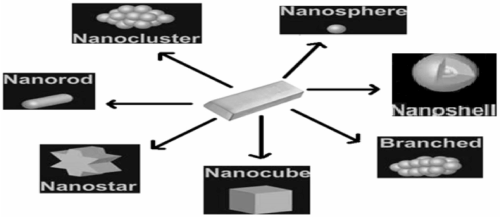
The discovery of nanoshell was made by Professor Naomi J. Halas and her team at Rice University in 2003 [1,2]. Nanotechnologies can be defined as design, characterization, production and application of structures, devices and systems by controlling shape and size at a nanometer scale [2,3]. Gold nanoparticles show different shapes as shown in Figure 1 [3].

Figure 1. Shapes of Gold Nanoparticles
Nanoshell particles comprise of special class of nanocomposite materials [4]. Gold nanoshells are spherical nanoparticles composed of a dielectric core which is covered by a thin gold shell with tunable optical resonances [5,6]. These nanoshells consist of a quasiparticle known as plasmon which is a collective excitation or quantum plasma oscillation in which the electrons simultaneously oscillate with respect to all the ions. Gold nanoshells possess an attractive set of optical, chemical and physical properties which help in cancer detection, treatment and medical biosensing [7]. Nanoshells can be varied across a broad range of light spectrum that spans the visible and near infrared regions. In the near infrared regions ‘tissue window’, light penetration into tissue is optimal. Nanoshells that are tuned to absorb near infrared region radiation are particularly useful as mediators of photo thermal cancer therapy because they are efficient enough to convert absorbed radiations into heat, and are stable at therapeutic temperatures. Moreover, nanoshells preferentially accumulate at tumor sites due to their nanoscale dimensions [7].
Advantages
- Biocompatibility
- Non-cytotoxicity
- Bio sensing application
- Protection of drugs from being degraded in the body before they reach their target site
- Enhance drug absorption into tumors and cancerous cells
- Prevention of drugs from interacting with normal cells, thus avoiding harmful effects [7,8]
How Nano shells work to kill cancerous cells
Gold nanoshells that are designed to absorb radiation at various frequencies absorb certain types of radiation .Once the nanoshells are attached to the cancerous cells, only laser light is needed to treat the cancer. Near infrared (NIR) light passes through the body and reaches the gold nanoshell The tuned gold nanoshell receives the NIR light and converts the light energy into heat, killing the cancer cells [8].
Mechanism
Near infrared light is a type of low-energy radiation not absorbed by living tissue. However, the nanoshells can be designed to absorb this light, which heats them up. A nanoshell uses surface plasmon resonance, or a wave-like excitation of electrons along the surface, to convert laser light into heat. Because nanoshells are metallic on the outside and small in size, the laser light interacts with the shell, causing surface plasmon resonance. Electrons in the gold prefer to have low energy, so they give off any extra energy as heat to the surrounding environment, including the attached cancer cell. This process causes the temperature of the cancer cell to increase after 4-6 minutes of laser light exposure. Nanoshells convert light to heat energy. The transferred heat is strong enough to destroy the cancer cell. Healthy cells then consume the dead cancer cells through phagocytosis. When the process is done, only healthy cells remain and there are no traces of cancer.
Surface plasmon resonance
Gold nanoshells exhibit unique optical properties because their interaction with the electromagnetic field is greatly intensified by a phenomenon known as localized surface plasmon resonance. It represents a collective oscillation of free electrons of metal at its surface. The surface plasmon resonance of gold nanoshells at visible and near infrared region wavelengths is possible because of two factors viz., nanoscale dimension and dielectric function of gold at optical wavelengths. Modification of the standard Drude dielectric approximation, by adding a single frequency dependent Drude-Lorentz oscillator term (L), to account for interband transitions of bound electrons, which are excited by wavelengths shorter than ∼ 550 nm. Mathematically, the Drude-Lorentz dielectric function is given as follows:

where,
ωp - Volume plasma frequency of bulk gold
ω - Angular frequency of electromagnetic field
Γ - Bulk collision frequency
The complex dielectric function of gold have a large, negative real component accounting for the reflectivity of bulk gold, and a small, positive imaginary component, which is associated with absorption [7,9,10,11].
Quasi - Static approximation (dipole approximation)
Quasi - static approximation is a first - order approach about the interaction between a gold nanoshell and electromagnetic radiation at optical frequencies. Equation for the electric field in the quasistatic approximation has been obtained by letting the wavelength tend to infinity relative to the particle size. The scattering, extinction and absorption cross-sections are

where,
c = Speed of light
ω = Angular frequency
Α = Complex polarizability of the particle

where,
∈core, ∈shell and ∈m = Dielectric functions of core, shell and medium that is surrounding
f = (rcore/rshell)1/3 is fraction of total particle volume occupied by core
Mie theory
Mie scattering theory provides complete analytical solution for scattering of electromagnetic radiations by particles with spherical or cylindrical symmetry. In Mie theory, harmonically oscillating electromagnetic fields are given in terms of a set of spherical vector basis and functions in such a way that each term in the expansion shows one of the resonances. Mie theory is a resourceful technique which is used for determining optical properties of nanoshells or other spherical particles of any dimension.
The Plasmon Resonance tunability of nanoshells can be predicted from Mie theory, but it does not intuitively explain why the position of plasmon resonance peak has to be selected by adjusting the core/shell ratio. The analysis is restricted to a dipole limit. In the hybridization picture, the geometry - dependent nature of plasmon resonance in a nanoshell comes from the interaction of individual sphere and cavity plasmons. The strength of interaction depends on the thickness of the gold shell, and therefore the core/shell ratio. The frequencies of bonding (ω-) and anti - bonding (ω+) plasmon modes decompose as spherical harmonics of order l are given by:

ωB = Bulk plasmon frequency
For bonding plasmon modes, a higher core/shell ratio produces lower - frequency plasmon modes, corresponding to plasmon resonance at longer wavelengths, which is consistent with Mie theory.
Near-field enhancement
Near - field enhancement takes place when large amounts of charge temporarily build up on the surface of a gold nanoshell exposed to light at resonant wavelengths.
Photo thermal material characteristics
The high efficient coupling of nanoshells to incident electromagnetic energy at surface plasmon frequencies give rise to intense absorption and, in turn, heating of gold nanoshells. Gold nanoshells display excellent thermal stability. The structure remains intact when exposed to light intensities, which is required for applications in imaging and photo thermal therapy [7].
Synthesis of gold nanoparticles
Gold nanoparticles are synthesized chemically; 3 ml ammonia with 50 ml absolute ethanol was stirred vigorously for 15 minutes. Tetraethyl Orthosilicate (TEOS) was added dropwise. Opaque white solution of silica nanoparticles was obtained. 3-Aminopropyltrimethoxy Silane (APTMS) was added with vigorous stirring and could react for 2 hours. APTMS coated silica nanoparticles obtained precipitated at the bottom and clear solution remained at the top. APTMS coated silica nanoparticles were purified by centrifugation (speed: 2000, 3500, 5500rpm) and redispersed in ethanol.
For preparation of colloidal gold nanoparticles, 1M NaOH and Tetrakis Hydroxymethyl Phosphonium Chloride (THPC) was added to HPLC grade water. Solution was stirred for 5 minutes. Hydrogen tetrachloraurate (HAuCl4) was added to the stirred solution. Brown colored solution of gold nanoparticles was produced within few seconds after addition of chloroauric acid.
Attachment of colloidal gold to silica nanoparticles: Silica nanoparticles dispersed in ethanol was added to colloidal gold in a tube and shaken for 5 minutes and left to settle down for 2 hours. The mixture was centrifuged (2000 rpm) and red coloured pellets precipitated at the bottom of the tube. Supernatant was removed and remaining red coloured pellets was redispersed in HPLC grade water (Solution A). (Solution B) Potassium carbonate was dissolved in water and kept for 10 minutes stirring after which Hydrogen tetrachloraurate (HAuCl4) was added. The solution was initially yellow and after 30 minutes the solution became colourless. Solution A was added to solution B and after addition of formaldehyde the colourless solution became purple. Purple colored nanoshells obtained (Figure 2) [8-11].

Figure 2. Transmission electron microscopy depicted picture of growth of nanoshell from core silica to gold covered nanoshell.
Transport mechanisms
If the host is tumor free nanoshell surface is not modified by antibodies or other protein binding ligands. Since normal blood vessels are highly permeable to particles, nanoshells will remain in circulation until they reach the spleen and liver, where they are scavenged by macrophages. If a tumor is present, a prominent feature of vascular transport in tumors is Enhanced Permeability and Retention (EPR effect), where particles with diameters of tens to hundreds of nanometers pass through the ‘leaky’ microvasculature and accumulate in the tumor interstitium. After accumulation into interstitial space, transport occurs via diffusion and convection [7].
Toxicity
No comprehensive studies evaluating long - term toxicity of gold nanoshells has been reported. All available proof shows that at physiological doses, gold nanoshells are not cytotoxic and do not cause any short-term health risks. Gold has been universally recognized as the most biologically inert of metals. Silica nanospheres have been shown to be nontoxic in a murine mouse model. The safety of gold nanospheres has been well documented; in experiments performed in vitro, a gold nanosphere incubated with macrophages was found to be both noncytotoxic and nonimmunogenic. A nanosphere was also found to reduce production of both Reactive Oxygen Species (ROS) and nitrite radicals, and does not stimulate secretion of inflammatory cytokines. Numerous in vivo studies that were conducted in mice have provided best evidence that nanoshells are not only nontoxic but also safe. Although a prolonged respiratory exposure to high doses of crystalline silica have been linked to lung cancer in epidemiological studies, the carcinogenic potential of gold nanoshells is minimal because silica used in nanoshells is completely obscured from the host by gold shell, which is not carcinogenic [7].
Biomedical applications
Due to their unique physical characteristics and benign toxicity profile, gold nanoshell has a number of biomedical applications. Stability, functional flexibility, low toxicity and ability to control the release of drugs offer gold nanoshells a number of possibilities for development of drug delivery systems. It has been shown great potential as integrated cancer targeting, imaging and therapy agents. As contrast agents, nanoshell bioconjugates has been used to detect and image individual cancer cells in vitro and in solid tumors in vivo. As photo thermal agents, nanoshells have been successfully used in animal studies to induce thermal necrosis of tumors. Besides cancer treatment, nanoshells have proven their use in a number of novel applications as biosensors for sensitive detection of biomarkers at the ng level
In vitro cancer detection and imaging
Detecting cancer in early stages is associated with positive patient outcomes, including reduced morbidity and improved survival rates. As cancer originates from a small number of malignant epithelial cells, the ability to detect low numbers of malignant or epithelial cells in vivo would represent a giant leap forward in the fight against cancer. A number of groups have successfully demonstrated in vitro single cancer cell detection, with exceptional contrast and specificity, using bioconjugated gold nanoparticles as molecular - specific contrast agents. General detection depends on conjugating nanoparticles to antibodies that target epithelial cell - surface receptors Epidermal Growth Hormone Receptor (EGFR) and human epidermal growth factor receptor 2 (HER2) (e.g. EGFR and HER2) that are generally overexpressed in cancer cells.

Figure 3. Applications of Gold nanoshells in various therapies.

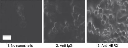
Figure 4. Dark - field images of SKBr3 cancer cells exposed to 1. No nanoshells, 2. Anti - IgG - conjugated nanoshells and 3. Anti - HER2 conjugated nanoshells.
The resultant high concentration of nanoparticles found on the surface of targeted cancer cells, combined with the high scattering cross - sections, thereby greatly facilitate imaging on the cellular level. Anti - HER2 - conjugated nanoshells has been used to detect and image HER2 - positive SKBr3 breast adenocarcinoma cells using dark - field microscopy in vitro. In this experiment, both SKBr3 and MCF7 (HER2 - negative) cancer cells were incubated with nanoshells at a concentration of 8 for one hour. Consequently, SKBr3 cells targeted with molecular specific anti-HER2 showed a marked (300%) increase in contrast over the nonspecific anti - IgG control group, whereby there is no appreciable differences in contrast were noted between HER2 - negative control cell groups, indicating that nanoshells targeted the HER2 receptor on SKBr3 cells with high specificity
In vivo cancer detection and imaging
Progress in detection and imaging of malignant cells in vitro have been followed up by in vivo studies, where bioconjugated gold nanoparticles have been successfully used to target and detect tumors in mice. Specialized gold nanospheres have been created for SERS imaging that are first stabilized with PEG - thiol and then conjugated to a Raman reporter (malachite green) and to anti -EGFR antibodies for active tumor targeting. In the experiment, nude mice with xenografted human head and neck cancer tumors (Tu686) were injected with specialized nanospheres through the tail vein. The tumors were approximately 3 mm in diameter. After 5 hours, NIR SERS spectra were obtained using a 785 nm excitation laser on a hand - held Raman system. The SERS spectra measured from an intramuscular tumor located approximately 1 cm below the surface were distinct from the background spectra, showing the effective detection of small tumors at depths of at least 1 cm. However, based on a favorable signal - to - noise ratio, it has been concluded that the maximum achievable penetration depth for SERS detection was likely in the 1-2 cm range. Nanoshells as contrast agents for in vivo Optical Coherence Tomography (OCT) imaging were demonstrated. BALBc mice with subcutaneous murine colon carcinoma tumors was injected with PEGylated nanoshells at 20 hours before OCT imaging, which were carried out using a commercially available OCT system. Due to their large resonant scattering cross - sections and ability to accumulate at tumor site, the nanoshells were found to significantly increase the optical contrast of the tumor compared to normal tissue. Therefore, in nanoshell - treated mice the integrated scattering intensity was 56% greater in tumor tissue than normal tissue, whereas in control mice the difference was only 16%. Thus, it shows that gold nanoshells represent excellent contrast agents, and are suitable for a wide range of imaging techniques.
Drug delivery
Because of small size, gold nanoshells are taken by cells where large particles would be excluded or cleared from the body A gold nanoshell carries the pharmaceutical agent inside its core, while its shell is functionalized with a ‘binding’ agent. Through the ‘binding’ agent, the ‘targeted’ nanoparticle recognizes the target cell. The functionalized nanoparticle shell interacts with the cell membrane. The nanoparticle is ingested inside the cell, and interacts with the biomolecules inside the cell. The nanoparticle particles breaks and the pharmaceutical agent is released [9-11].
Gold nanoshells vs. alzheimer [10]
Alzheimer and other degenerative diseases are caused by the clustering of amyloidal beta (Aβ) protein. Gold nanoparticles can be functionalized to specifically attach to aggregates of this protein (amyloidosis). The functionalized gold nanoparticles specifically attach to the aggregate of amyloidal protein. The microwaves of certain frequency are irradiated on the sample. Resonance with the gold nanoshells increases the local temperature and destroys the aggregate (Figure 5).

Figure 5. (a) Before irradiation (b) After irradiation
Diagnosis and sensing
Diseases can be diagnosed through the simultaneous detection of a set of biomolecule characteristic to a specific disease type and stage (biomarker). Each cell type has a unique molecular signature that distinguishes between healthy and sick tissues. Similarly, an infection can be diagnosed by detecting the distinctive molecular signature of the infecting agent. A nanoparticle can be functionalized in such a way that it specifically targets a biomarker. Thus, the detection of the nanoparticle is linked to the detection of the biomarker, and to the diagnosis of a disease [11].
Metal nanoshells for biosensing applications
Biosensors are analytical tools that help to detect the presence of several chemical and biological agents like viruses, drugs, or bacterium in biological or ecological systems. Nanoshell biosensors work by emitting a signal that is characteristic of the virus, toxin, or bacteria to be measured, thus identifying the presence or absence of the material. The ratio between the thicknesses of the gold nanoshell to the non-conductive, silicon core determines the signal to be emitted. Gold is used because of its non-reactive properties and conductive nature, allowing the nanoparticle to absorb specific frequencies of infrared or ultraviolet light [11].
Ultrasonic delivery of silica gold nanoshells for photothermolysis of sebaceous glands in humans
Through in vitro studies and in vivo human pilot clinical studies, the use of inert, inorganic silica-gold nanoshells for the treatment of a widely prevalent, yet poorly treated disease of acne. Used ∼150nm silica-gold nanoshells, tuned to absorb near-IR light and near-IR laser irradiation to thermally disrupt overactive sebaceous glands in the skin which define the etiology of acne-related problems. Low-frequency ultrasound was used to facilitate deep glandular penetration of the nanoshells. Upon delivery of the nanoshells into the follicles and glands, followed by wiping of superficial nanoshells from skin surface and exposure of skin to near-infrared laser, nanoshells localized in the follicles absorb light, get heated, and induce focal thermolysis of sebaceous glands. Pilot human clinical studies confirmed the efficacy of ultrasonically-delivered silica-gold nanoshells in inducing photothermal disruption of sebaceous glands without damaging collateral skin [11].
Because of unique features and extensive potential for several biomedical applications, gold nanoshells and other gold nanoparticles represent a major attainment in nanotechnology. Targeted nanoshells offer a light of hope for the fight against cancer. An ideal gold nanoshells: detects, diagnoses and attacks tumorous cells. Side effects of conventional therapies have been minimized by conjugation with gold nanoshells as they provide enormous sensitivity, throughput and flexibility to increase the quality life of patients. The ultimate goal is that nanoparticle-based agents can allow efficient, specific in vivo delivery of drugs without systemic toxicity, and the dose delivered as well as the therapeutic efficacy can be accurately measured noninvasively over time.
- S Jain, D G Hirst, J M O’sullivan (2012) Gold nanoparticles as novel agents for cancer therapy. The British Journal of Radiology 101-113. [Crossref]
- ASTM International (2006) E 2456-06 Terminology for nanotechnology. West Conshohocken, PA: ASTM International.
- AK Khan, R Rashid, G Murtaza, A Zahra (2014) Gold Nanoparticles: Synthesis and Applications in Drug Delivery. Tropical Journal of Pharmaceutical Research 13: 1169-1177.
- Suchita Kalele SW, Gosavi J Urban, SK Kulkarni (2006) Nanoshell particles: Synthesis, properties and applications. Current Science 91: 1038-1052.
- Jadhav Nikhilesh, Karpe Manisha, Kadam Vilasrao (2014) Gold Nanoshells: A Review on Its Preparation Characterisation And Applications. World Journal of Pharmacy and Pharmaceutical Sciences 3: 1679-1691.
- S Farjami Shayesteh, Matin Saie (2015) The effect of surface plasmon resonance on optical response in dielectric (core)–metal (shell) nanoparticles. PRAMANA- Journal of Physics 85: 1245-1255.
- Averitt RD, Westcott SL, Halas NJ (1999) Linear optical properties of gold nanoshells. Journal of the Optical Society of America B 16: 1824-32.
- Mohammad E. Khosroshahi, Mohammad Sadegh Nourbakhsh, Lida Ghazanfari (2011) Synthesis and Biomedical Application of SiO2/Au Nanofluid Based on Laser-Induced Surface Plasmon Resonance Thermal Effect. Journal of Modern Physics 2: 944-953.
- Bradley Duncan, Chaekyu Kim, Vincent M. Rotello (2010) Gold nanoparticle platforms as drug and biomacromolecule delivery systems. Journal of Controlled Release 122-127.
- Ghosh P, Han G, De M, Kim CK, Rotello VM (2008) Gold nanoparticles in delivery applications. Adv Drug Deliv Rev 60: 1307-1315. [Crossref]
- Diego A. Gomez-Gualdron (2010) Potential Application of Nanoparticles in Medicine: Cancer Diagnosis and Therapy. Texas A&M University.


