Aim: Nowadays, functional MRI is widely used in the study of brain functions. If it has the advantage of being non-invasive and allows delimiting the zones that are activated at each stimulus in 3D, it presents several deficiencies. On the psychotic studies, the zones which activate induce the analyst in error in view of the complexity and the differences between individuals and their states of anxiety. Even minimal movements of the head influence the result; the response of the vascular system signal is delayed after the stimulus...etc. A heavy numerical processing in particular in statistical analyzes is necessary to refine the images. Despite this, difficulties persist.
Method: In this study, a fuzzy logic system in this analysis is proposed. Viewing the complexity of the system, the variables that define the constructed image are considered as inaccurate variables and therefore fuzzy variable. The motor and emotional stimulus, the parasites movements, the anxiety state are considerate as inputs system. The quality of image constructed is the output system. The data base constructed permits adjusting input variables for optimal image.
Conclusion: The proposed system allows defining the optimal interaction of the different factors for an optimal image. Considering the variables of input and output as fuzzy variables thus imprecise, this makes it possible to overcome the deficiencies of the system
fMRI, BOLD, cerebral activity, magnetic field, blood oxygenation, Fuzzy logic
IRMF is a non-invasive technique of brain function. For several years now, IRFF has been used in different fields of research; however, the translation of IRFF into cognitive neuroscience as a basis for clinical investigation is recent. Today, several difficulties are being solved [1]. Functional magnetic resonance imaging (fMRI) is the result of the blood oxygenation level (BOLD). This technique has become a widely used tool for the exploration of brain function; nevertheless, the neuro-physiological bases are still ill-defined [2]. Functional magnetic resonance imaging (fMRI) indirectly measures the increase in blood flow resulting from the neuron activation using the property of oxygen-deprived hemoglobin (Deoxy-hemoglobin) which is to disrupt a magnetic field. The BOLD signal (Blood Oxygenation Level Dependent) gives access to a section of the brain. Blood oxygenation contrast (BOLD) is a technique often used in functional magnetic resonance imaging (MRI).
This allows the study of neuronal activity. The problem is that it reflects variations in the volume of blood and its oxygenation and not neuronal activity as such. The BOLD signal represents indirectly the activity of neural networks. Thus, it does not directly reflect its activity.
In recent years several studies have been devoted to the analysis of behaviors based on magnetic resonance imaging, in particular functional magnetic resonance imaging (MRI).
These techniques are based on the contrast of images resulting from the level of oxygenation of the blood. From a spatial resolution of the BOLD contrast image of the fIRM, it becomes easy to locate the active brain areas and their boundaries. However, the response of the vascular system signal is delayed by one to two seconds after the stimulus and sometimes even up to five seconds [3,4].
For the purpose of understanding and physiological interpretation of the BOLD signal, it is then necessary to separate the signal resulting from the transformation of microscopic vascular dilatations and oxygenation. That is to say each of them individually to different sequences of pulses and intensities of the magnetic field.
This is not a slim task because it is very difficult to quantify the oxygenation of blood in individual compartments during brain activity [5].
Different factors are involved in the formation of the image. These factors are very complex and difficult to analyze given the interconnection between them and the complexity of the neural network. Statistical analysis models are used. This is mainly to select the statistically significant pixels to form the three-dimensional image of the activated brain areas. Motor or emotional external parameters interfere with the stimulus to be studied and thus produce an imprecision in the formation of the image obtained.
The proposed system makes it possible to link the stimuli and parasitic factors as input variables of the system to the quality of the obtained image considered as output variable. Viewing the nature of the environment, these variables are considered fuzzy. A rule base is established to link inputs to output from multiple clinical records. The system will then fix the optimal variables at the input for a precise image.
The first difficulty in functional MRI analysis using BOLD contrast is that it is the result of the oxyHb / deoxyHb ratio expressing functional neuronal hemodynamic activation, whereas these are two phenomena that do not overlap exactly. This practically generates an error of identification of the activated zone. Also, even the smallest movements such as breathing alter the result and can produce distortions of the magnetic signal and even its loss at the interfaces of the bones, air, hematomas ... etc.
Despite the repetition of the stimulus several times according to the mode of activation sensory, motor or emotional, deficiencies persist [6]. This limits the spatial resolution as well as the quality of the image either of the physiological MRI or that of the RMF using the echo planar imaging [7].
On the psychometric level, non-concordant questions can generate the activation of certain brain areas which are not due, for example, to the responses given. This makes interpretation difficult.
Different techniques are used to overcome these deficiencies. One can notice the Angio-MRI, the inclusion of a pulse of echo of rotation and the increase of the magnetic force, the diffusion and perfusion MRI as well as the parallel techniques [8].
The most commonly used technique for achieving maximum accuracy is the establishment of the theoretical curves of the hemodynamic response and the BOLD signal. From these curves, the time lag between the measured hemodynamic response and the neuronal activation is treated in the case of repeated tasks. A heavy statistical processing is used in the analysis of the data in particular the generalized linear model (GLM). This model will be used to detect pixel by pixel those whose signal variation in time is related to the sequence of the different activation tasks. From there, the high-resolution morphological image is obtained only from the pixels that are statistically significant.
In the generalized linear model (GLMA) application, the pre-processing of acquired images uses the raw MRI signal and it must be numerically processed. The images are phased one after the other. It is also necessary to correct the effects of movements of the head of the subject by realigning the cerebral volumes between them. A step of normalization of the images, of spatial smoothing is necessary to improve the signal-to-noise ratio and possibly a temporal smoothing. These various steps of processing images can give rise to errors, in particular as regards correction of the effects of movements [9].
Fuzzy analysis is an imitation of human reasoning. In case the variables are characterized by their inaccuracy and uncertainty, the application of this mode of reasoning becomes very adequate. The time that the stimulus lasts is also a factor to be taken into account as an input variable.
In this case, the input variables (motor or emotional stimulus, parasitic disturbances such as anxiety state or involuntary movements of the patient and the stimulus duration) are imprecise and therefore fuzzy. The reaction is not the same in all patients. Also in the same subject, the consequences vary with time. The capture of several successive images is necessary. The quality of the constructed image (from the most significant pixels in the statistical methods) is also considered fuzzy. This involves defining the limits of the latter (the activated cerebral area) and from what threshold we judge the good quality of the image.
Each input or output variable is fuzzy. In this phase, the numerical variables are translated into linguistic variables. Thereafter, it is necessary to establish a database where the input variables are linked to the output variable from the observations recorded on several received images. This basis of rules is established by the human specialist. A fuzzy inference analysis [10] offers the possibility of readjustment and its adaptation to each case of study.
The result which defines the quality of the image is obtained after defuzzyfication by adopting the method of Mamdani [11].
The output variable (cerebral image quality of the activated zone) is therefore a function of the input variables (motor stimulus, emotional stimulus, motor disturbance, psychometric disturbance, stimulus duration).
Output = f (M,E,m,p)
Where: M (Motor stimulus)
E (Emotional stimulus)
m (motor disturbance)
p (psycho disturbance)
p (psycho disturbance)
d (stimulus duration)
Proposed system
Using Matlab 10, the proposed system is composed of five inputs variables and one output variable that A variable that expresses the quality of the image Figure 1.
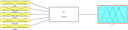
Figure 1. System architecture.
Fuzzyfication of input and output variables
The input variables are:
· M (Motor stimulus) is fuzzyfied in three fuzzy intervals (small, medium and big)
· E (Emotional stimulus) is fuzzyfied in three fuzzy intervals (low, medium and high)
· m (motor disturbance) is fuzzyfied in three fuzzy intervals (small, medium and big)
· p (psycho disturbance) is fuzzyfied in three fuzzy intervals (low, medium and high)
· d (stimulus duration) is fuzzyfied in three fuzzy intervals (short, medium and long)
The output variable (image quality) is fuzzyfied in three fuzzy intervals (bad, medium and good)
System]
Name='fMRI'
Type='mamdani'
Version=2.0
NumInputs=5
NumOutputs=1
NumRules=28
AndMethod='min'
OrMethod='max'
ImpMethod='min'
AggMethod='max'
DefuzzMethod='centroid'
[Input1]
Name='Motor'
Range= [0 4]
NumMFs=3
MF1='Smal':'trimf', [0 1 2]
MF2='Medium':'trimf', [1 2 3]
MF3='Big':'trimf', [2 3 4]
[Input2]
Name='Emotional'
Range= [0 4]
NumMFs=4
MF1='mf1':'trimf', [4 8 12]
MF2='Medium':'trimf', [1 2 3]
MF3='Low':'trimf', [0 1 2]
MF4='High':'trimf', [2 3 4]
[Input3]
Name='Mot.Dist'
Range= [0 4]
2021 Copyright OAT. All rights reserv
NumMFs=3
MF1='Small':'trimf', [0 1 2]
MF2='Medium':'trimf', [1 2 3]
MF3='Big':'trimf', [2 3 4]
[Input4]
Name='Psycho.Dist'
Range= [0 4]
NumMFs=3
MF1='Low':'trimf', [0 1 2]
MF2='Medium':'trimf', [1 2 3]
MF3='High':'trimf', [2 3 4]
[Input5]
Name='Duration'
Range= [0 4]
NumMFs=3
MF1='Short':'trimf', [0 1 2]
MF2='Medium':'trimf', [1 2 3]
MF3='Long':'trimf', [2 3 4]
[Output1]
Name='Quality'
Range= [0 4]
NumMFs=3
MF1='Bad':'trimf', [0 1 2]
MF2='Medium':'trimf', [1 2 3]
MF3='Good':'trimf', [2 3 4]
Base rules
A rule base is established that connects the input variables to the output variables. Correspondence between input and output spaces is based on real recorded values. The general syntax of the rules is of the form (IF ... THEN). The rule base must contain all possible combinations.
Once the rules are established, the system allows the result to be read instantly at the output in linguistic and numerical terms (example of application Figure 2).
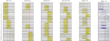
Figure 2. Application example.
By fuzzyfication, the system takes into account all uncertainties related to the nature of the variables. The result is the contribution of all factors to the input from the real values recorded during the performed tests.
It is also possible to predict the image quality the variation of the input parameters of the system.
The recorded image relating the cerebral activity is the result of the blood volume in the vessels. However, the magnetic field is very sensitive to perturbations during magnetic resonance. Sometimes the zones that are activated vary according to stimulus and are difficult to delimit. Sometimes other zones activate and do not correspond to the planned zones. The system is very complex. The proposed system, which treats all these variables as fuzzy variables, makes it possible to overcome these shortcomings. The fuzzy input variables are expressed in linguistic terms. This comes close to human reasoning. In addition, fuzzy areas are introduced between two neighboring intervals. This compensates for the vagueness associated with the linguistic valuesintroduced. In order to avoid the cumbersome nature of mathematical calculations and numerical processing mainly the statistical models used in the processing of these images, the proposed method has the advantage of simplicity and precision.
The basis of the rules being established by the human expert on the basis of the results obtained from the various successive images recorded allows laying the foundations of software that takes care of all the possibilities and the combinations encountered.
The established software makes it possible to fix values at the input to instantly read the result at the output. This may constitute a tool to assist in the radiologist of the use of fMRI by predicting the image quality and optimal parameters.
This work was carried out in collaboration between all authors. Author B.I. designed the study and wrote the protocol. Author B.K. wrote the first draft of the manuscript and managed the analyses of the study. B.S. performed intelligent analysis. M.H.C. managed the literature searches. All authors read and approved the final manuscript.
Authors declare that there are any financial competing interests (political, personal, religious, ideological, academic, intellectual, commercial or any other) in relation to this manuscript.
- Mohammad RD, Mehdi B (2012) Clinical and Research Applications. OMICS J Radiology 1:4.
- Gagnon L, Sakadžić S, Lesage F, Musacchia JJ, Lefebvre J, et al. (2015) Quantifying the Microvascular Origin of BOLD-fMRI from First Principles with Two-Photon Microscopy and an Oxygen-Sensitive Nanoprobe. J Neurosci 35: 3663-3675. [Crossref]
- Chow MSM, Wu SL, Webb SE, Gluskin K, Yew DT (2017) Functional magnetic resonance imaging and the brain: A brief review. World J Radiol 9: 5-9 [Crossref]
- Logothetis NK (2008) What we can do and what we cannot do with fMRI. Nature 453: 869-878. [Crossref]
- Buxton RB (2010) Interpreting oxygenation-based neuroimaging signals: the importance and the challenge of understanding brain oxygen metabolism. Front Neuroenergetics 2:8. [Crossref]
- Huettel SA, Song AW, McCarthy G (2009) Functional Magnetic resonance imaging. 2nd ed. Sunderland, Mass: Sinauer Associates, 1-501.
- Sung-Hong P, Paul KH, Seung HC (2015) Physiological and Functional Magnetic Resonance Imaging Using Balanced Steady-state Free Precession. Korean J Radiol 16: 550-559. [Crossref]
- Carr VA, Rissman J, Wagner AD (2010) Imaging the human medial temporal lobe with high-resolution fMRI. Neuron 65: 298-308. [Crossref]
- Freire L, Manguin JF (2001) Motion correction algorithms may create spurious brain activations in the absence of subject motion. Neuroimage 14: 709-722. [Crossref]
- Numata K (1991) General purpose fuzzy reasoning library. Version 3.0. Tokyo, Japan: Tokyo University.
- Nguyen HT, Walker EA (2006) A First Course in Fuzzy Logic. Chapman & Hall/CRC, Boca Raton.