Cardiac involvement is one of the extra-pulmonary presentations of COVID19. We describe a patient with COVID19 who had a devastating cardiac involvement without any pulmonary presentation. The cardiac presentations of COVID19 are myocardial injury, fulminant myocarditis, and myocardial infarction. Pulmonary involvement does not always appear, and clinical deterioration is unexpected.
COVID-19 is a respiratory virus of the beta-coronaviruses family [1], causing a variety of respiratory clinical manifestations from minor cough to severe respiratory failure with bi-lateral pneumonia and acute respiratory distress syndrome (ARDS) [2]. The COVID-19 virus binds to cells expressing angiotensin-converting enzyme 2 (ACE2) cells which is expressed by both the lungs and the heart [3]. Accordingly, myocardial involvement was found to be acommon extra pulmonary manifestations of COVID-19, including cardiac arrythmia, acute myocardial injury, heart failure and myocarditis [4]. Myocardial injury is associated with increased risk of mortality among COVID-19 patients [5]. Although myocardial involvement can be a devastating complication, little is known about the clinical implications and risk factors, most of which were described in myocarditis case reports.
Furthermore, severe viral infections expose patients to acute coronary events, especially among patients with cardio-vascular risk factors, who represent up to 40% of the severe ill COVID-19 patients [6].
The purpose of this current case presentation and literature review was to evaluate the clinical, electrocardiographic and electrocardiogram findings among patients with COVID-19 infection and myocarditis and discuss the differential diagnosis with acute coronary syndrome (ACS).
Case description
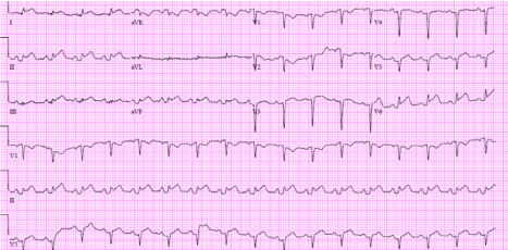
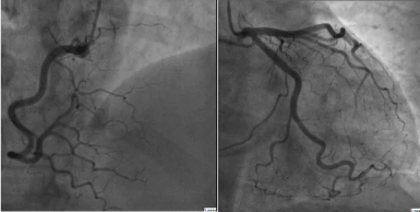
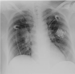
A 50 years old female with systemic scleroderma and hypertension was admitted to our medical center on March 21st with fever and weakness after exposure to a known COVID19 carrier. On arrival patient was hemodynamically and respiratory stable. Discussion was made whether or not the patient needs to be admitted to the hospital or to be sent to home care and isolation. Finally, a decision was made to admit the patient due to her past medical history and current methotrexate treatment. Few hours post hospitalization the patient became tachycardic (109 BPM) and hypotensive (87/60) which responded well to fluids. Saturation was 96% on room air, with a body temperature of 36.0. Laboratory results showed mild leukocytosis (13.7 K/micoL), normal renal function (Cr 0.98 mg/dl), hyponatremia (122 meq/L), and only mildly elevated Di-dimer (718 ng/ml) but with high sensitive Troponin I of 9297 ng/L (upper normal limit 12). The electrocardiogram showed ST elevation in inferior and posterior leads with PR depressions (Figure 1). Echocardiography (video) revealed a globally depressed left ventricular systolic function with inferior wall akinesis. After carful considerations patient was transmitted to the catheterization unit where she had invasive coronary angiography which revealed angiographically normal coronary arteries (Figure 2). The following day the patients’ condition deteriorated, and she was intubated and was started on Noradrenaline. High-sensitive Troponin levels rose to > 26,500 ng/L. Chest X-ray (Figure 3) showed pulmonary congestion without the “classical” signs of COVID-19 pneumonia or ARDS. Over the next 12 hours the patient continued to deteriorate hemodynamically and multiorgan failure occurred. The patient was put on an extracorporeal membrane oxygenation (ECMO) and Intravenous immunoglobulin treatment was initiated. Despite a previous scleroderma crisis as a result of steroids treatment a discussion was made, and the patient received pulse steroids treatment.
Figure 1. Electrocardiogram on admission
Figure 2. Cardiac catheterization
Figure 3. Chest X-ray
COVID-19 and cardiac involvement
The coronavirus family have all been associated with significant cardiovascular involvement and complications [4]. It was found that COVID-19 can cause a variety of cardiovascular complications. Acute cardiac injury, cardiogenic shock and arrythmias have all been described in varies frequencies [3]. Moreover, prior cardiovascular disease is a significant risk factor for cardiac deterioration and ACS. As in other viral diseases, infection can cause an imbalance between cardiac demand and coronary flow and cause a previously stable coronary artery disease to become uncompensated. In addition, inflammation can cause a previously stable plaque to rupture and cause type 1 myocardial infarction [4].
He XW et al. compared in 54 patients who admitted with severe conditions of COVID-19. The prevalence of myocardial injury was high among the critically ill COVID-19 patients. In-hospital mortality was significantly higher in patients with myocardial injury than in patients without myocardial injury. A recent paper by Huang [7] described 41 patients who were admitted with a confirmed COVID19 infection. Five of them had myocardial injury expressed as increased high-sensitivity cardiac troponin I, four of whom required intensive care unit. Both high levels of Troponin and LDH were associated with increased inflammation and higher mortality rates [5]. Likewise, myocarditis is a well-known cardiac sequala of many viral infections [8]. The differential diagnosis between myocarditis with viral involvement versus myocardial injury and ST-elevation myocardial infarction can be a difficult and complicated task. We here describe the three main cardiac involvement of COVID-19: myocarditis, myocardial injury, and ACS.
COVID-19 and myocardial injury
Jia-Hui Zeng et al. described [9] a 63 years old male admitted to the hospital due to COVID-19 infection. Laboratory results showed high Troponin I, myoglobin and n-terminal brain natriuretic peptide levels. Electrocardiogram showed sinus tachycardia without ST-T changes, and echocardiography showed enlarged left ventricle, diffuse myocardial dyskinesia and reduced ejection fraction of the left ventricle (32%). Treatment included Methylprednisolone and immunoglobulin. On day 11 of hospitalization the patient’s hemodynamic condition was poor and he was put on an ECMO. Fortunately, his heart gradually recovered, and the left ventricle and wall thickness returned to the normal range.
COVID-19 and myocarditis
A 37 years old male who was hospitalized due to chest pain and dyspnea was diagnosed with COVID-19 infection [10]. X-ray and chest computed tomography revealed an enlarged heart with pleural effusion. Electrocardiogram showed ST-segment elevation in inferior leads, while echocardiography showed enlarged heart with marked decrease in ventricular systolic function (ejection fraction 27%). A coronary computed tomography angiography (CCTA) found no coronary stenosis. Treatment included pulse steroids, immunoglobulin, noradrenaline, milrinone and antibiotics. One week after hospitalization echocardiography showed normal size and function of the heart. As opposed to a healthy 37 years old male, our patient was a 50 years old female with cardio-vascular risk factors. In addition, her echocardiography showed regional wall motion abnormalities. As such, anatomical clearance had to be made as soon as possible.
COVID-19 and acute coronary syndrome
Chor-Cheung Frankie et al compared the course of 7 STEMI patients during the COVID-19 outbreak to 108 patients with STEMI treated with PPCI in the prior year. The study shows numerically longer median times in all components including the time from symptom onset to first medical contact, but also in door-to-balloon and cath lab arrival to device in the COVID-19 era. The largest time difference was derived large delays in seeking medical help but also in evaluating patients with STEMI after hospital arrival. Although CCTA can replace invasive coronary angiography as a diagnostic tool, among patients with high suspicious for ACS it is a time consuming and expensive tool that exposes unnecessary medical staff to COVID-19 infection. Furthermore, since patients with COVID-19 infection and myocardial involvement are more likely to have cardio-vascular risk factors and are exposed to ACS due to high inflammatory response, invasive angiography may be the advisable tool that is cost effective, offering treatment if need be as well as diagnostic tool. However, this issue is needs to be further evaluate and at the current era a decision should be case by case.
Conclusion
In conclusion, COVID-19 is a new, fast-moving pandemic virus with cardiac involvement. On the one hand this virus seems to have myocardial affinity and cause fulminant myocarditis with severe left ventricular dysfunction and cardiogenic shock. On the other hand, the vast majority of the severely ill patients have high risk profile for ACS which may be triggered by an ongoing inflammation response. Patients with COVID-19 related myocarditis may benefit from anti-inflammatory agents such as glucosteroids and immunoglobulin, while those with ACS should be treated with anti-platelets therapy and cardiac catheterization. To the best of our knowledge, this is the first case of a patient with proved infection of COVID-19 who had cardiac catheterization and complex hospital course without manifestations of pulmonary COVID-19.
References
Zu ZY, Jiang M Di, Xu PP, Chen W, Qian Ni, et al. (2020) Coronavirus Disease 2019 (COVID-19): A Perspective from China. Radiology 296: E15-E25. [Crossref]
Lai CC, Shih TP, Ko WC, Tang HJ, Hsueh PR (2020) Severe acute respiratory syndrome coronavirus 2 (SARS-CoV-2) and coronavirus disease-2019 (COVID-19): The epidemic and the challenges. Int J Antimicrob Agents 55: 105924. [Crossref]
Wang D, Hu B, Hu C, Zhu F, Liu X, et al. (2020) Clinical Characteristics of 138 Hospitalized Patients with 2019 Novel Coronavirus-Infected Pneumonia in Wuhan, China. JAMA 323: 1061–1069. [Crossref]
Xiong T-Y, Redwood S, Prendergast B, Chen M (2020) Coronaviruses and the cardiovascular system: acute and long-term implications. Eur Heart J 41: 1–3. [Crossref]
Wu C, Hu X, Song J, Du C, Xu J, et al. (2020) Heart injury signs are associated with higher and earlier mortality in coronavirus disease 2019 (COVID-19).
Huang C, Wang Y, Li X, Ren L, Zhao J, et al. (2020) Clinical features of patients infected with 2019 novel coronavirus in Wuhan, China. Lancet 395: 497–506. [Crossref]
Andréoletti L, Lévêque N, Boulagnon C, Brasselet C, Fornes P (2009) Viral causes of human myocarditis. Arch Cardiovasc Dis 102: 559–568. [Crossref]
Zeng JH, Liu Y-X, Yuan J, Wang FX, Wu WB, et al. (2020) First Case of COVID-19 Infection with Fulminant Myocarditis Complication: Case Report and Insights. Infection 1-5. [Crossref]
Hu H, Ma F, Wei X, Fang Y (2020) Coronavirus fulminant myocarditis saved with glucocorticoid and human immunoglobulin. Eur Heart J ehaa190. [Crossref]
Editorial Information
Editor-in-Chief
Shigeo Masuda
University of Tokyo, Japan
Article Type
Case Report
Publication history
Received: August 28, 2020
Accepted: September 08, 2020
Published: September 15, 2020
Berkovitch A, Wasserstrum Y, Mayan H, Vatury O, Maor A, et al (2020) Fulminant myocarditis mimicking acute myocardial infarction in a 50 years old female patient with COVID19 proven infection. J Cardio Case Rep 3. DOI: 10.15761/JCCR.1000148.
Corresponding author
Anat Berkovitch
Division of Cardiology, Chaim Sheba Medical Center, Tel Hashomer 52621, Israel.