Hyperbaric oxygen therapy (HBO2) is approved for number of different diagnoses including chronic wounds. Frostbite represents a unique type of wound that disrupts tissue due to ice crystals forming followed by ischemic/reperfusion damage as the circulation returns during rewarming.
The benefit of HBO2 for treating frostbite results from the mechanisms of how this treatment works. The increased oxygen delivery helps to resolve the ischemia and increases the antioxidants to deal with the potential oxidative damage.
A case study is presented of a 52-year-old man who suffered frostbite while mountain climbing in Pakistan.
cold injury, frostbite, hyperbaric oxygen
Frostbite is a cold-induced injury and the term used to indicate damage to the skin and other tissues caused by freezing. There is a wide spectrum of cold injury that can vary from minimal tissue loss with mild sequelae, to major necrosis of the limbs resulting in amputations [1]. “Frostnip” is a mild form of cold injury where ice crystal form in the tissues, but no destruction occurs and the crystals dissolve as soon as the skin is warmed. Frostnip affects areas such as the earlobes, cheeks, nose, fingers, and toes and is noted when skin turns pale and numbness or tingling in the affected part is experienced until warming begins. Frostnip is considered the mildest cold exposure injury that occurs at freezing temperatures but only affects superficial layers of the skin. The injury results in skin blanching and numbness with no damage to the dermis or deeper tissues. It is completely reversible [2]. The term “chilblains” represents a more severe form of cold injury than frostnip and occurs after exposure to cold and moisture for 3-6 hours but has little or no residual consequences [3]. Immersion or “trench” foot affects the sympathetic nerves and blood vessels in the feet and is seen in those whose feet have had prolonged exposure to wet, but not freezing conditions [2]. The severity of injury is related to the temperature gradient at the skin surface and the duration of exposure [4] and wind chill [1]. The lower the temperature, of course, the faster the tissue will freeze. However, the degree of irreversible damage appears to be related to the length of time the tissue is frozen. As such, if there is prolonged exposure or a delay in seeking medical help, chances of a successful outcome are diminished [4].
Historically, frostbite concerns were primarily of interest to the military as those personnel had the greatest risk of cold weather injury [5,6]. Homeless individuals, those with psychiatric illness and excessive alcohol consumption are at risk for frostbite [7]. Smoking is also an adverse factor as it reduces the synthesis of nitric oxide [6], a vasodilator, and can potentiate the development of thrombosis by increasing fibrinogen levels and platelet activity [2].
In general, there is consensus that frostbite injury is caused by two separate processes: (1) freezing of the tissue that leads to cellular death and (2) the reperfusion injury that occurs during rewarming [8]. The formation of ice crystals begins when tissue temperature falls below 28o F (-2.2 oC) and occurs in both intra- and extracellular spaces causing direct mechanical damage to cell walls. Ice crystals in the intravascular spaces result in the second mechanism of injury that damages the endothelium and causes inflammation resulting in vascular stasis, thrombosis, and tissue ischemia [9]. The rewarming and reperfusion cause endothelial injury which results in progressive edema and intravascular thrombosis [10]. The direct injury leading to cellular destruction appears irreversible. Tissue can often appear viable initially on rewarming, however, the necrosis that follows is due to microcirculatory collapse leading to the indirect or secondary injury that occurs with reperfusion. The injury at this point may or may not be reversible depending on the mechanism of injury [11]. The mechanism(s) of indirect injury tend(s) to be the focus of frostbite research and the reason for the development of potential adjunctive therapies.
The evidence-based approach is to immerse the body part with frostbite in a warm water bath at a temperature of 37oC (98.6oF) for at least 30 min. More warm water should be added to maintain the optimal temperature range. Since rewarming is painful, ibuprofen should be started. This can provide some pain relief as well possible supporting tissue viability by decreasing the production of thromboxane and other inflammatory mediators [12].
In the early stages (> 24 h) imaging with technetium 99m pertechnetate [13] or magnetic resonance angiography [14] can be helpful in predicting the potential levels of tissue viability for amputation. This should be done after tissue rewarming [12]. Other diagnostic tests have been used to help predict the severity and assist in the prognosis of the injury. These have included radiographs, infrared thermography, angiography, triple phase bone scanning, laser Doppler studies, digital plethysmography and magnetic resonance imaging/magnetic resonance angiography [1].
Thrombolytic therapy in frostbite injury is used to address microvascular thrombosis. Bruen, et al. determined that the digital amputation rates were reduced from 41% to 10% in patients who received tPA within 24 h of injury [15]. However, there are potential risks of tPA that need to be evaluated prior to use such as compartment syndrome and failure to salvage tissue [12].
Vasodilators have been used as primary and adjunctive therapies in treating frostbite to increase vasodilatation and prevent platelet aggregation. However, Iloprost is the only vasodilator that is supported with scientific evidence [16], but it is not currently available in the United States [12].
Other studies indicate that during this second phase of frostbite injury, progressive ischemia leading to tissue necrosis, results from a cellular inflammatory-mediated process is similar-to the seen following ischemic/reperfusion injury [17]. The ischemic/reperfusion injury has been described by as the cessation of blood flow followed by vascular reflow and subsequent microvascular damage [18]. During the vascular reflow, an inflammatory reaction occurs with neutrophilic aggregation and adherence to endothelial cells leading to the production of free radicals [11]. Manson, et al. also suggested that the ischemia/reperfusion cycle generates free radicals that induce damage leading to tissue necrosis and has been shown to contribute to frostbite injury. The successful use of superoxide dismutase and deferoxamine in a rabbit ear model of frostbite confirmed that a mechanism of tissue injury in frostbite is mediated by free radicals generated during reperfusion [19].
Hyperbaric oxygen therapy (HBO2) initially, seems counterintuitive for use with an injury such as frostbite where tissue damage results from ischemia, because high levels of oxygen are linked to vasoconstriction and reduced blood flow. This conclusion was drawn from a report that, in healthy volunteers, vasoconstriction and reduction of blood flow occurred during HBO2 [4]. However, HBO2 reverses vasoconstriction in ischemic tissue and the resulting hyperoxia, from oxygen dissolved in plasma, surpasses any potential reduction in blood flow [20]. In addition to the hyperoxygenation, another mechanism of HBO2 therapy is an increased diffusion distance thus improving oxygenation in hypoxic tissue [21]. This allows marginal tissue to be maintained until revascularization has been established. Angiogenesis is also a process stimulated by HBO2 [22]. Early research into the mechanisms of HBO led to the observation that HBO reduced edema and necrosis in hypoxic skeletal muscle [23]. The detrimental effects of oxygen free radicals [24] might also be raised as an objection of HBO2, however, the intermittent administration of HBO2 results in significant increases in antioxidant enzymes [25]. After an incidence of frostbite, it can take 1 to 3 months for complete demarcation of tissue necrosis [12]. Early debridement increases the risk of removing tissue that may have survived if allowed at least 4 weeks to recover [26]. It has been determined that the use of HBO2 delineates viable from necrotic tissues [27].
The mechanism(s) involved in the delayed tissue damage following cold injury represent a complex cascade of factors. Contributing to the treatment difficulties is the fact that the injured tissues are likely to be at different stages of injury making it apparent that a combination of interventions should be used rather than a single treatment. The similarities of frostbite to thermal burns and ischemia reperfusion injury suggest that including the use of HBO2 as part of the treatment regimen is an area of investigation that should be pursued von Heimburg, et al. suggests that with the low risk associated with HBO2 and its potential benefit, it should be recommended as adjunct therapy in the treatment of deep frostbite [28].
8-11-2010
Figure 1 Patient was a 52-year-old man with a history of hypercholesterolemia but otherwise, with no significant past medical history. Two weeks prior he had been mountain climbing in Pakistan when he encountered bad weather and was caught in a snow storm and had to hike through the snow. Upon reaching camp and removing his shoe, he noticed that most of his right foot had turned a dark purple. He was treated locally for frostbite with a slow warming technique, but it was recommended that he seek treatment with hyperbaric oxygen to prevent further complications. The patient was referred to the hyperbaric unit at Texas health Presbyterian Hospital, Dallas. The patient reported that his food had more discoloration initially but today it involved mostly the distal digits. While pain was present it was controlled. The wounds were weeping, and he had not been applying any dressing but had been doing foot soaks. Recommendations were made to discontinue the foot soaks and begin wound care. The patient has dry gangrenous changes to the tips of the great toe, middle toe, and fifth digit with minimal changes on the second and fourth toes. The gangrene appears to have a good line of demarcation and no evidence for proximal cellulitis at that time.
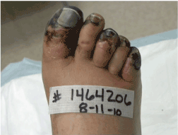
Figure 1. Patient was a 52-year-old man with a history of hypercholesterolemia but otherwise, with no significant past medical history.
Assessment indicated acute peripheral ischemia with possible micro emboli secondary to frostbite and rewarming. The patient was considered a good candidate for hyperbaric oxygen therapy (HBO2) to attempt to spare limb and digits by increasing oxygen delivery to the tissues and stimulate healing in the tissues. This treatment also helps in fighting infection and provides direct toxicity to anaerobic organisms. The treatment plan was to begin HBO2 the same day at 2.4 atmospheres absolute (ATA) for 90 min once daily with air breaks every 30 min. Alginate dressings between the toes would begin to keep them separated and help manage drainage and application of betadine to the gangrenous areas covered with an ABD pad would be applied and changed daily.
8-13-2010
Figures 2A and 2B The patient returned for continued care with no new complaints. Slightly more redness on the 3rd toe was observed but the foot was generally better. The HBO2 treatments continue but staphylococcus aureus with E. Coli was cultured. Augmentin was prescribed as an oral medication. Examination of the foot shows that improvement continues. On the 1st toe there are 2 areas of eschar left, one is lateral and considerably reduced in size. A small amount of eschar is left, perhaps 70% is removed. Sharp, excisional debridement was done with a curette through the subcutaneous tissue through the wound margins, removing the eschar. There is still some eschar on the tip of the 1st toe (1.5 cm). The toenail that was cut back is improving.
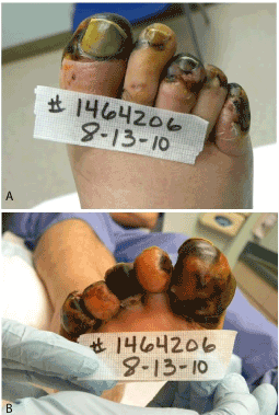
Figure 2. Figure 2A and 2B. The patient returned for continued care with no new complaints. Slightly more redness on the 3rd toe was observed but the foot was generally better. The HBO2 treatments continue but staphylococcus aureus with E. Coli was cultured. Augmentin was prescribed as an oral medication.
On the 3rd toe, sharp excisional debridement was done with a curette and scissors to remove about 70% of the thick eschar on the distal toe and the smell is better. The wound has closed over the bone. Acute peripheral arterial insufficiency continues with frostbite cellulitis of the foot, wounds to 1st, 3rd and 5th toes. The plan is to continue with HBO2 through the week and use Bactroban on the non-open areas that have had the wounds to keep bacteria down. The Acticoat foam will also be continued on the foot.
9-17-2010
30 HBO2 Tx have been completed at this time and while patient’s wounds have done very well, he will continue to be followed for wound care.
10-18-2010
The patient came in for continued wound care and the medial wound on the 1st toe is healing slowly. It has granulation over the bone that is superficial. The 5th toe is healed and the 3rd toe is healed except for the left tip of the toe. On the right 1st toe distally, sharp excisional debridement was done with a curette through the subcutaneous tissue through the wound margins, removing devitalized tissue, some fibrin and slough. No significant bleeding, minimal discomfort. On the right medial 1st toe, sharp excisional debridement was done with a curette thought the subcutaneous tissue, through the wound margins, particularly through the wound margins, removing devitalized tissue and some slough. There was quite a bit of fibrin in the base with some bleeding along the wound edges which may help initiate healing. Collagen powder and Prisma were used daily on these areas and efforts made to offload the toe more than previously.
10-25-2010
Figure 3 Distally on the 1st toe on the right foo has a lot of granulation and is still opened about 0.3 x 0.7 cm but the wound is superficial and probably less than that. Sharp excisional debridement was done with a curette through the subcutaneous wound margins removing devitalized tissue and Altrazeal with a wound veil dressing was applied.
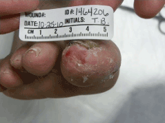
Figure 3. . Distally on the 1st toe on the right foo has a lot of granulation and is still opened about 0.3 x 0.7 cm but the wound is superficial and probably less than that. Sharp excisional debridement was done with a curette through the subcutaneous wound margins removing devitalized tissue and Altrazeal with a wound veil dressing was applied.
11-1-2010
After nearly losing his toes, they are healed except for a persistent are on the lateral 1st toe. The wound measures 0.6 x 0.3 with unlimited depth. Sharp excisional debridement was done with a curette through the subcutaneous tissue removing devitalized tissue and some fibrin and slough. There was some minimal bleeding. Distally, on the 1st toe, that area was found to be healed underneath, below the area that was cleaned off. Foam dressing applied to be changed a couple of times a week.
11-8-2010
Patient’s toes are healed except for the lateral aspect of the 1st toe that is very close to being healed. Sharp excisional debridement was done with a curette through the subcutaneous, removing devitalized tissue and some fibrin slough. There was no significant bleeding. Altrazeal and a wound veil with collagen enhancing dressing was placed over the area.
11-29-2010
Figure 4 The medial as well as the distal area of the 1st toe has now healed. There is obvious subcutaneous tissue loss that happened with the frostbite. He has areas on the 3rd and 5th toes that also are significant. He has lost lot of the distal subcutaneous tissue particularly on the 3rd toe, but these are healed. As patient resumes activity he is encouraged to find a soft foam that will protect his foot so that he doesn’t not get any ulcers. Lyrica had been offered for neuropathic pain but declined as not felt to be needed by patient. Cautions were made regarding susceptibility to frostbite and cold injury and patient will be followed as needed.
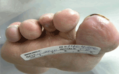
Figure 4. The medial as well as the distal area of the 1st toe has now healed. There is obvious subcutaneous tissue loss that happened with the frostbite. He has areas on the 3rd and 5th toes that also are significant. He has lost lot of the distal subcutaneous tissue particularly on the 3rd toe, but these are healed.
The authors declare that they have no competing interests.
- Imray C, Grieve A, Dhillon S, Caudwell Xtreme Everest Research Group (2009) Cold damage to the extremities: frostbite and non-freezing cold injuries. Postgrad Med J 85: 481-488. [Crossref]
- Golant A, Nord RM, Paksima N, Posner MA (2008) Cold exposure injuries to the extremities. J Am Acad Orthop Surg 16: 704-715. [Crossref]
- Prasad BK (2011) ENT morbidity at high altitude. J Laryngol Otol 125: 188-192. [Crossref]
- Murphy JV, Banwell PE, Roberts AH, McGrouther DA (2000) Frostbite: pathogenesis and treatment. J Trauma 48: 171-178. [Crossref]
- DeGroot DW, Castellani JW, Williams JO, Amoroso PJ (2003) Epidemiology of U.S. Army cold weather injuries, 1980-1999. Aviat Space Environ Med 74: 564-570. [Crossref]
- Ervasti O, Juopperi K, Kettunen P, Remes J, Rintamäki H, et al. (2004) The occurrence of frostbite and its risk factors in young men. Int J Circumpolar Health 63: 71-80. [Crossref]
- Yousif J, Saitta P, Grekin SK (2014) Frostbite on the hand of a homeless man. Cutis 94: E5-E6. [Crossref]
- Jurkovich GJ (2007) Environmental cold-induced injury. Surg Clin North Am 87: 247-267, viii. [Crossref]
- Wagner C, Pannucci CJ (2011) Thrombolytic therapy in the acute management of frostbite injuries. Air Med J 30: 39-44. [Crossref]
- Folio LR, Arkin K, Butler WP (2007) Frostbite in a mountain climber treated with hyperbaric oxygen: case report. Mil Med 172: 560-563. [Crossref]
- Zook N, Hussmann J, Brown R, Russell R, Kucan J, et al. (1998) Microcirculatory studies of frostbite injury. Ann Plast Surg 40: 246-253. [Crossref]
- McIntosh SE, Opacic M, Freer L, Grissom CK, Auerbach PS (2014) Wilderness Medical Society practice guidelines for the Prevention and treatment of frostbite: 2014 update. Wilderness Environ Med 25: S43-S54.
- Cauchy E, Chetaille E, Marchand V, Marsigny B (2001) Retrospective study of 70 cases of severe frostbite lesions: a proposed new classification scheme. Wilderness Environ Med 12: 248-255. [Crossref]
- Barker JR, Haws MJ, Brown RE, Kucan JO, Moore WD (1997) Magnetic resonance imaging of severe frostbite injuries. Ann Plast Surg. 38: 275-279.
- Bruen KJ, Ballard JR, Morris SE, Cochran A, Edelman LS, et al. (2007) Reduction of the incidence of amputation in frostbite injury with thrombolytic therapy. Arch Surg 142: 546-551. [Crossref]
- Groechenig E (1994) Treatment of frostbite with iloprost. Lancet 344: 1152-1153. [Crossref]
- Mohr WJ, Jenabzadeh K, Ahrenholz DH (2009) Cold injury. Hand Clin 25: 481-496. [Crossref]
- Zamboni WA, Roth AC, Russell RC, Graham B, Suchy H (1993) Morphologic analysis of the microcirculation during reperfusion of ischemic skeletal muscle and the effect of hyperbaric oxygen. Plast Reconstr Surg 91: 110-1123.
- Manson PN, Jesudass R, Marzella L, Bulkley GB, Im MJ, et al. (1991) Evidence for an early free radical-mediated reperfusion injury in frostbite. Free Radic Biol Med 10: 7-11. [Crossref]
- Zdeblick TA, Field GA, Shaffer JW (1988) Treatment of experimental frostbite with urokinase. J Hand Surg Am 13: 948-953. [Crossref]
- Ninikoski J, Hunt TK (1972) Measurement of wound oxygen with implanted Silastic tube. Surgery 71: 22-26. [Crossref]
- Sander AL, Henrich D, Muth CM, Marzi I, Barker JH (2009) In vivo effect of hyperbaric oxygen on wound angiogenesis and epithelialization. Wound Repair Regen. 17: 179-184.
- Strauss MB, Hargens AR, Gershuni DH, Greenberg DA, Crenshaw AG (1983) Reduction of skeletal muscle necrosis using intermittent hyperbaric oxygen in a model compartment syndrome. J Bone Joint Surg Am. 65: 656-662.
- Buras JA, Reenstra WR (2007) Endothelial-neutrophil interactions during ischemia and reperfusion injury: basic mechanisms of hyperbaric oxygen. Neurol Res 29: 127-131.
- Harabin AL, Braisted JC, Flynn ET (1990) Response of antioxidant enzymes to intermittent and continuous hyperbaric oxygen. J Appl Physiol 69: 328-335. [Crossref]
- Roche-Nagle G, Murphy D, Collins A, Sheehan S (2008) Frostbite: management options. Eur J Emerg Med 15: 173-175. [Crossref]
- Lu J, Elmarsafi T, Chrisovalantis L, Sher SR, Attinger C, et al. (2017) Septic shock following prostate biopsy: aggressive limb salvage for extremities after pressor-induced ischemic gangrene. Plast Reconstr Surg Global Open 5: 1-4.
- Von Heimburg D, Noah EM, Sieckmann UP, Pallua N (2001) Hyperbaric oxygen treatment in deep frostbite of both hands in a boy. Burns 27: 404-408. [Crossref]