Abstract
Introduction: Extravasation is an uncommon but potentially severe complication that can occur in patients being administered parenteral nutrition (a hyperosmolar solution in excess of 1000 mOsm/L in concentration) either via central venous catheters (CVC) or peripheral lines.
Case presentation: A 66-year-old male Caucasian patient who was receiving parenteral nutrition via a central venous catheter suddenly developed neck swelling (16.5 × 11.5cm) during an infusion. We discuss the clinical course taken by the patient along with a detailed review of the literature available which affected the patient's management.
Conclusion: The approach to the treatment of extravasated parenteral nutrition can be conservative, or surgical in nature. However, there is currently a lack of clearly defined evidence-based protocols for the definitive management of parenteral nutrition extravasations. Further research and implementation of sound, well-researched protocols are required for the effective management of this complication in order to avoid potentially deleterious consequences.
Keywords
Extravasation, parenteral nutrition, total parenteral nutrition, management, treatment
Introduction
Extravasation refers to the process by which substances (including fluids or medications) escape into the extravascular space, either by leakage from a vessel into surrounding tissue or by direct infiltration [1-4]. This may occur through the administration of fluids via central venous catheters (CVC) or peripheral lines [3]. Extravasates are broadly divided into irritants or vesicants, based on their potential for local toxicity [2]. The extravasation of irritants can cause an inflammatory reaction accompanied by warmth, erythema, and tenderness in the extravasated area [4,5]. Vesicants, on the other hand, are agents which can potentially result in blistering, sloughing of the skin, and deep tissue damage because they are inherently toxic [4,5].
The extravasation of total parenteral nutrition (TPN), largely an irritant, has most commonly been reported in newborns in the intensive care setting [6]. However, there are only a few case reports of this occurring in adults with the vast majority of reports over a decade old [3,6-10] (Table 1). TPN is a complex mixture of a wide variety of substances including amino acids, dextrose, lipids, vitamins, electrolytes and trace elements [4]. Very often, the solution is hyperosmolar (in excess of 1000 mOsm/L) compared with serum osmolarity of approximately 285 mOsm/L [4]. Although the exact mechanism of tissue toxicity caused by extravasated TPN is not clear at this stage, it has been previously suggested that the tissue toxicity could be related to the hyperosmolarity, acidic pH and local ions present in the parenteral nutrition [4,5,7].
Authors |
Year |
Gil ME, Mateu J. |
1998 |
Davies J, Gault D, Buchdahl R. |
1994 |
MacCara ME. |
1983 |
Gault DT. |
1993 |
Upton J, Mulliken JB, Murray JE. |
1979 |
O'Reilly C, McKay FM, Duffty P, Lloyd DJ. |
1988 |
Table 1. List of studies in citation.
Case
A 66-year-old male Caucasian patient was admitted to the Intensive Care Unit with severe pneumonia, renal failure, and gastrointestinal bleeding. During this time, he received parenteral nutrition via a subclavian CVC. This was inserted on admission three days prior, via Seldinger technique without difficulty. Correct placement was confirmed with plain film radiography. He developed a neck swelling approximately two hours after the commencement of his parenteral nutrition (1100 mOsm/L, with lipids) infusion. He complained of tightness in his neck and became progressively dyspnoeic with increasing oxygen requirements. He was subsequently intubated and ventilated. On examination of his neck, there was a soft fluctuancy (16.5 × 11.5cm) suspected to be an extravasation injury, which was not amenable to aspiration under ultrasound guidance (Figure 1). It was felt he had developed. The infusion was ceased as soon as the infiltrate was detected. He was managed conservatively with his head up at 30°, and the area of the swelling was marked for daily monitoring, and his CVC was left in place. TPN was provided via a new CVC on the contralateral side. The size of the swelling gradually reduced daily. Three days after the initial injury, it was no longer visible (Figure 2). He eventually recovered without any complications from the extravasation injury.
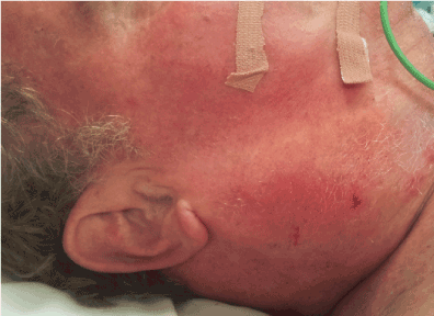
Figure 1: The initial evidence of extravasation injury.
Figure 2: Complete resolution of extravasation injury.
Discussion
The treatment of TPN extravasation should include early recognition of extravasation, with immediate discontinuation of the infusion [4,5,7]. Conservative measures such as elevation of the affected limb or the application of heat or cold have not shown any benefit [7,11-14]. It has also been recommended that the cannula is left in-situ as it may be used as a route to either administer an antidote or to aspirate the extravasated drug [7,11]. However, the passage for aspiration is often blocked [11]. Synthesis of what little literature available to us led us to adopt a conservative approach to managing this patient – watchful waiting and ceasing the infusion. The use of topically administered drugs in the treatment of extravasations is controversial [9,11]. Various “antidotes” (including glucocorticoids, antihistamines, sodium bicarbonate, heparin, and lidocaine) have been injected or topically applied to sites of chemotherapeutic extravasation injuries and have been found to be ineffective in treating such injuries [15]. The use of subcutaneously administered hyaluronidase has been previously advocated for the extravasation of parenteral nutrition [7]. However, its use is currently only recommended for the extravasation of plant alkaloids [5].
The majority of research which has been conducted with regards to the approaches to extravasation injuries examines the treatment of extravasated chemotherapeutic agents (especially cytotoxic agents) [5]. Scuderi and Onesti found that the local injection of varying amounts of normal saline solution (20-90mL, depending on the area of injury) into extravasation sites of cytostatic agents of 40 patients was sufficient to avoid tissue necrosis in all patients [16]. A variation of this technique combining saline lavage with suction has been which was performed on a patient with doxorubicin extravasation injury [17]. The saline flush-out technique described by Gault was used in the neonatal intensive care setting to treat two newborns with TPN extravasation [6,8]. Both infants healed with minimal scarring and no functional defect [6]. This technique has been proposed to flush out extravasated vesicants [5,6,18,19]. The limited success of saline lavage techniques has been mainly attributed to dilution and removal of extravasated vesicants in the tissues, with the procedure optimally performed within 6 hours of extravasation injury [18]. However, this is a both a time and labour-intensive surgical intervention which still leaves the possibility of residual extravasate remaining in the tissue [20].
The majority of the literature surrounding extravasation injuries is extremely old and is largely not specific to parenteral nutrition extravasation injuries in the neck. Further research is necessary to evaluate its true efficacy in the treatment of parenteral nutrition extravasations.
Conclusions
Extravasation of parenteral nutrition solution is potentially hazardous and can cause tissue damage due to osmotic factors and the presence of ions [7]. Prevention still remains the preferred treatment for iatrogenic injuries. However, when extravasation does occur, it is important to recognize and treat it promptly [3-5]. There is currently a paucity of literature on the principles of management of extravasation injuries, with no clear guidelines with respect to validated treatment recommendations for TPN extravasation.
References
- Fischer D, Knobf M, Durivage H (1997) The Cancer Chemotherapy Handbook: Mosby.
- Jones L, Coe P (2004) Extravasation. Eur J Oncol Nurs 8: 355-358. [Crossref]
- Gil ME, Mateu J (1998) Treatment of extravasation from parenteral nutrition solution. Ann Pharmacother 32: 51-55. [Crossref]
- Hannon MG, Lee SK (2011) Extravasation injuries. J Hand Surg Am 36: 2060-2065. [Crossref]
- Schulmeister L (2011) Extravasation management: clinical update. Semin Oncol Nurs 27: 82-90. [Crossref]
- Davies J, Gault D, Buchdahl R (1994) Preventing the scars of neonatal intensive care. Archives of disease in childhood. Fetal and neonatal edition. 70: F50-1. [Crossref]
- MacCara ME (1983) Extravasation: a hazard of intravenous therapy. Drug Intell Clin Pharm 17: 713-717. [Crossref]
- Gault DT (1993) Extravasation injuries. Br J Plast Surg 46: 91-96. [Crossref]
- Upton J, Mulliken JB, Murray JE (1979) Major intravenous extravasation injuries. Am J Surg. 137: 497-506. [Crossref]
- O'Reilly C, McKay FM, Duffty P, Lloyd DJ (1988) Glyceryl trinitrate in skin necrosis caused by extravasation of parenteral nutrition. Lancet. 2: 565-566. [Crossref]
- Dorr RT (1990) Antidotes to vesicant chemotherapy extravasations. Blood Rev 4: 41-60. [Crossref]
- Brown AS, Hoelzer DJ, Piercy SA (1979) Skin necrosis from extravasation of intravenous fluids in children. Plast Reconstr Surg. 64: 145-150. [Crossref]
- Hastings-Tolsma M, 2021 Copyright OAT. All rights reservear signs, no clear treatment? RN 57: 34-38. [Crossref]
- Beason R (1990) Antineoplastic vesicant extravasation. J Intraven Nurs. 13: 111-114. [Crossref]
- Wickham R, Engelking C, Sauerland C, Corbi D (2006) Vesicant extravasation part II: Evidence-based management and continuing controversies. Oncol Nurs forum. 33: 1143-1150. [Crossref]
- Scuderi N, Onesti MG (1994) Antitumor agents: extravasation, management, and surgical treatment. Ann Plast Surg. 32: 39-44. [Crossref]
- Vandeweyer E, Deraemaecker R (2000) Early surgical suction and washout for treatment of cytotoxic drug extravasations. Acta chirurgica Belgica. 100: 37-38. [Crossref]
- Giunta R (2004) Early subcutaneous wash-out in acute extravasations. Ann Oncol 15: 1146. [Crossref]
- Heitmann C, Durmus C, Ingianni G (1998) Surgical management after doxorubicin and epirubicin extravasation. J Hand Surg Br. 23: 666-668. [Crossref]
- Langer S, Sehested M, Jensen P (2009) Anthracycline extravasation: a comprehensive review of experimental and clinical treatments. Tumori. 95: 273-282. [Crossref]