Background: Emphysematous osteomyelitis (EOM) remains a rare entity, and only a few case reports have been published so far. Gas-forming organisms cause it, and radiological assessment plays a significant role in diagnosis. We add another case report of EOM in the sacrum.
Case presentation: We present the case of a 60-year-old woman with a septic shock and accompanying renal failure. An outside native computed tomography (CT) scan revealed multiple abscesses in the autochthonous back muscles and a tubo-ovarian abscess. In addition, a significant gas accumulation in the first sacral vertebra was noticed. The patient was subsequently transferred to our hospital. The abscess was surgically cleaved, and the patient was transferred to intensive medical care. Hemodialysis, as well as a calculated intravenous antibiotic regimen, was started for treatment. The patient's condition improved over time, and a significant decrease in intraosseous gas was observed on follow-up CT.
Conclusion: We add another case of emphysematous osteomyelitis. Short-term follow-up imaging showed the rapid resolution of the gas accumulations.
emphysematous, osteomyelitis, spine, CT
Emphysematous osteomyelitis is a rare disease with high mortality rates and is caused by gas-forming microorganisms. Only a few cases have been described in the literature, mainly in the spine, pelvis, and lower extremities [1,2,4,5,7,8]. CT is the imaging of choice, and the detection of an infectious intraosseous gas collection is essential for a prompt diagnosis. We present another case of emphysematous osteomyelitis in the first sacral vertebra.
A 60-year-old female patient was admitted to an outside hospital due to septic shock and accompanying renal failure. Diabetes, hypertension, and obesity are pre-existing conditions. First, a native magnetic resonance imaging (MRI) of the lumbar spine was obtained and showed multiple fluid collections in the autochthonous back muscles and the psoas muscle.
Besides, edema zones could be detected in the sacrum (Figure 1). A native computed tomography (CT) scan of the abdomen was obtained for further clarification and search of infection focus. Here, a slightly inhomogeneous fluid collection in the left retroperitoneum was detected. It showed a close positional relationship to the ovary and the sacrum. Adjacent to this, extensive intraosseous punctate gas accumulations were seen in the first sacral vertebra, mainly on the right side (Figure 2). The spinal canal and adjacent soft tissues showed no gas accumulation, and no cortical destruction could be detected. Also, an unclear foreign body was detected in the cervix (not shown).

Figure 1: Sagittal (a) STIR-image (short-tau-inversion-recovery) shows edema in the sacrum as well as fluid collections in the Back musculature. Transversal T2-image (b) shows left-sided fluid collections in the back musculature and psoas muscle.
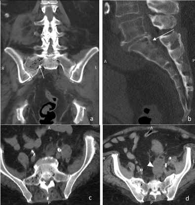
Figure 2: Coronal (a), sagittal(b), and axial(c) CT-slices highlight the presence of an extensive gas collection in sacral vertebral (white arrows), mainly on the right side. Axial CT slice of the abdomen (d) shows the intraosseous gas and the ovarian abscess formation in the left retroperitoneum (white arrowhead).
A tubo-ovarian abscess, most likely due to a residual intrauterine device (IUD), was suspected. The patient was subsequently transferred to our hospital ((Nuremberg South Hospital) and underwent surgery. The diagnosis was confirmed intraoperatively, the "forgotten" IUD was removed, and the abscess was cleaved. Emphysematous osteomyelitis was suspected, but a biopsy was not performed due to the patient's poor condition. Calculated antibiotic therapy with meropenem, linezolid, and cefazolin was started. Haemodialysis was performed to treat acute renal failure. An intraoperative vaginal swab detected colonization by Streptococcus constellatus.
Follow-up CT four days after the initial CT-scan showed regression of abscess formation and the sacral air accumulation significantly decreased (Figure 3). The patient's condition gradually improved, and she could be discharged to outpatient care in the course.
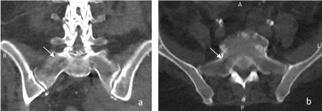
Figure 3: Coronal (a) and axial (b) CT-slices show only a minor gas accumulation in the sacral vertebra (white arrows).
Emphysematous osteomyelitis is a rare finding. The pathognomonic gas accumulations are caused by gas-forming bacteria and are usually seen in critically ill patients, as in our case [1,2,4-8]. These patients, including ours, often have predisposing conditions like Diabetes mellitus [1,5-8]. About 50 cases have been described so far, mainly in the lower spine, pelvis, and lower extremities [1,2,6,7]. Our case showed emphysematous osteomyelitis findings in the first sacral vertebra, probably due to a tubo-ovarian abscess.
Correct radiological diagnosis plays a key role since gas accumulations are a frequent finding in the radiological routine. They are usually observed in the spine and pelvis. In the intervertebral spaces and iliosacral joints, they are predominantly due to osteochondrotic changes with nitrogen gas formation. In the vertebra, they often correspond to pneumatocysts. Besides, small intraosseous air inclusions may also be detected after surgery [2,3,5].
In our case, the gas accumulations were mottled and partially confluent. No cortical bone destruction could be detected. This resembles a pattern already described in the literature, the "pumice-stone pattern" [2]. Due to this pattern and close positional relationship to the tuboovarian abscess, the gas accumulation in our case could be identified as emphysematous osteomyelitis.
In the follow-up-CT scan four days later, the gas accumulation had almost entirely resolved. This correlated to the patient's symptoms improve. As mentioned in other case reports, aggressive management, including surgery and intravenous antibiotics, was mandatory to control the infection and ensure patient survival [1,2,4,6].
We add another case report of emphysematous osteomyelitis in the sacrum. The patient recovered, and short-term follow-up imaging showed the rapid resolution of the gas accumulations.
We want to thank Prof. Tomczak for kindly providing us with the images.
- Abdelbaki A, Bhatt N, Gupta N, Li S, Abdelbaki S, Kumar Y (2017) Emphysematous osteomyelitis of the forefoot. Proc (Bayl Univ Med Cent) 31:100-101. [CrossRef]
- Small JE, Chea P, Shah N, Small KM (2018) Diagnostic Features of Emphysematous Osteomyelitis. Curr Probl Diagn Radiol S0363-0188(18)30119-1. [CrossRef]
- Feng SW, Chang MC, Wu HT, Yu JK, Wang ST, Liu CL (2011) Are intravertebral vacuum phenomena benign lesions? Eur Spine J 20:1341-8. [CrossRef]
- Luey C, Tooley D, Briggs S (2012) Emphysematous osteomyelitis: A case report and review of the literature. International journal of infectious diseases. Int J Infect Dis 16. e216-20. [CrossRef]
- Khanduri S, Singh M, Goyal A, Singh S (2018) Emphysematous osteomyelitis: Report of two cases and review of literature. Indian J Radiol Imaging 28: 78-80. [CrossRef]
- Elshikh A, Gowda N, Glass L, Maximos RB (2020) Emphysematous osteomyelitis of the clavicle: a pleural process? BMJ Case Rep13: e235764. [CrossRef]
- McDonnell O, Khaleel Z (2014) Emphysematous Osteomyelitis. JAMA Neurol 71: 512. [CrossRef]
- Kivrak AS, Sumer S, Demir NA, Aydin BK (2013) The life-saving little tip: intraosseous gas. BMJ Case Rep 2013: bcr2013201648. [CrossRef]