Abstract
Objectives: In overhead throwing sports, traditional pre-throwing programs have focused primarily on the upper extremity. However, training the total body and utilizing both the lower and upper extremity in dynamic overhead movements is becoming more popular and advised. Thus, it was the purpose of this study to examine muscles about the lumbopelvic-hip complex (LPHC) and scapula during selected exercises that could possibly be utilized in a pre-throwing conditioning program.
Design: A controlled laboratory study.
Methods: Twenty-one healthy, active individuals (171.1 ± 13.0 cm; 75.5 ± 14.8 kg; 25.3 ± 5.5 years), regardless of sex, volunteered. Surface EMG was utilized to measure muscle activation of the biceps femoris, semitendinosus, bilateral gluteus medius, gluteus maximus, erector spinae, latissimus dorsi and lower trapezius while performing four total body exercises (lungeW, kettlebell swing, kettlebell deadlift, gluteal/hamstring raise).
Results: A nonparametric Friedman Test revealed significantly different muscle activations as a factor of exercise for the biceps femoris (χ2(3) = 21.18, p < .001), gluteus maximus (χ2(3) = 39.17, p < .001), right gluteus medius (χ2(3) = 21.21, p < .001), left gluteus medius (χ2(3) = 11.02, p = .012), erector spinae (χ2(3) = 28.47, p < .001), and lower trapezius (χ2(3) = 29.84, p < .001).
Conclusion: The four exercises successfully elicited moderate to high muscle activation in all musculature, except the lower trapezius. These results imply that these four exercises could be utilized as a warm-up/pre-throwing protocol to achieve LPHC as proximal scapula muscle activation.
Key words
Core stability; Core strength; LPHC control; Muscle activation; Pre-throwing programs; Total body exercises
Introduction
The overhead throwing motion is a dynamic movement that requires coordinated sequencing of body segments via the kinetic chain, in effort to transfer energy from the lower extremity to the upper extremity for efficient ball delivery [1]. The kinetic chain denotes the segmental linkage from the most proximal segments of the foot, lower leg, upper leg to the more distal segments of the lumbopelvic-hip complex, upper arm, forearm, and hand. Efficiency of the kinetic chain requires the entire musculoskeletal-system to work synergistically in a proximal to distal manner. When the ultimate goals of overhead throwing are speed and accuracy, efficient kinetic chain function is vital.
With coordination of the kinetic chain required for dynamic overhead throwing, body segments must move interdependently in a proximal to distal manner with adequate and efficient muscle activations in attempt to transfer energy to the most proximal segment of the hand and onto the ball [2]. When examining the musculature of the kinetic chain in throwing, it is known that the legs and trunk serve as the major force generators [3], with approximately 50% of the energy generated from the lower extremity [4-6], and a 20% reduction in lower extremity forces leads to a 34% increase in forces on the shoulder [7]. With the notion that the most distal segments, the upper extremity, should be a funnel for energy transfer, [8] focus of developing and conditioning proximal segmental musculature of the lower extremity and lumbopelvic-hip complex (LPHC) for force production is paramount [9].
The inability to utilize the body as an efficient kinetic chain with proximal stability for distal mobility when performing a repetitive movement such as throwing, often leads to upper extremity injury [1,3,9]. Frequently, these types of injuries sustained by overhead throwing athletes are habitually insidious in nature and a result of a combination of cumulative overload and poor throwing mechanics [1,3,9,10]. It has been found that pitching with fatigue is the most significant risk factor, with pitch counts, pitch types, pitch velocities and pitching mechanics also being associated with the onset of overuse injury [9,11-13]. Additionally, it has been recommended that training, pre-throwing and rehabilitation programs focus on LPHC and scapular stability [13-15].
Upon examination of dynamic throwing, such as the baseball pitch, it has been found that bilateral hamstring and gluteal musculature exhibit moderate to high activation in the roles of LPHC rotation as well as stability for efficient energy transfer to the upper extremity [16]. Additionally, stability of the LPHC as well as the scapula is warranted in repetitive throwing in attempt for upper extremity injury prevention [5,6]. Thus with the known importance of both lower and upper extremity muscle activation, as well as the importance of stability about the LPHC and scapula, it was the purpose of this study to examine muscles about the LPHC and scapula during selected exercises that could possibly be utilized in a pre-throwing warm-up. It was hypothesized that muscles about the LPHC and scapula would elicit moderate to moderately high activations thus allowing for additional variety when selecting pre-throwing warm-up exercises.
Methods
The aim of this study was to quantitatively describe muscle activations of LPHC and scapular stabilizing musculature during four common rehabilitative exercises that could be utilized as in a total body conditioning for pre-throwing. Musculature selected were the following: biceps femoris (lateral hamstring), semimembranosus (medial hamstring), gluteus medius, gluteus maximus, erector spinae, latissimus dorsi and lower trapezius. For each of exercises tested, participants were instructed on performance and then were allowed to practice prior to testing. The same investigator gave exercise instruction and verbal cue feedback for all participants throughout the exercise practice and testing. All EMG data were normalized as a percent of the participant’s maximum voluntary isometric contraction (%MVIC).
Twenty-one healthy, active individuals (171.1 ± 13.0 cm; 75.5 ± 14.8 kg; 25.3 ± 5.5 years), regardless of sex, volunteered. Healthy was determined by answering NO to all questions on the Physical Activity Readiness Questionnaire (PAR-Q) and having no history of upper or lower extremity injury within the past 6 months. Active was defined as 30 minutes of physical activity most days of the week. The Institutional Review Board of the University approved all testing protocols. All exercises were explained prior to the testing procedures and informed consent was obtained from each participant prior to data collection.
Participants reported to testing prior to engaging in any physical activity that day. Location of muscle belly for EMG electrode placement was identified through palpation. The muscle bellies of all muscles were abraded and cleaned using 70% isopropyl alcohol solution prior to electrode placement. Bipolar surface electrodes (inter electrode distance: 10mm) were placed over the muscle bellies parallel to the muscle fibers using previously established standards [1,9,17,18]. The use of surface electrodes was chosen because they have been deemed to be a noninvasive technique that is able to reliably detect surface muscle activity [19-21].
The biceps femoris electrode placement was identified as the mid-point of the line from ischial tuberosity to lateral epicondyle of the tibia, while semitendinosus was the mid-point from the ischial tuberosity to the medial epicondyle of the tibia [20]. Gluteus medius placement was the proximal third of the distance from iliac crest to greater trochanter, anterior to the gluteus maximus [21,22]. The midpoint between the sacral vertebrae and greater trochanter was the placement location of the gluteus maximus [21,22]. Latissimus dorsi electrode placement was oblique, below the inferior tip of the scapula, approximately half the distance between the spine and lateral torso and parallel to the muscle fibers. Erector spinae placement was two finger widths from the spinous process of lumbar vertebra one (L1) [21,22]. Lower trapezius electrode placement was placed obliquely upward and laterally between the spine of the scapula and vertebral border of scapula and seventh thoracic spinous process [22].
Manual muscle testing (MMT) was performed following electrode placement to determine baseline maximum voluntary isometric contraction (MVIC) to which all EMG data were normalized [20,22]. The same trained investigator performed all MMTs. Inter-class correlation coefficients for the investigator were the following semitendinosus = 0.763, p < 0.001; biceps femoris = 0.658, p = 0.031; right gluteus maximus = 0.836, p = 0.001; gluteus medius = 0.796, p = 0.003, left gluteus medius = 0.985, p < 0.001; erector spinae = 0.991, p < 0.001; latissimus dorsi = 0.872, p < 0.001; lower trapezius = 0.958, p < 0.001.
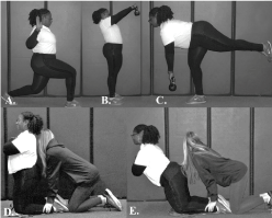
An eight channel Noraxon TeleMyo DTS (Noraxon USA, Inc, Scottsdale Arizona) was used to collect all EMG data. The signal was smoothed using a root mean square with a moving window at 100 milliseconds; data were sampled at a rate of 1000 Hz and notch filtered at frequencies of 59.5 and 60.5 Hz, respectively [23]. Following the MMTs, participants were given proper technique instruction for the four exercises. Order of exercises performed were randomized and the investigator provided verbal cues to the participants throughout all warmup practice and testing. Typical warmup practice time for each of the exercises was one minute. The common verbal cues used by the investigator were regarding postural control corrections of pelvic neutral and scapular retraction. Pelvic neutral was instructed to contract gluteals and draw stomach into spine, while scapular retraction was to pull back and squeeze together shoulder blades. Once an exercise was completed, participants were allowed a maximum of two minutes before beginning the next exercise to control for any effects of fatigue. The four exercises performed were the following: lungeW, kettlebell swing, kettlebell deadlift and gluteal/hamstring raise (Figure 1). Participants were instructed to perform a lunge by stepping forward with non-dominant arm side leg, lowering hips until both hip and knee were at 90 degrees. Participants then performed scapular retraction and humeral external rotation to make a “W” [24]. To encourage proper execution, participants were told to keep upper body straight, shoulder back and maintain pelvic neutral. The lunge exercise was performed for five repetitions.

Figure 1. A. LungeW exercise. B. Kettlebell Swing exercise. C. Kettlebell Deadlift exercise. D. Gluteal/hamstring Raise exercise.
Participants were instructed to stand with feet shoulder width apart and perform a two-hand kettlebell swing with a 4.54 kg kettlebell for the kettlebell swing exercise. Participants held the kettlebell with palms facing their body. To maintain a stable pelvis, participants were instructed and keep their back flat and neck straight then drive with hips to propel the kettlebell forward. Participants were instructed to not allow the kettlebell to go higher than their line of sight. Participants performed five consecutive swings.
For the kettlebell deadlift, participants were instructed to stand with their feet shoulder width apart and perform a kettlebell deadlift with a 4.54 kg kettlebell. Participants held the kettlebell in their non-dominant hand. Participants were instructed to keep feet parallel, flex the trunk forward and push hips back, and then lift contralateral leg off the grown. Participants then returned to the starting position for the next repetition until all five repetitions were completed.
The gluteal/hamstring raise required the participants to kneel on a cushion with knees hip width apart and cross arms over chest in an “X” position. Participants’ legs were anchored by the investigator at the base of the calves with feet plantar flexed. Participants were instructed to lean their trunk forward to a point of comfort without flexing at the hip and then return to the start position. Participants performed five consecutive repetitions.
Data were organized using a customized MATLAB (MATLAB R2010a, MathWorks, Natick, MA, USA) script. EMG data were collected for five repetitions for each of the four exercises and the third repetition recordings were averaged and used for analysis. All statistical analyses were performed using IBM SPSS Statistics 22 software (IBM Corp., Armonk, NY) with an alpha level set a priori at ? = 0.05. Prior to analysis Shapiro-Wilk Tests of Normality were run and determined data were non-normal. We ran a non-parametric Friedman test followed by a post-hoc Wilcoxon signed-rank test. For comparison purposes, low muscle activity was considered to be between 0-20% MVIC while moderate activity was 21-40%, high muscle activity was 41-60%, and very high activity was >60% [17,18,25,26].
Results
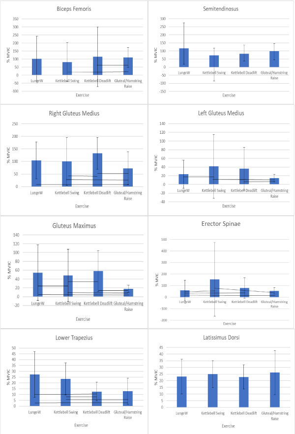
A nonparametric Friedman Test for each muscle by a factor of exercise revealed significant activation difference between exercises for the biceps femoris (χ2(3) = 21.18, p < .001); gluteus maximus (χ2(3) = 39.17, p < .001); right gluteus medius (χ2(3) = 21.21, p < .001); left gluteus medius (χ2(3) = 11.02, p = .012); erector spinae (χ2(3) = 28.47, p < .001), and lower trapezius (χ2(3) = 29.84, p < .001). There was no significance found for activation difference between the semitendinosus (χ2(3) = 7.26, p = .064) or latissimus dorsi (χ2(3) = 2.46, p = .484). Afterward, we ran a post-hoc Wilcoxon signed-rank test for each muscle. Results from the post-hoc Wilcoxon signed-rank test can be found in Table 1. Means and standard deviations can be found in Figure 2.
Table 1: Results from a post-hoc Wilcoxon signed ranks test.
Biceps Femoris |
LungeW |
Kettlebell Swing |
Kettlebell Deadlift |
LungeW |
- |
|
|
Kettlebell Swing |
-0.47 |
- |
|
Kettlebell Deadlift |
-1.19 |
-0.90 |
- |
Glute/Ham Raise |
-1.75 |
-2.50* |
-1.96* |
Right Glute Med |
LungeW |
Kettlebell Swing |
Kettlebell Deadlift |
LungeW |
- |
|
|
Kettlebell Swing |
-1.63 |
- |
|
Kettlebell Deadlift |
-1.77 |
-2.66* |
- |
Glute/Ham Raise |
-2.74* |
-2.33* |
-3.07* |
Left Glute Med |
LungeW |
Kettlebell Swing |
Kettlebell Deadlift |
LungeW |
- |
|
|
Kettlebell Swing |
-2.90* |
- |
|
Kettlebell Deadlift |
-1.38 |
-0.06 |
- |
Glute/Ham Raise |
-0.44 |
-2.48* |
-3.19* |
Glute Maximus |
LungeW |
Kettlebell Swing |
Kettlebell Deadlift |
LungeW |
- |
|
|
2021 Copyright OAT. All rights reserv
Kettlebell Swing |
-2.28* |
- |
|
Kettlebell Deadlift |
-1.44 |
-2.28* |
- |
Glute/Ham Raise |
-4.02* |
-3.81* |
-4.02* |
Erector Spinae |
LungeW |
Kettlebell Swing |
Kettlebell Deadlift |
LungeW |
- |
|
|
Kettlebell Swing |
-2.87* |
- |
|
Kettlebell Deadlift |
-3.33* |
-0.96 |
- |
Glute/Ham Raise |
-1.96* |
-2.32* |
-1.22 |
Lower Trapezius |
LungeW |
Kettlebell Swing |
Kettlebell Deadlift |
LungeW |
- |
|
|
Kettlebell Swing |
-1.15 |
- |
|
Kettlebell Deadlift |
-3.75* |
-4.11* |
- |
Glute/Ham Raise |
-3.10* |
-2.97* |
-0.37 |
Note: z scores are reported inside the table. *Indicates significance at p ≤ 0.05.

Figure 2: Muscle activation represented by %MVIC. Connecting lines show statistical significance of p < 0.05 as determined by a post-hoc Wilcoxon signed ranks test.
Discussion
The aim of this study was to describe muscle activations about the LPHC and scapula during four selected total body exercises that could possibly be utilized in a pre-throwing warm-up. The muscles selected have been documented in the literature for their contribution to LPHC and scapular stability and or mobility. Overhead throwing requires efficient movement of segments interdependently from proximal to distal with adequate muscle activations for energy generation and transfer. With the need for proximal stability through the lower extremity in throwing, strength and conditioning programs should implement exercises, such as the ones presented in this study, that could potentially assist with proximal stability for distal mobility.
The exercises examined were selected because they activated both musculature of the lower and upper extremity. Additionally, the exercises required the participants to have postural control through maintaining pelvic neutral as well as scapular retraction. Performing exercises with pelvic neutral and scapular retraction are key components of shoulder rehabilitation [27]. Of particular importance in shoulder rehabilitation is developing and maintaining LPHC and scapular stability [17,18]. Thus it was the purpose of this study to examine total body exercise that could possibly be implemented into a pre-throwing regimen in attempt to activate the musculature of the LPHC and scapula.
The current study revealed that all the four exercises (LungeW, kettlebell swing, kettlebell deadlift, and gluteal/hamstring raise) elicited moderate (21-40%MVIC) to very high (>60%MVIC) muscle activation for all selected LPHC and scapular musculature. However, low activation of the lower trapezius was demonstrated when performing the kettlebell deadlift and gluteal/hamstring raise. This finding is not too surprising when considering the two aforementioned exercises. In the execution of the kettlebell deadlift and gluteal/hamstring raise, scapular posture was not as much a part of the exercise performance as it was in the other two exercises (lungeW and kettlebell swing), even though verbal cues regarding scapular posture were given to the participants.
Upon examination of the exercises, it was evident that the total body exercises selected for this investigation were able elicit moderate to very high activation of all musculature minus the lower trapezius. Examining muscle activation further, muscle activation of the hamstrings (semitendenosis & biceps femoris) was the highest (115.02 %MVIC) when performing the lungeW. When performing the kettlebell deadlift the gluteal musculature produced the highest at 77.40 %MVIC (maximus & bilateral medius), while the kettlebell swing was able to elicit the greatest erector spinae activation (154.82 %MVIC). Examination of the scapular stabilizers (latissimus dorsi and lower trapezius) the lunge W resulted in averaged moderate activation (26.73 %MVIC) while the gluteal/hamstring raise resulted in a combined averaged of 26.16 %MVIC. Previously it has been documented that moderate muscle activation of 20-30 %MVIC is effective for muscle strengthening [16,28]. Thus, based on the importance of LPHC and scapular stability, performing exercises that have the propensity to elicit moderate activation of the aforementioned musculature, would be beneficial in any training as well as injury prevention regimen for overhead throwing athletes. Therefore, the exercises examined in this study would be good additions to an overhead athlete’s programming.
Conclusion
Overhead throwing athletes are at greater risk of upper extremity injury due to the repetitive nature of the sport. Utilization of the total body, both lower and upper extremity for efficient energy production for effective performance and injury prevention is imperative. With the need for overhead athletes utilizing both their lower and upper extremity in performance, incorporating total body exercises into a traditional upper extremity focused pre-throwing conditioning program will assist training and conditioning professionals working with throwing athletes. The total body exercises presented allow coaches, strength and conditioning, as well as sports medicine personnel alternative exercises that are capable of producing LPHC and scapular musculature activation and can effectively be utilized in a pre-throwing program.
Acknowledgements
The authors would like to acknowledge the assistance of all the members of the Sports Medicine and Movement lab for assisting with data collection.
References
- Chu SK, Jayabalan P, Kibler WB, Press J (2016) The Kinetic Chain Revisited: New Concepts on Throwing Mechanics and Injury. PM R 8: S69-S77. [Crossref]
- Putnam CA (1993) Sequential motions of body segments in striking and throwing skills: descriptions and explanations. J Biomech 26: 125-135. [Crossref]
- Kibler WB, Press J, Sciascia A (2006) The role of core stability in athletic function. Sports Med 36: 189-198. [Crossref]
- Burkhart SS, Morgan CD, Kibler WB (2003) The disabled throwing shoudler: Spectrum of pathology. Part III: the SICK scapula, scapular dyskinesis, the kinetic chain, and rehabilitation. Arthroscopy 19: 641-661. [Crossref]
- Campbell BM, Stodden DF, Nixon MK (2010) Lower extremity muscle activation during baseball pitching. J Strength Cond Res 24: 964-971. [Crossref]
- Oliver GD, Keeley DW (2010) Gluteal muscle group activation and its relationship with pelvis and torso kinematics in high-school baseball pitchers. J Strength Cond Res 24: 3015-3022. [Crossref]
- McMullen J, Uhl TL (2000) A kinetic chain approach for shoulder rehabilitation. J Athl Train 35: 329-337. [Crossref]
- Kibler WB, Chandler TJ, Shapiro R, Conuel M (2007) Muscle activation in coupled scapulohumeral motions in the high performance tennis serve. Br J Sports Med 41: 745-749. [Crossref]
- Kibler WB, Wilkes T, Sciascia A (2013) Mechanics and pathomechanics in the overhead athlete. Clin Sports Med 32: 637-651. [Crossref]
- Lintner D, Noonan TJ, Kibler WB. Injury patterns and biomechanics of the athlete's shoulder. Clin Sports Med. 2008;27(4):527-551. [Crossref]
- Liebenson C, Crenshaw K, Shaw N (2008) Warm-up and training exercises for the overhead athelte. J Bodyw Mov Ther 12: 290-292. [Crossref]
- Fleisig GS, Andrews JR (2012) Prevention of elbow injuries in youth baseball pitchers. Sports Health 4: 419-424. [Crossref]
- Lyman S, Fleisig GS, Andrews JR, Osinski ED (2002) Effect of pitch type, pitch count, and pitching mechanics on risk of elbow and shoulder pain in youth baseball pitchers. Am J Sports Med 30: 463-468. [Crossref]
- Olsen SJ 2nd, Fleisig GS, Dun S, Loftice J, Andrews JR (2006) Risk factors for shoulder and elbow injuries in adolescent baseball pitchers. Am J Sports Med 34: 905-912. [Crossref]
- Fleisig GS, Andrews JR, Cutter GR, Weber A, Loftice J, et al. (2011) Risk of serious injury for young baseball pitchers: a 10-year prospective study. Am J Sports Med 39: 253-257. [Crossref]
- Oliver GD, Weimar WH, Plummer HA (2015) Gluteus medius and scapula muscle activations in youth baseball pitchers. J Strength Cond Res 29: 1494-1499. [Crossref]
- Oliver GD, Plummer HA, Gascon SS (2016) Electromyographic analysis of traditional and kinetic chain exercises for dynamic shoulder movements. J Strength Cond Res 30: 3146-3154. [Crossref]
- Oliver GD, Washington JK, Barfield JW, Gascon SS, Gilmer G (2017) Quantitative analysis of proximal and distal kinetic chain musculature during dynamic exercises. J Strength Cond Res 1.
- Oliver GD, Stone AJ, Plummer H (2010) Electromyographic examination of selected muscle activation during isometric core exercises. Clin J Sport Med 20: 452-457. [Crossref]
- Basmajian JV, Deluca CJ (1985) Apparatus, detection, and recording techniques. In: Muscles alive, their functions revealed by electromyography. Baltimore MD: Williams and Wilkins.
- Cram JR, Kasman GS, Holtz J (1998) Electrode placement. In: Introduction to Surface Electromyography. Gaithersburg, MD.
- Muscles SEftN-IAo (2017) Recommendations for sensor locations on individual muscles.
- Kendall F, McCreary EK, Provance PG, Rodgers MM, Romani W (1993) Muscles: Testing and Function. 4th ed. Baltimore, MD: Lippincott Williams &Wilkins.
- Blackburn JT, Padua DA (2009) Sagittal-plane trunk position, landing forces, and quadriceps electromyographic activity. J Athl Train 44: 174-179. [Crossref]
- Escamilla RF, Andrews JR (2009) Shoulder muscle recruitment patterns and related biomechanics during upper extremity sports. Sports Med 39: 569-590. [Crossref]
- Digiovine NM, Jobe FW, Pink M, Perry J (1992) An electromyographic analysis of the upper extremity in pitching. J Shoulder Elbow Surg 1: 15-25. [Crossref]
- Kibler WB, McMullen J, Uhl TL (2001) Shoulder rehabilitation strategies guidlines, and practice. Orthop Clin North Am 32: 527-538. [Crossref]
- Wilk KE, Arrigo CA, Hooks TR, Andrews JR (2016) Rehabilitation of the Overhead Throwing Athlete: There Is More to It Than Just External Rotation/Internal Rotation Strengthening. PM R 8: S78-S90. [Crossref]


