Ebola hemorrhagic fever disease affects humans and primates; and is produced by five ebola virus of the Filoviridae family: Zaire virus, Sudan, Bundibugyo, Ivory Coast (Taï forest virus) and Reston. The disease has a high mortality rate, ranging from 25% to 90% of those infected with the virus. During 2014, more than 10,000 suspected cases and numerous deaths have been reported.
The virus can be acquired by coming into contact with blood or other body fluids of an infected human or other animal. The symptoms begin 2 to 21 days after contracting the virus, with fever, sore throat, muscle pain and headaches. They are followed by vomiting, diarrhea, rashes, impaired liver and kidney function and subsequently, bleeding.
Outbreak control requires community participation, case management, surveillance and follow-up of contacts, appropriate laboratory services and adequate treatment of remains through cremation or burial. There is no specific treatment for the disease. In laboratories where, diagnostic tests are carried out, level 4 containment biosecurity is required. Forced isolation (or quarantine) and contact tracking are considered important to contain an outbreak
Ebola virus, hemorraghic fever, health alert
Occasionally there are new outbreaks of the Ebola virus. In the outbreak of 2014 the WHO reported that Guinea, Liberia, Nigeria and Sierra Leone were the most affected countries. Its estimate of the number of people affected rose to more than 12,000 suspected cases and numerous deaths.
The disease usually manifests with the sudden onset of fever, severe weakness, muscle pain, headache and sore throat, symptoms usually followed by vomiting, diarrhea, petechiae, kidney and liver failure, and in some cases internal and external bleeding. The protection of health personnel is essential, especially the ones who perform invasive procedures such as placing intravenous catheters, or those who handle blood and other fluids. All hospital workers should wear full biological protection suits: gown, gloves, mask and goggles. Non-disposable protective equipment should not be reused unless properly sterilized. In the same way, garments and bedding of infected patients should be sterilized [1-3].
Marburg and Ebola are RNA viruses whose particles, visualized by electron microscopy, have an elongated filamentary structure giving the name to the family to which they belong, Filoviridae, from the Latin root. These viruses are considered to be among the most virulent pathogens for humans and are one of the causes of hemorrhagic viral fever (FVH) (Figure 1) [1-5].
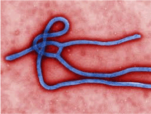
Figure 1. El virus Ebola
All Marburg virus isolates belong to the same species, while the Ebola virus genus comprises five different species: Zaire, Sudan, Côte d'Ivoire, Bundibugyo and Reston. Marburg hemorrhagic fever was first described after simultaneous outbreaks that occurred in 1967 in Marburg (Germany) and in Belgrade (Yugoslavia). These outbreaks were associated with contact with African laboratory monkeys (Cercopithecus aethiops) imported from Uganda. Subsequently, isolated cases were identified in South Africa in 1975 (possibly acquired in Zimbabwe), and in 1980 and 1987 in Kenya, probably acquired in Kitum, in Mount Elgon Park. Between the years 1998 and 2000 the Democratic Republic of the Congo recorded the first major outbreak under natural conditions of Marburg haemorrhagic fever, with 154 cases, of which 128 died (83% mortality). Subsequent studies identified several strains of the virus introduced into human populations from unknown environmental sources. In 2004 and 2005, the most important outbreak in Angola was registered with 374 cases (329 deaths, 88% mortality). In 2007, in Uganda, three cases of Marburg infection were detected, and later, in the summer of 2008, a Dutch tourist who had traveled to Uganda and visited a cave of bats died of Marburg hemorrhagic fever. In the USA in the same year, the serum of a patient who had had a fever with coagulopathy and hepatitis was retrospectively re-visited on a trip where she had visited the same cave in Uganda, showing evidence of past infection with Marburg virus [1,2].
The first recorded case of Ebola virus was in 1976, in a western equatorial province of the Sudan and in a neighboring region of Zaire (now the Democratic Republic of the Congo), causing an epidemic after which there was a second outbreak in Sudan in the same year. This outbreak corresponded to a new variety called Ebola-Sudan, causing the death of 340 people in 550 identified cases. The first cases were diagnosed as malaria because they had very similar symptoms and due to poor treatment [3,4]. In 1989, the Ebola Reston subtype was identified in Macaca fascicularis monkeys from a laboratory in Reston, USA, and several outbreaks were recorded in the USA and Italy between 1989 and 1996, from monkeys imported from the Philippines, with few associated asymptomatic infections in humans.
In 1994, a case of Ebola hemorrhagic fever of the Côte d'Ivoire subtype was detected. In Gabon, cases of Ebola were diagnosed for the first time in 1994 and in 1996, and in 1995 another important epidemic was declared in the Republic of the Congo. In 2000 the Sudan Ebola subtype reappeared in Uganda with 425 cases (224 deceased), the most important Ebola outbreak to date. Between 2001 and 2003, the Zaire subtype caused several outbreaks in Gabon and the Democratic Republic of the Congo. In Uganda, the Ebola Bundibugyo virus was identified in 2007 as the cause of an outbreak of hemorrhagic fever with a lower mortality rate (approximately 30%) than that associated with outbreaks by the Zaire and Sudan subtypes. Genetic sequencing has shown that Bundibugyo is more like the Côte d'Ivoire subtype that has been identified as the only causative agent of a non-lethal human infection so far. A study of a swine outbreak of Ebola Reston in 2008 identified asymptomatic cases in humans. In total, since the Ebola virus was discovered there have been about 1,850 cases (more than 1200 deaths, global mortality greater than 65%, between 30-88%, depending on the virus species). With the exception of isolated cases, all cases of hemorrhagic fever due to filovirus were recorded in sub-Saharan Africa. The total number of cases does not exceed 3,000 but in the last decades the number of outbreaks has increased. Due to its pathogenicity and virulence, Ebola is classified as a level 4 pathogenic biological agent (biosafety level 4), according to the US CDC. Those agents that threaten life and that are highly contagious, airborne viruses and inhaled aerosols, need to be handled at a level 4 of biosecurity. Some of the viruses that cause hemorrhagic fevers are also classified at this level; like the Marburg viruses; Lassa and Junin. Other viruses classified at this level are Hanta viruses that cause pulmonary syndrome in humans, and encephalitis-causing viruses such as Hendra virus and Summer-Russia virus. Viruses classified at biosafety level 3 are bacteria that cause anthrax and prions that cause degenerative diseases of the brain. Biosafety level 2 viruses are HIV-AIDS and hepatitis A, B and C viruses. Level 1 biosecurity is established for those viruses that do not cause highly lethal diseases and are of low pathogenicity [6].
The case-fatality rate of the Marburg FVH is 85%. The overall case fatality rate of the Ebola FVH is 65%, but with large differences depending on the type of virus and outbreak (30%-85%). There is no specific treatment. There are several treatments and vaccines still in an experimental phase, so case management is based on support treatment. The isolation of patients and the manipulation of samples/secretions to avoid secondary transmission are particularly important. If a case of filovirus infection is suspected, consulting the official guidelines regarding the isolation and management of these cases is recommended [5,6].
The Ebola virus is very aggressive and has a high rate of lethality, due to not knowing its effective treatment, as well as to the serious consequences in the population, especially with reference to any type of illness similar to the Ebola and the same.
The transmission mechanism of the Ebola virus is the close contact with people or non-human primates who suffer from the disease, such as monkeys, health personnel or relatives, because the virus is found in body fluids. Another form of transmission is through sexual contact. Patients who have recovered from the disease caused by the virus are not very likely to spread the infection. However, the virus can be present in genital secretions for up to 7 weeks [7].
Although a source of human infection has been non-human primates, it is not believed that these are the source. Like humans, these primates are infected by natural sources directly or through a chain of transmission originated in natural sources. The natural source is not known despite numerous studies that have been conducted, but a type of fructivorous bat is suspected. Laboratory observations have shown that bats experimentally infected with Ebola virus do not die, and this has led to the assumption that these mammals can play the role of keeping the virus in tropical forests [5,8].
The incubation period varies from 2 to 21 days. The period of transmissibility persists while the blood and secretions contain viruses. Among people with direct contact cases were presented between 5 and 15% [6,7].
Most patients arrive for treatment dehydrated, apathetic and disoriented, but the clue symptom is the extraordinary amount of internal and external bleeding. Death occurs within 7 to 16 days. The pathology of Ebola produces lesions in the spleen and kidney. Shock is produced by severe blood loss. As the disease progresses, hemorrhages occur in the gastrointestinal tract, pleural and peritoneal spaces. Coagulation abnormalities occur in the final stage of the disease
Diagnosis is reached by analyzing blood samples in specialized laboratories with a level of biological security type 4. Handling samples supposes extreme biological danger and takes place in conditions of maximum biological containment. There are also new techniques using non-invasive methods, with analysis of saliva and urine samples, and analysis of non-active samples that allow rapid laboratory diagnostics.
There are rapid diagnostic techniques based on the detection of antigens by ELISA or RNA by RT-PCR. The confirmation diagnosis is obtained after the identification of viral particles obtained by cell culture. These techniques are only available in highly specialized, level 4 biosafety laboratories.
We must proceed toward an early disclosure of disease activity and its extension in order to carry out adequate containment measures. Multidisciplinary surveillance teams, supervised by epidemiologists, must be equipped with protection equipment to collect appropriate samples and send them to reference laboratories for confirmation of a suspected diagnosis, and they must have the authority to prescribe emergency isolation measures. These initiatives must have standardized forms for the evaluation of control and monitoring reports, adequate transportation and rapid means of communication by means of telecommunications technology. Active surveillance is a prerequisite for the rational measurement that must be taken at various levels.
The creation of adequate treatment centers in suspected endemic areas is essential for controlling the virus, because the treatment of patients with Ebola virus infections requires isolation facilities and special equipment for health centers and their workers.
The international movement of populations, especially those from an epidemic area, should be monitored and controlled, with evacuation flights specially pre-arranged in agreement with health authorities of the receiving country. Contingency planning by reception centers is very important. Informing the general population about the nature of the disease and the measures necessary to contain outbreaks —including those related to the prompt and safe burial of corpses— is essential [5-9].
There are currently several vaccines in development or in experimental phase. The vaccine called rVSV-ZEBOV has been analyzed in a trial involving 11,841 people in Guinea. Among the 5,837 participants who received the vaccine there were no cases of Ebola ten days after vaccination, while there were 23 cases ten days or more after vaccination among those who did not receive the vaccine. The manufacturer of the vaccine is Merck, Sharpe & Dohme.
The trial was conducted in the coastal region of Basse-Guinée, the area of Guinea that was still experiencing new cases of Ebola when the trial began in 2015. The trial used a design called "ring vaccination" method that was already used for eradicating smallpox. When a new case of Ebola was diagnosed, all people who might have been in contact with that case in the previous three weeks were tracked, such as those who lived in the same home, or were visited by the patient, or were in close contact with the patient or with his clothes. A total of 117 groups or rings were identified, each consisting of an average of 80 people. Initially, the rings were randomly assigned to receive the vaccine immediately or at 3 weeks. Only adults older than 18 could be vaccinated. In addition to showing high efficacy among the vaccinated, the study revealed that unvaccinated people were indirectly protected against the Ebola virus through the vaccination method indicated [10-17].
The authors declare no conflict of interest.
- Towner JS, Amman BR, Sealy TK, Carroll SA, Comer JA, et al. (2009) Isolation of genetically diverse Marburg viruses from Egyptian fruit bats. PLoS Pathog 5: e1000536. [Crossref]
- Centers for Disease Control and Prevention (CDC) (2009) Imported case of Marburg hemorrhagic fever- Colorado, 2008. MMWR Morb Mort Wkly Rep 58: 377-1381.
- Gétaz L, Abbas M, Loutan L, Schrenzel J, Iten A, et al. (2011) Fatal Acute melioidosis in a tourist returning from Martinique Island, November 2010. Euro Surveill 16: 19758. [Crossref]
- Towner JS, Sealy TK, Khristova ML, Albariño CG, Conlan S, et al. (2008) Newly discovered Ebola virus associated with hemorrhagic fever outbreak in Uganda. PLoS Patog 4: e1000212. [Crossref]
- World Health (2014) Ebola haemorrhagic fever - Global Alert and Response (GAR). http://www.who.int/csr/disease/ebola/en/.
- Peters J (1995) Biosafety and emerging infections: Key issues in the prevention and control of viral hemorrhagic fevers Clarence special pathogens branch/division of viral and rickettsial diseases national center for infectious diseases/centers for disease control and prevention. Proceedings of the 4th National Symposium on Biosafety 1600 Clifton Road Atlanta, GA 30333, USA.
- European Center for Disease Prevention and Control (2014) Outbreak of Ebola haemorrhagic fever in Guinea. Rapid Risk Assessment.
- European Center for Disease Prevention and Control (2010) ECDC fact sheet: Ebola and Marburg fever: ECDC.
- Organización Mundial de la Salud (OMS) (2014) Sitio de Información de Eventos. Reglamento Sanitario Internacional.
- Huttner A, Agnandji ST, Combescure C, Fernandes JF, Bache EB, et al. (2018) Determinants of antibody persistence across doses and continents after single-dose rVSV-ZEBOV vaccination for Ebola virus disease: an observational cohort study. Lancet Infect Dis 18: 738-748. [Crossref]
- Metzger WG, Vivas-Martínez S (2018) Questionable efficacy of the rVSV-ZEBOV Ebola vaccine. Lancet 391: 1021. [Crossref]
- Longini IM, Røttingen JA, Kieny MP, Edmunds WJ, Henao-Restrepo AM (2018) Questionable efficacy of the rVSV-ZEBOV Ebola vaccine - Authors' reply. Lancet 391: 1021-1022. [Crossref]
- Gsell PS, Camacho A, Kucharski AJ, Watson CH, Bagayoko A, et al. (2017) Ring vaccination with rVSV-ZEBOV under expanded access in response to an outbreak of Ebola virus disease in Guinea, 2016: an operational and vaccine safety report. Lancet Infect Dis 17: 1276-1284. [Crossref]
- Henao-Restrepo AM, Camacho A, Longini IM, Watson CH, Edmunds WJ, et al. (2017) Efficacy and effectiveness of an rVSV-vectored vaccine in preventing Ebola virus disease: final results from the Guinea ring vaccination, open-label, cluster-randomised trial (Ebola Ça Suffit!). Lancet 389: 505-518. [Crossref]
- Henao-Restrepo AM, Longini IM, Egger M, Dean NE, Edmunds WJ, et al. (2015) Efficacy and effectiveness of an rVSV-vectored vaccine expressing Ebola surface glycoprotein: interim results from the Guinea ring vaccination cluster-randomised trial. Lancet 386: 857-866. [Crossref]
- Ledgerwood JE (2015) Use of low dose rVSV-ZEBOV: safety issues in a Swiss cohort. Lancet Infect Dis 15: 1117-1119. [Crossref]
- Huttner A, Dayer JA, Yerly S, Combescure C, Auderset F, et al. (2015) The effect of dose on the safety and immunogenicity of the VSV Ebola candidate vaccine: a randomised double-blind, placebo-controlled phase 1/2 trial. Lancet Infect Dis 15: 1156-1166. [Crossref]