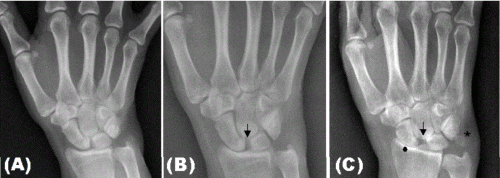
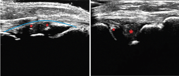
The patient, a 45-year-old white woman, presented with long lasting arthritis of the wrists and inflammatory arthralgia of knees, ankles and elbows. An extensive laboratory investigation including antinuclear antibodies, rheumatoid factor, and cyclic citrullinated peptide antibody was negative. X-ray images of the hands and wrists made in January, 2011 showed no remarkable findings (Figure 1, A). A diagnosis of seronegative rheumatoid arthritis was made based on clinical findings. The patient received treatment with prednisone and a combination of DMARDs (methotrexate, leflunomide, and hydroxychloroquine), without exhibiting clinical response. A new X-ray made in April, 2012 (Figure 1, B) showed narrowing of the wrist joint space and increase in the separation between scaphoid and semilunar bones (arrow). Eventually, anti-TNF agents (etanercept and adalimumab, one at a time) were added to the scheme. After several unsuccessful treatments, the X-ray was repeated in March, 2015 (Figure 1, C) and the following findings were observed: scapholunate dissociation (also known as Terry Thomas sign1, Figure 1, C, arrow), chondrocalcinosis of the triangular fibrocartilage (Figure 1, C, asterisk), and pressure erosion at distal end of the radius (Figure 1, C, circle). X-ray of knees and pelvis were normal. We diagnosed probable calcium pyrophosphate dihydrate crystal deposition (CPPD) disease, but an investigation of endocrine and metabolic diseases did not reveal a specific etiology. Colchicine 0.5 mg twice a day was initiated with limited therapeutic response. Ultrasonography (US) confirmed features suggestive of CCPD (intraarticular calcifications in right wrist and second metacarpophalangeal [MCP] joints bilaterally; calcification of triangular cartilage in left wrist and knee menisci bilaterally; absence of articular erosions; osteophytes in MCP and metatarsophalangeal joints). Abnormalities suggesting calcifications in the left wrist on US are shown in Figure 2.

Figure 1. Evolution of radiographic signs of chondrocalcinosis over 4 years in the left wrist.

Figure 2. A- Dorsal longitudinal scan of left wrist showing grade 2 synovial proliferation and intrarticular hyperecoic images (small asterisks).
B- Lateral longitudinal scan of the left wrist showing hyperecoic enhancement of the triangular cartilage (large asterik).
The authors declare no conflict of interest.
- Tischler BT, Diaz LE, Murakami AM, Roemer FW, Goud AR, et al. (2014) Scapholunate advanced collapse: a pictorial review. Insights Imaging 5: 407-417. [Crossref]
2021 Copyright OAT. All rights reserv
Editorial Information
Editor-in-Chief
Andras Perl
State University of New York
Article Type
Mini Review
Publication history
Received date: September 16, 2017
Accepted date: October 05, 2017
Published date: October 09, 2017
Copyright
© 2017 Machado J. This is an open-access article distributed under the terms of the Creative Commons Attribution License, which permits unrestricted use, distribution, and reproduction in any medium, provided the original author and source are credited.
Citation
Machado J (2017) Emergence of radiographic signs of chondrocalcinosis over 4 years of follow-up. Rheumatol Orthop Med, 2: DOI: 10.15761/ROM.1000130
Corresponding author
Markus Bredemeier
Serviço de Reumatologia do Hospital Nossa Senhora da Conceição, Avenida Francisco Trein, 596, sala 2048, Porto Alegre, RS, 91350-200, Brazil
E-mail : bhuvaneswari.bibleraaj@uhsm.nhs.uk


