Abstract
Aim: The purpose of this study was to evaluate the effects of chamomile tea consumption inflammatory markers and insulin resistance in subjects with type 2 diabetes mellitus (T2DM).
Methods: This single-blind randomized controlled clinical trial was conducted on 64 individuals with T2DM (males and females) aged between 30 and 60 years. The intervention group (n=32) consumed chamomile tea (3g/150 mL hot water) 3 times per day immediately after meals for 8 weeks. The control group (n=32) followed a water regimen for the same intervention period. Fasting blood samples, anthropometric measurements and 3-day, 24-h dietary recalls were collected at the baseline and at the end of the trial. Data were analysed by independent t test, paired t test, Pearson correlation test, and analysis of covariance.
Results: Analysis of covariance showed that tumour necrosis factor-α (TNFα) and high sensitive C-reactive protein (hs-CRP) levels and homeostatic model assessment for insulin resistance (HOM-IR) were significantly decreased by 4.87%, 57.20% and 23.60% respectively in chamomile group compared with these variables in control group at the end of the intervention (all p<0.05). There was significant positive correlation between the changes of serum TNFα and hs-CRP with HOMA-IR (r=0.567, p<0.001 and r= 0.520, p<0.001, respectively) in chamomile tea group at the end of the study.
Conclusion:
Short term intake of chamomile tea has beneficial effects on inflammatory markers and insulin resistance in patients with T2DM. Higher larger sample population and a longer intervention period may be required to show significant clinical improvements.
Keywords
type 2 diabetes mellitus, chamomile, inflammation, insulin resistance
Introduction
Type 2 diabetes mellitus (T2DM) is one of the most common chronic diseases worldwide [1]. It is estimated that six people die every minute from the diabetes worldwide, a figure that will soon make T2DM one of the world’s most common causes of preventable mortality [2].
Chronic hyperglycaemia increases circulating levels of inflammatory biomarkers such as tumour necrosis factor-α (TNF-α), C-reactive protein (CRP) and IL-6. TNFα is a pro inflammatory cytokine that play a major role in metabolic disorders, such as obesity and insulin resistance (IR) [3]. Hotamisligil, et al. [4] and Karasik, et al. [5] first showed that TNF-α was able to induce IR, which is a key component of T2DM. TNF-α and IL-6, as the main cytokines, initiate inflammatory responses and lead to the production of CRP as an acute-phase reactant [6]..
CRP, a nonspecific marker of the inflammatory response [7], is most consistently related to the development of T2DM [8-10]. CRP is a cytokine that is mainly produced by the liver. High concentrations of CRP have been associated with obesity, diabetes, coronary heart disease (CHD) and a sedentary lifestyle [11].
In view of the increased risk of CVD associated with diabetes, it is important to investigate the ability of nutritional therapies to attenuate the effect of modifiable risk factors associated with CVD in this high-risk patient group.
Chamomile is a medicinal plant that has been used for centuries in many human cultures to treat various inflammatory conditions such as eczema, ulcers, gout, neuralgia and rheumatic pains [12,13]. It is consumed at a rate of more than a million cups per day [14,15]. Substantial part of the pharmacological effects is determined by the biologically active chemical constituents that have been identified, namely the sesquiterpenic and the phenolic compounds [13,16].
The anti-inflammatory and antihyperglycemic effect of aqueous and ethanolic extracts of chamomile was evaluated in experimental studies [12,14-18]. In a study by Shipochliev, et al. the aqueous extract of chamomile suppressed both the inflammatory effect and leukocyte infiltration induced by a simultaneous injection of carrageenan and prostaglandin E1. The inhibitory effects of chamomile aqueous extracts were similar to those elicited by 10 mg/kg of the anti-allergenic agent oxatomide in Wistar albino rats [19]. Natarajan, et al. indicated that Chamomile treatment inhibited LPS-induced nitric oxide (NO) production and significantly blocked IL-1β, IL-6 and TNFα-induced NO levels in RAW 264.7 macrophages [20].
Although some experimental studies have reported effects of different extracts of chamomile on inflammation biomarkers, the possible effects on IR and inflammatory parameters of patients with diabetes have not been investigated. Therefore, we initiated a study to evaluate the effects of chamomile tea consumption on IR, TNF-α and hs-CRP levels in subjects with T2DM.
Methods
Subjects
Sixty-four patients with type 2 diabetes (male and female) aged 30-60 years with a body mass index (BMI) lower than 37 kg/m2 were recruited for this study from the endocrinology clinic, Imam Hossien Hospital in Tehran, Iran from March 2013 to June 2013. Diagnosis of T2DM was assessed at least 6 months prior to our examination. Exclusion criteria included insulin treatment, smoking, alcoholism, consumption of any dietary supplements, green tea and other herbal infusion with the past 3 months or during the study, history of diseases including liver, kidney and cardiovascular diseases, thyroid disorders, gastrointestinal problems, cholesterol-lowering or anti-hypertension treatment, using corticosteroids, cyclosporine, non-steroidal anti-inflammatory or immunosuppressive drugs, warfarin and anti-epileptic medications, pregnancy or breast-feeding, following a specific diet and regular exercise (>2/weeks ), having allergy to plants of ragweed species and having jobs that need high level of consciousness .
The study consisted of a single blinded randomized, controlled clinical trial with treatment and control groups running in parallel for a period of 8 weeks. Ethical Committee of Tabriz University of Medical Sciences approved the study protocol and was registered on the Iranian Registry of Clinical Trials website (available at: http:// www.irct.ir, identifier: IRCT2013012712299N1). The study was conducted in accordance with the guidelines of the Declaration of Helsinki principles. All subjects gave written informed consent before clinical trial enrolment.
Study design
The sample size was determined based on the primary information obtained from the study by Kato, et al. for blood glucose [16]. For an α value equal to 0.05 and a power of 80%, the sample size was computed as 23 per group. This number was increased to 32 per group to accommodate the anticipated dropout rate. The participants were randomly allocated in two groups using a block randomization procedure (of size 4) with matched subjects in each block based on sex, age and BMI. The random sequence was generated using random allocation software by statistician of the study. Endocrinologist randomly assigned participants to an intervention group or a control group. Whereas patients and Endocrinologist allocated to the intervention group were aware of the allocated arm, outcome assessors and statistician were kept blinded to the allocation.
A general questionnaire was completed for each subject. Body weight was measured using a scale (Seca, Germany), without shoes and wearing light clothing. Height was measured using a mounted tape without shoes. BMI was calculated as the weight in kilogram divided by the height in meters squared. Information about daily energy and macronutrient intakes was obtained by 24-hour recall method for 3 days, including 2 week day and 1 weekend. Three day average of energy and macronutrient intakes of all subjects was analysed by Nutritionist 4 software (First Databank Inc., Hearst Corp., San Bruno, CA).
Intervention
Chamomile was obtained as homogenous chamomile tea bags (finished product) from the Iranian Institute of medicinal plants, Karaj Iran. The tea bag, containing approximately 3 g of chamomile tea, was manufactured on March 2013. These tea-bags were the same commercially available product. The intervention group (n=32) consumed one cup of chamomile tea infusate (1 chamomile tea bag infused for 10 min [21] in 150 mL hot water without milk or sugar) three times a day immediately after meals (breakfast, lunch, and dinner) for 8 weeks [16,22]. The control group (n=32) consumed an equivalent volume of warm water during the 8-week period (Figure 1). Subjects were asked to keep a record of all beverages consumed during the clinical trial and maintain their usual dietary intake and physical activity and to avoid any changes in medication, if possible. The compliance of the volunteers with the study protocol was monitored by telephone interviews once a week and counting returned tea bags in person every 2 weeks.
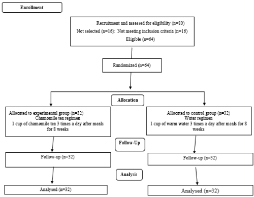
Figure 1. Flow diagram showing trial profile and study design
Blood sampling and biochemical assays
Venous blood samples (5 mL) from each subject were collected between 7:00 to 9:00 AM after an overnight fast at the beginning of trial. The serum samples were separated from whole blood by centrifugation at 3500 rpm for 10 min (Avanti J-25, Beckman, Brea, CA, USA). The serum and whole blood samples were frozen immediately at −70°C. Serum glucose was measured using the standard enzymatic methods with commercially available Pars Azmun kit (Karaj, Iran). Glycosylated haemoglobin (HbA1C) was measured in the whole blood by cation exchange chromatography with a Nycocard HbA1C kit (Norway). The serum Insulin level was measured by ELISA method using Monobind kit (USA) and insulin resistance was determined by Homeostasis Model Assessment (HOMA) index with formula: HOMA-IR=Fasting insulin (μU/mL)×fasting glucose (mg/dl)/405 [23]. The concentration of hs-CRP was measured by spectrophotometer method using pars azmun kit. Serum TNF-α was measured by platinum enzyme-linked immunosorbent assay (ELISA) kits (Bender Med System, eBioscience, Vienna, Austria).
All anthropometric, dietary intakes, blood sampling and biochemical measurements were assessed again at the end of intervention period in both groups.
Statistical analysis
Data were analysed using SPSS (version 16; SPSS Inc., Chicago, IL) and the results are expressed as means ± SD. The normal distribution of variables was tested and confirmed by Kolmogorov- Smirnov test. The baseline measurements and dietary intakes of subjects in two groups were compared using independent samples t-test and chi-square test for quantitative and qualitative variables respectively. ANCOVA was used to identify any differences between the two groups at the end of study, adjusting for baseline values and covariates. The changes in anthropometric measurements, energy and macronutrient intakes, serum levels of hs-CRP, TNF-α and HOMA-IR between the beginning and end of the study were compared by paired samples t-test. The percentage of changes in variables after intervention was determined by formula: [(after values–before values)/before values]×100
Results with P<0.05 were considered as statistically significant.
Results
All of the patients (32 patients in chamomile tea group and 32 patients in placebo group) completed the study (Figure 1). Compliance was good, with more than 97% of the tea bags being consumed in a prescribed manner during the study period. Participants did not report any adverse effects or symptoms with the chamomile tea consumption during the study.
The baseline measurements and dietary intakes of subjects in two groups were compared using independent samples t-test and chi-square test for quantitative and qualitative variables, respectively. ANCOVA was used to identify any differences between the two groups at the end of the study, adjusting for baseline value, gender, intake of OHA, duration of diabetes and changes of weight and calorie intake during the study.
Anthropometric characteristics and dietary intakes of participants at the beginning and end of the study are shown in Table 1. Each group consisted of 26 (81%) women and 6 (9%) men. There were no significant differences between two groups in gender (p=0.999) at the beginning of the study (data not shown). No significant differences between and within groups in weight and BMI were observed at the beginning of the study and after 8 weeks of intervention. There were no significant differences in age, energy and other dietary intakes between two groups at baseline. Total energy and nutrient intakes also did not change significantly in any of the groups during the study.
Table 1. General characteristics and dietary intakes of diabetic patients at baseline and after 8 weeks of intervention
Variable |
Measurement Period |
Chamomile tea group
(n=32) |
Control group (n=32) |
Age (yr) |
Baseline |
50.19 ± 7.08 |
51.97 ± 6.42 |
Height(cm) |
Baseline |
160.00 ± 6.50 |
162.12 ± 6.34 |
Weight (kg) |
Baseline |
76.00 ± 6.71 |
79.65 ± 6.62 |
After intervention |
74.53 ± 6.48 |
79.87 ± 6.80 |
BMI (kg/m2) |
Baseline |
29.48 ± 2.79 |
30.38 ± 2.6 |
After intervention |
29.12 ± 3.61 |
30.48 ± 2.72 |
Energy (kcal/day) |
Baseline |
1988.2 ± 201.2 |
1958.3 ± 217.1 |
After intervention |
2011 ± 221 |
1979.1 ± 248.00 |
Carbohydrate (g/day) |
Baseline |
298.20 ± 40.90 |
298.8 ± 53.6 |
After intervention |
303.24 ± 38.22 |
293.01 ± 54.78 |
Protein (g/day) |
Baseline |
74.43 ± 15.82 |
74.79 ± 14.11 |
After intervention |
76.32 ± 13.06 |
80.65 ± 14.49 |
Total Fat (g/day) |
Baseline |
57.6 ± 11.8 |
53.3 ± 10.4 |
After intervention |
57.51 ± 15.26 |
55.84 ± 11.89 |
BMI: body mass index. The results are described as mean ± Standard Deviation (SD)
Serum levels of glucose, HbA1C, insulin, HOMA-IR, TNFα and hs-CRP levels of subjects at baseline and after 8 weeks of intervention are shown in Table 2. Baseline values of hs-CRP and HbA1C were not different between two groups. Significant differences were seen between the two groups in serum levels of glucose, insulin, TNFα and HOMA-IR at baseline. Results of analysis of covariance showed statistically significant differences between two studied groups in serum HbA1C (p=0.023) , TNFα (p<0.001), hs-CRP (p<0.001) and HOMA-IR (p<0.001) levels at the end of the study, adjusted for baseline value, intake of OHA, duration of diabetes, gender and changes of weight and calorie intake during the study. Consumption of chamomile tea decreased HOMA-IR by 23.60%, levels of TNFα by 4.87% and hs-CRP by 57.20% compared with these variables in control group. As shown in Table 2, HOMA-IR and serum levels of TNFα and hs-CRP significantly reduced in the chamomile tea group (by 39.76%, 23.70% and 46.04%, respectively) at the end of the study.
Table 2. Glycemic and inflammatory indices of patients with diabetes at baseline and after 8 weeks of intervention
Variable |
Measurement period |
Chamomile tea group (n=32) |
Control group (n=32) |
MD (95% CI), P value |
Glucose (mmol/L) |
Baseline |
10.01 ± 3.44 |
8.46 ± 1.86 |
-1.55 (-2.94, -0.17), 0.028a |
| |
After intervention |
8.88 ± 3.69 |
8.73 ± 1.55 |
1.34 (0.42, 2.25), 0.081c |
| |
MD (95% CI), P valueb |
-1.13 (-1.87, -0.39), 0.004 |
0.27 (-0.23,0.77), 0.282 |
|
HbA1c (%) |
Baseline |
7.91 ± 1.76 |
7.48 ± 1.09 |
-0.43 (-1.17, 0.301), 0.243a |
| |
After intervention |
7.48 ± 1.59 |
7.50 ± 0.92 |
0.28 (0.01, 0.56), 0.03c |
| |
MD (95% CI), P valueb |
-0.43 (-0.61, -0.24), <0.001 |
0.01 (0.17, 0.2), 0.842 |
|
Insulin (µIU/dL) |
Baseline |
15.96 ± 1.71 |
13.93 ± 0.81 |
-2.02 (-2.69, -1.35), <0/001a |
After intervention |
10.68 ±1.41 |
14.28 ± 0.90 |
4.30 (3.51,5.09), < 0/001c |
MD (95% CI), P valueb |
-5.27 (-5.97, -4.58), <0.001 |
0.34 (0.19, 0.50), <0.001 |
|
HOMA-IR |
Baseline |
7.05 ± 2.34 |
5.24 ± 1.23 |
-1.81 (-2.74, -0.87), <0/001a |
After intervention |
4.24 ± 1.95 |
5.55 ± 1.12 |
2.26 (1.49, 3.03), < 0.001c |
MD (95% CI), P valueb |
-2.81 (-3.42, -2.19), <0.001 |
0.31 (-0.01,0.64), 0.06 |
|
TNFα (Pg/ml) |
Baseline |
61.70 ± 16.93 |
44.14 ± 10.09 |
-17.56 (-24.56, -10.56), <0/001a |
| |
After intervention |
46.42 ± 14.98 |
48.80 ± 10.70 |
13.43 (7.71, 19.16), < 0/001c |
| |
MD (95% CI), P valueb |
-15.27 (-19.45, -11.09), <0.001 |
4.66 (2.35, 6.96), <0.001 |
|
Hs-CRP (mg/L) |
Baseline |
1.93 ± 1.11 |
2.09 ± 1.76 |
0.16 (-0.58, -0.90), 0.665a |
| |
After intervention |
1.04 ± 0.73 |
2.43 ± 1.88 |
1.17 (0.73,1.61), < 0/001c |
|
MD (95% CI), P valueb |
-0.88 (-1.11, 0.65), <0.001 |
0.34 (0.30, 0.65), 0.03 |
|
HbA1c: Glycated Hemoglobin A1c; HOMA: Homeostasis Model Assessment Index; MD: Mean Difference
The results are described as mean ± Standard Deviation (SD)
aMD (95%CI), p value is reported based on the analysis of Independent sample t-test
bMD (95%CI), p value is reported based on the analysis of paired sample t-test
cMD (95%CI), p value is reported based on the analysis of covariance
There was significant positive correlation between the changes of serum TNFα and hs-CRP with HOMA-IR (r=0.567, p<0.001 and r= 0.520, p<0.001, respectively) in chamomile tea group at the end of the study (data not shown).
Discussion
Based on our literature review, this trial is the first report about the effects of chamomile tea consumption on inflammatory markers and IR in patients with T2DM.
Our findings indicated that drinking of chamomile tea by subjects with T2DM significantly decreased serum level of TNFα and hs-CRP compared to control group. These inhibitory effects of chamomile on inflammatory markers are agreement with findings of various experimental studies [20, 24-26]. In a study by Curra, et al. topical aqueous chamomile extract reduced the tissue levels of TNF-α and IL-1β, thereby demonstrated anti-inflammatory action in oral mucositis in hamsters [24]. In the study by Natarajan, et al. aqueous extract of chamomile treatment significantly blocked TNFα, IL-1β and IL-6 -induced NO levels in RAW 264.7 macrophages. In their study chamomile caused reduction in LPS-induced iNOS mRNA and protein expression. Inhibitory effect of chamomile on iNOS gene expression suggests that this is one of the mechanisms responsible for its anti-inflammatory properties [20]. Shipochliev, et al. reported that the aqueous extract of chamomile suppressed both the inflammatory effect and leukocyte infiltration induced by a simultaneous injection of carrageenan and prostaglandin E1. These results were similar to those elicited by 10 mg/kg of the anti-allergenic agent oxatomide [19]. In another study by Janmejai, et al. aqueous chamomile extract treatment inhibited the release of LPS-induced prostaglandin E (2) in RAW 264.7 macrophages. This effect was found to be due to inhibition of COX-2 enzyme activity by chamomile, with a mechanism of action similar to that attributed to NSAID [25]. Several studies also demonstrated anti-inflammatory effects of active components of chamomile including flavonoids (apigenin, luteolin and quercetin) and essential oils (α-bisabolol, bisabolol oxides A and B, chamazulene and α- and β-farnesene) [26-28,19]. Abe, et al. indicated that chamomile essential oil tested at 0.1% concentration suppressed TNF-α-induced neutrophil adherence [27]. Chamazulene carboxylic acid, a sesquiterpenic compound of chamomile, presents a chemical structure similar to profens [28], known as anti-inflammatory compounds that inhibit the key enzymes of prostaglandin metabolism [15].
Based on result of this study chamomile tea consumption significantly decreased serum level of glucose in the intervention group compared to its baseline values and declined levels of serum insulin and HOMA-IR compared to control group. Glucose lowering effect of chamomile was demonstrated in some previous studies [16-18]. Similar to our results a recent study demonstrated that ethanolic chamomile extract significantly reduced fasting plasma insulin levels by 13% and insulin resistance by 23% in mice after 2 weeks of treatment [22]. According to our obtained findings it was possible that improvement in IR in chamomile group contributed to their lower blood glucose level during the study trial.
It was suggested that antihyperglycemic activity of chamomile might be due to stimulating of peripheral glucose utilization, especially in muscle and adipose tissue, and/or inhibition of key enzymes involved in the gluconeogenesis and glycogenolysis pathways. Modulation of PPARs and other factors by chamomile extract may be another possible related mechanism(s) in this regard [16-18,22].
Other underlying mechanisms for favourable effects of chamomile on blood glucose parameters and IR in present study might also be through serum TNFα and hs-CRP reductions which were demonstrated in our treated group (Table 2). In current study there was a significant direct correlation between serum levels of TNFα and hs-CRP with HOMA-IR in chamomile group.
TNFα belongs to the TNF family, which is produced by macrophages, natural killer cells and T cells [4]. TNF-α may alter insulin sensitivity in different ways. A strong candidate mechanism is attenuating insulin receptor signalling path-ways. TNFα induces an increase in Ser/Thr phosphorylation of insulin receptor substrate 1 (IRS-1) or IRS-2. This may be quite important because increased IRS-1 Ser/Thr phosphorylation is strongly correlated with IR in insulin responsive cells or tissues, and because other perturbations that can provoke an increase in Ser/Thr phosphorylation of IRS-1 (such as hyperinsulinemia) also attenuate insulin signalling.
Another potential mechanism is decreasing glucose transporter-4 (GLUT4) in adipocytes by TNFα [29]. TNF-α has been shown to inhibit preadipocyte differentiation and it might also induce in vitro dedifferentiation of 3T3-L1 adipocytes [30]. These negative effects on adipocyte differentiation correlate with suppression of other adipocytes genes, such as those encoding adipocyte fatty acid binding protein (aP2), adipsin and the insulin-responsive glucose transporter [30-32]. Thus, downregulation of GLUT4 or other cellular components that mediate the metabolic effects of insulin represents a plausible mechanism for how TNF-a might mediate insulin resistance.
Another involved mechanism of TNFα-induced IR is suppression of adiponectin [33]. Adiponectin is an adipose-specific plasma protein with insulin-sensitizing effects [34]. The proinflammatory cytokine TNFα reduce adiponectin expression and throw this way induced IR.
Numerous studies have indicated that chronic, modest elevations in CRP are associated with the development of IR in humans [35-37]. CRP is measured by a high-sensitivity assay (hs-CRP). In agreement with our results in the study by Leinonen, et al. HOMA IR was positively correlated with CRP in subjects with T2DM [38]. In another study by Yudkin, et al. in 107 nondiabetic subjects, CRP levels were related to IR [39]. Tanigaki, et al. demonstrated that modest elevations in CRP cause insulin resistance in mice by decreasing skeletal muscle glucose delivery and that the process is due to attenuated endothelial insulin action mediated by FcgRIIB, which is abundant in skeletal muscle microvascular endothelium [40].
It should be note that in the present study, the subjects were asked to maintain their former diet during the study and data indicated that the population diet did not change significantly during the intervention trial. Thus, dietary factors could not be considered as confounding factors in the interpretation of studied biochemical variables. It was also expected that the randomized design could balance eventual bias between the study and control groups. However, our study had some limitations, including short study duration of 8 weeks and discrepancy at the gender issue. In addition, effects of chamomile components on inflammatory markers and IR may be dose dependent [18]. Therefore, the interpretation of the results of current study may not be applicable for using other amounts of chamomile tea or other intervention periods. Moreover, understanding the effects of chamomile tea on other inflammatory factors and especially related possible mechanisms warrants further studies.
Conclusion
In conclusion, the present study showed that short term intake of chamomile tea had some beneficial effects on inflammation and IR in patients with T2DM. Higher larger sample population and a longer intervention period may be required to show significant clinical improvements.
Funding
We thank The Research Vice-Chancellor and Nutrition Research Center of Tabriz University of Medical Sciences, Tabriz, Iran for the financial support
Conflict of interest
No conflict of interest exits in the submission of this manuscript, and this manuscript is approved by all authors for publication.
References
- Shaw JE, Sicree RA, Zimmet PZ (2010) Global estimates of the prevalence of diabetes for 2010 and 2030. Diabetes Res Clin Pract 87: 4-14. [Crossref]
- Wild S, Roglic G, G2021 Copyright OAT. All rights reservl prevalence of diabetes: estimates for the year 2000 and projections for 2030. Diabetes Care 27: 1047-1053. [Crossref]
- Kern PA, Saghizadeh M, Ong JM, Bosch RJ, Deem R, et al. (1995) The expression of tumour necrosis factor in human adipose tissue. Regulation by obesity, weight loss, and relationship to lipoprotein lipase. J Clin Invest 95: 2111-2119.
- Hotamisligil GS, Shargill NS, Spiegelman BM (1993) Adipose expression of tumour necrosis factor-a: direct role in obesity-linked insulin resistance. Science 259: 87-91.
- Feinstein R, Kanety H, Papa MZ, Lunenfeld B, Karasik A (1993) Tumour necrosis factor-a suppresses insulin-induced tyrosine phosphorylation of insulin receptor and its substrates. J Biol Chem 268: 26055-26058.
- Spranger J, Anja Kroke MM, Kurt H, Hoffmann K, Bergmann MM, et al. (2003) Inflammatory cytokines and the risk to develop type 2 diabetes results of the prospective population-based European Prospective Investigation into Cancer and Nutrition (EPIC)-Potsdam Study. Diabetes 52: 812-817.
- Pepys MB, Hirschfield GM (2003) C-reactive protein: A critical update. J Clin Invest 111: 1805-1812. [Crossref]
- Nakanishi S, Yamane K, Kamel N, Okubo M, Kohno N (2003) Elevated C-reactive protein is a risk factor for the development of type 2 diabetes in Japanese Americans. Diabetes Care 26: 2754-2757.
- Thorand B, Löwel H, Schneider A, Kolb H, Meisinger C, et al. (2003) C-reactive protein as a predictor for incident diabetes mellitus among middle-aged men: results from the MONICA Augsburg cohort study, 1984-1998. Arch Intern Med 163: 93-99. [Crossref]
- Duncan BB, Schmidt MI, Pankow JS, Ballantyne CM, Couper D, et al. (2003) Low-grade systemic inflammation and the development of type 2 diabetes: The Atherosclerosis risk in communities study. Diabetes 52: 1799-1805.
- Santos MG, Pegoraro M, Sandrini F, Macuco EC (2008) Risk factors for the development of atherosclerosis in childhood and adolescence. Arq Bras Cardiol 90: 276-283. [Crossref]
- McKay DL, Blumberg JB (2006) A review of the bioactivity and potential health benefits of chamomile tea (Matricaria recutita L.). Phytother Res 20: 519-530. [Crossref]
- Srivastava JK, Gupta S (2007) Antiproliferative and apoptotic effects of chamomile extract in various human cancer cells. J Agric Food Chem 55: 9470-9478.
- Oehler Ch, Bensdorf K, Gust R, Imming P (2009) Chamavioline antiedematous, but not a constituent of Matricaria recutita. Phytochem Lett (2009) 2: 171-175.
- Ramadan M, Goeters S, Watzer B, Krause E, Lohmann K, et al. (2006) Chamazulene carboxylic acid and matricin: a natural profen and its natural prodrug, identified through similarity to synthetic drug substances. J Nat Prod 69: 1041-1045.
- Kato A, Minoshima Y, Yamamoto J, Adachi I, Watson AA, et al. (2008) Protective effects of dietary chamomile tea on diabetic complications. J Agric Food Chem 56: 8206-8211. [Crossref]
- Eddouks M, Lemhadri A, Zeggwagh NA, Michel JB (2005) Potent hypoglycaemic activity of the aqueous extract of Chamaemelum nobile in normal and streptozotocin-induced diabetic rats. J Diabetes Res Clin Prac (2005) 67: 189-195.
- Cemek M, Kaga S, Simsek N, Buyukokuroglu ME, Konuk M (2008) Antihyperglycemic and antioxidative potential of Matricaria chamomilla L. in streptozotocin-induced diabetic rats. J Natur Med 62: 284-293.
- Shipochliev T, Dimitrov A, Aleksandrova (1981) Anti-inflammatory action of a group of plant extracts. Vet Med Nauki 18: 87-94.
- Bhaskaran N, Shukla S, Srivastava JK, Gupta S (2010) Chamomile, an anti-inflammatory agent inhibits inducible nitric oxide synthase expression by blocking RelA/p65 activity. Int J Mol Med 26: 935.
- Harbourne N, Jacquier JC, O’Riordan D (2009) Optimisation of the extraction and processing conditions of chamomile (Matricaria chamomilla L.) for incorporation into a beverage. Food Chem 115: 15-19.
- Christopher W, Sylvia JW, Morten R, Freiwald A, Kodelja V, et al. (2013) Antidiabetic effects of chamomile flowers extract in obese mice through transcriptional stimulation of nutrient sensors of the peroxisome proliferator-activated receptor (PPAR) family. J Plos One 8: 1-16.
- Matthews DR, Hosker JP, Rudenski AS, Naylor BA, Treacher DF, et al. (1985) Homeostasis model assessment: insulin resistance and beta-cell function from fasting plasma glucose and insulin concentrations in man. J Diabetologia 28: 412-419.
- Curra M, Martins MAT, Lauxen IS, Pellicioli AC, Sant'Ana Filho M, et al. (2013) Effect of topical chamomile on immunohistochemical levels of IL-1ß and TNF-α in 5-fluorouracil-induced oral mucositis in hamsters. Cancer Chemother Pharmacol 71: 293-299.
- Srivastava JK, Pandey M, Gupta S (2009) Chamomile, a novel and selective COX-2 inhibitor with anti-inflammatory activity. Life Sci 85: 663-669. [Crossref]
- Smolinski AT, Pestka JJ (2003) Modulation of lipopolysaccharide-induced proinflammatory cytokine production in vitro and in vivo by the herbal constituents apigenin (chamomile), ginsenoside Rb1(ginseng) and parthenolide (feverfew). Food Chem Toxicol 41: 1381-1390.
- Abe S, Maruyama N, Hayama K, Ishibashi H, Inoue S, et al. (2003) Suppression of tumour necrosis factor-alpha-induced neutrophil adherence responses by essential oils. Mediators of inflammation. 12: 323-328.
- Imming P, Goeters S, Pawlitzki G, Hempel B (2001) Absolute stereochemistry of guaianolides, of matricin and its epimers, of yarrow proazulenes, and of chamazulene carboxylic acid. Chirality 13: 337-341.
- Stephens JM, Lee J, Pilch PF (1997) Tumour necrosis factor-a induced insulin resistance in 3T3-L1 adipocytes is accompanied by a loss of insulin receptor substrate-1 and GLUT4 expression without a loss of insulin receptor- mediated signal transduction. J Biol Chem 272: 971-976.
- Zhang B, Berger J, Hu E, Szalkowski D, White-Carrington S, et al. (1996) Negative regulation of peroxisome proliferator-activated receptor-g gene expression contributes to the anti-adipogenic effects of tumour necrosis factor-a. Mol Endocrinol 10: 1457-1466.
- Szalkowski D, White-Carrington S, Berger J, Zhang B (1995) Antidiabetic thiazolidinediones block the inhibitory effect of tumour necrosis factor-alpha on differentiation, insulin-stimulated glucose uptake, and gene expression in 3T3-L1 cells. Endocrinology 136: 1474-1481.
- Stephens JM, Pekala PH (1991) Transcriptional repression of the GLUT4 and c/EBP genes in 3T3-L1 cells by tumour necrosis factor-alpha. J Biol Chem 266: 21839-21845.
- Wang B, Trayhurn P (2006) Acute and prolonged effects of TNF-a on the expression and secretion of inflammation-related adipokines by human adipocytes differentiated in culture. P flugers Arch 452: 418-427.
- Kadowaki T, Yamauchi T, Kubota N, Hara K, Ueki K, et al. (2006) Adiponectin and adiponectin receptors in insulin resistance, diabetes, and the metabolic syndrome. J Clin Invest 116: 1784-1792.
- Haffner SM (2003) Insulin resistance, inflammation, and the prediabetic state. Am J Cardiol 92: 18J-26J. [Crossref]
- Pradhan AD, Manson JE, Rifai N, Buring JE, Ridker PM (2001) C-reactive protein, interleukin 6, and risk of developing type 2 diabetes mellitus. JAMA 286: 327-334.
- Freeman DJ, Norrie J, Caslake MJ, Gaw A, Ford I, et al. (2002) C-reactive protein is an independent predictor of risk for the development of diabetes in the West of Scotland Coronary Prevention Study. Diabetes 51: 1596-1600.
- Leinonen E, Hurt-Camejo E, Wiklund O, Hultén LM, Hiukka A, et al. (2003) Insulin resistance and adiposity correlate with acute-phase reaction and soluble cell adhesion molecules in type 2 diabetes. Atherosclerosis 166: 387-394.
- Yudkin JS, Stehouwer C, Emeis J, Coppack S (1999) C-reactive protein in healthy subjects: associations with obesity, insulin resistance, and endothelial dysfunction a potential role for cytokines originating from adipose tissue? Arterioscler Thromb Vasc Biol 19: 972-978.
- Tanigaki K, Vongpatanasin W, Barrera JA, Atochin DN, Huang PL, et al. (2013) C-reactive protein causes insulin resistance in mice through Fc? receptor IIB–mediated inhibition of skeletal muscle glucose delivery. Diabetes 62: 721-731.