Introduction: Low-dose Whole Body Multi-Detector Computed Tomography (MDCT) has been established as an alternative to conventional X-ray imaging for Multiple Myeloma (MM) diagnosis. During an MDCT scan two dose indices are displayed on the monitor to account for the dose delivered to the patient: the volume computed tomography dose index (CTDIvol) and the dose length product (DLP). Both parameters though, are not sufficient in estimating the actual dose on their own. Two methods are proposed to promptly evaluate the scan dose, based on the two indices displayed: an effective dose evaluation through the DLP (Huda et al, 2008) and the Size-Specific Dose Estimate (SSDE), which also takes into account the patient’s size, based on the CTDIvol (AAPM Report 204).
Material and method: In this study a standardized protocol was developed and data from a good number of clinical examinations were collected. Effective dose was calculated based on the scanner displayed DLP. SSDE calculations were based on the scanner displayed values for the CTDIvol. SSDE is the averaged patient dose within the scan volume corrected for patient size. Dose is estimated using both methods for a set of 85 patients, examined for MM, for the torso body part. Although these indices are quite different in principle, they both present a rough but fast and prompt evaluation of the delivered dose. The results of the two methods are presented and evaluated.
Result: Calculated ED and SSDE values were found to present a weak correlation. Pairwise comparisons showed that the dose values through the two methods differed significantly (P < 0.001) by (0.92 ± 0.79) mSv. The Bland-Altman plot showed that the 95% LOA is 3.1 mSv wide, yelding a relatively poor agreement between the two methods.
Conclusion: The two methods of evaluation of the CT scan dose indices, ED and SSDE, based on DLP and CTDIvol correspondingly, provide an easily applicable dose estimation of a CT scan, but their values are found to present a notable difference. This means that they can not be used interchangeably clinically, but the most appropriate one should be used accordingly. ED relies on standardized phantoms and therefore has shortcomings with respect to its ability to reflect any individual patient effective dose. The SSDE is a good tool for estimating the average radiation dose for a given patient depending on the input parameters and the dimensions of the specific person in question but does not incorporate any organ/tissue weighting factors. It is recommended that when examined patients deviate significantly from the reference person by ICRP, dose should be estimated through the SSDE method. It is also proposed that tissue weighting factors would be incorporated with the SSDE methodology to provide a more refined estimate of risk.
multiple myeloma imaging, torso, SSDE, effective dose, low dose CT
Multiple Myeloma Imaging
Multiple Myeloma (MM) is a disease originating in the bone marrow and possibly extending into soft tissue or the peripheral blood, characterized by a proliferation of malignant plasma cells [1]. Imaging is required both for the disease staging and for bone fractures anticipation [1].
A series of plain X-ray images was the standard procedure for both staging and follow up [1,2]. Conventional radiography until recently was used as the standard for staging newly diagnosed myeloma, partly due to its wide availability and low costs [3]. However, this procedure cannot refrain from being painful and tiring for the patient. Apart from conventional radiography, a variety of newer imaging modalities including whole-body low-dose-CT, whole-body MRI and 18F-FDG PET/CT may be used for imaging and detection of MM disease. Detection of lytic bone lesions is crucial in the workup for multiple myeloma and often in asymptomatic patients defines the decision to start treatment.
Given the high intrinsic contrast of bone tissue, even a low-dose CT protocol produces images of good quality, suitable for diagnosis, with a significant radiation dose saving. A low dose whole body CT protocol can be used by lowering significantly the tube current. In their pioneer work Horger M, et al. [2] explore protocols of whole-body low-dose multi-detector row-CT(MDCT) in the diagnosis of MM that lower the dose of radiation necessary to image the skeletal system while preserving sensitivity and image detail.
As far as the required contrast is concerned, CT is superior in detecting focal bone destructions [3] and intramedullary lesions surrounded by fat but might be proven more challenging when diffuse bone marrow infiltration and intra-vertebral lesions are examined. These cases are better imaged with MRI modalities [4].
Whole-body low-dose CT is increasingly used due to its higher sensitivity for the detection of osseous lesions and its ability to diagnose extra osseous lesions. MM population can be favored of the CT investigation features, of increased diagnostic accuracy, comfort of the patient and balance between high image quality and absorbed dose [5].
New whole-body low-dose CT protocols allow for collection of higher quality images and details of the skeleton at doses of radiation not much higher than conventional radiography. They have a superior detection rate for lytic bone lesions compared with whole-body X-ray, for a possible restaging of the disease Furthermore, rapid acquisition time, superior image quality without need for i.v. contrast agents due to the intrinsic contrast of bone and utility in planning for image-guided biopsy and surgical interventions are some features that have contributed to the growing use of whole-body low-dose CT protocols [6].
Due to the high intrinsic contrast of bone the tube current can be lowered to a range of values 50mAs to 100mAs depending on the size of the patient [7].
These protocols are increasingly used in MM patients imaging in many European institutions. Our institution has applied the whole-body low-dose CT protocol since 2013 in all MM patients.
Dose Evaluation in MDCT scans
Volume CT Dose Index (CTDIvol) and Dose Length Product (DLP): During a MDCT scan both the Volume CT Dose Index (CTDIvol) and the Dose Length Product (DLP) are displayed, to account for the dose delivered to the patient.
When measuring or communicating the radiation output of a CT scanner, CTDI-based calculations are the reference standard [8], even though some weaknesses of the CTDI as a dose index have been debated [9,10]. CTDIvol was established to provide a standardized method to compare the scanner output between different scanners, using a reference phantom. This reference phantom is made of PMMA, is cylindrical and has a diameter of either 16 cm or 32 cm, accounting for either the patient’s head or body, respectively.
The dose received by a patient during a CT scan is affected by both the scanner radiation output and the patient size. CTDIvol provides information only on the scanner output and cannot therefore estimate, exclusively, patient dose [11].
As far as the DLP is concerned, it is calculated as the product of CTDIvol (mGy) and scan length (cm) and can be used to quantify the total amount of radiation received by a patient during a scan [12]. DLP however has the drawback of not being comparable with dose metrics used with other imaging modalities.
Both the CTDIvol and the DLP are sensitive to scan parameters (tube voltage, gantry rotation time, tube current, pitch, bow-tie filter), but are independent of patient size [13].
A more detailed analysis of all the dose indices mentioned can be found in the referenced literature [13,14].
Effective Dose (ED)
Effective Dose (ED) is a weighted average of organ doses. By taking into account the tissues receiving the radiation dose it attempts to reflect the equivalent whole-body dose that results in a stochastic risk equivalent to the risk of the actual absorbed dose of the partial body irradiation, such as a CT scan [14]. It provides a standardized dose estimate, comparable along all imaging modalities, as well as a quantification of the induction of genetic effects due to radiation [15].
A study by Huda et al. [16] has published data on the ratio of ED to DLP, for 16-section CT scanners, from four vendors, for typical scan lengths encountered at head and body CT examinations. In this way, a direct conversion of the DLP displayed on the scanner monitor to ED is allowed. This study [16] used commercially available CT dosimetry software (ImPACT CT Patient Dosimetry Calculator, version 0.99x; ImPACT, London, England) to calculate patient organ dose and ED from CT scans, using the Monte Carlo dose data sets published by the National Radiological Protection Board (NRPB) [17].
The equation of the ED estimation, through the proposed Conversion Factors (CF), is the following:
The study [16] also examines the possible alterations in the conversion factors due to different long-axis scan location, x-ray tube voltage and tube current, as well as a comparison between different dosimetry software packages.
Size-Specific Dose Estimate (SSDE)
The Size-Specific Dose Estimate (SSDE) is a recently proposed dose index to quantify the dose received by a patient during a scan, by taking into account the patient’s size as well [13]. It is based on a series of published conversion factors by the AAPM that takes into account the patient’s size and is applied to the displayed CTDIvol. Transverse CT images should be used to estimate patient size since localizer radiographs generally overestimate patient size due to magnification. Measurement of patient size can be obtained from the mid-slice location on the transverse CT image series [18].
The conversion factors are divided in those concerning either head scans (based on the 16 cm diameter head dosimetry phantom) or body scans (based on the 32 cm diameter body dosimetry phantom) [13]. The patient size is taken into account by considering one of four possible measurements: the patient’s lateral (LAT) dimension (left-to-right dimension of the body part being scanned), the anterior-posterior (AP) dimension (thickness of the body part being scanned), the sum of the LAT and AP dimensions or the effective diameter. The effective diameter (Eff-D) can be thought as the diameter of the circle whose area is the same as that of the patient cross section [13]. It can be calculated as:
By knowing the reference phantom used (32cm or 16cm diameter reference phantom) and the patient’s Eff-D, the appropriate conversion factor is derived from the AAPM 204 tables or is calculated by using the exponential equation as described in Appendix A [12]. By multiplying this factor with the displayed CTDIvol the SSDE (in mGy) of a certain CT scan is calculated.
The general form of the conversion is the following:
for the 32 cm and the 16 cm reference body phantoms respectively. The fB and fH factors are calculated based on the effective diameters and the scanning parameters. For example, when a tube of 120 kV is used, they are calculated as:
fB = 3.704369*exp(-0.03671937*Eff-D)
fH = 1.874799*exp (-0.03871313* Eff-D)
for the 32 cm and the 16 cm reference body phantoms respectively.
Dose Units
As already stated, SSDE is measured in mGy whereas ED in mSv. The numerical comparison between the two methods though is possible because of the type of radiation used in CT. The dose equivalent (mSv) is equal to the product of the absorbed dose (mGy) and the radiation quality factor Q; since Q equals 1 for radiations used in CT (and in diagnostic radiology in general), absorbed dose in mGy and dose equivalent in mSv are numerically equal and are many times used interchangeably [18].
Patient sample, CT system and scan parameters
A set of 85 adult patients, MM diagnosed, was examined. These patients underwent CT scan examinations between November 2013 and May 2014 in the 1st Radiology Department of the University of Athens.
All CT scans were performed on a Philips CT Brilliance 16 scanner, using the helical whole-body low dose protocol of our department, common in many other European Institutes for MM imaging. Patient’s weight varied between 50 – 85 kg, for adults above the age of 40. The parameters used were a tube voltage of 120 kV, a tube current - time product of 60 mAs and a collimation of 16 × 1.5 mm. In every scan the patient’s weight, the anterior posterior (AP) dimension, the lateral dimension at the patient’s largest area, the CTDIvol and the DLP were documented.
All patients were positioned supine, head first, with the arms above their head. Scan lengths varied between 1.05 – 1.54 m, measuring from the top of the head to the level beneath the knees (Figures 1 and 2).
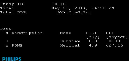
Figure 1. CTDIvol and the DLP values are displayed on the CT screen, useful in evaluation of absorbed dose delivered per MDCT scan performed on the Philips CT Brilliance 16 scanner in our Radiology department.
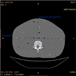
Figure 2. The documented dimensions from a CT scan, needed to calculate the Effective Diameter (Eff-D) of each patient. Rando Phantom transverse slice is displayed to show the AP and LAT measurements
This whole-body protocol was used for both imaging the body and the head, i.e. the 32 cm reference phantom, leading to a possible miscalculation of head and legs dose. Moreover, both the ED and SSDE conversion factors concern specific body parts.
For these reasons the head and legs parts were omitted from calculations and only the body part (trunk) was examined. This was done by calculating the percentage of the total scan length occupied by the trunk. It was found to have an average value of 47.9%. By multiplying this percentage to the DLP value, the body DLP was extracted. DLP values for the trunk varied between 240 - 340 mGy *cm and effective diameters between 20 – 36 cm. The part examined can be seen on a scanogram in figure 3.
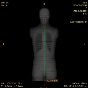
Figure 3. A scanogram showing the area under examination, accounting for an average of 58.9% of the total scan length
Effective Dose calculation
Firstly, estimations of the ED were done, based on the displayed DLP and the conversion factors (CF) introduced by Huda et al [16]. A direct conversion of the displayed on the scanner monitor DLP (in mGy *cm) to ED (in μSv) is possible this way, as presented in the Introduction section.
ED = CF * DLP
The representative value of 18 μSv/mGy *cm, for a body scan of ED/DLP at 120 kV, was used as a CF in our calculations, since a whole-body protocol was used [15]. The CF accounting for our system in specific examinations are 18, 17 and 20 μSv/mGy *cm for chest, abdominal and pelvic examinations respectively [15].
SSDE index calculation
Secondly, calculations of the SSDE indices were done, by multiplying the CTDIvol of the scanner for a reference phantom (32 cm) and the size dependent conversion factors introduced by the AAPM Report 204.
The factors were calculated as described in Appendix A [13], for the 32 cm reference body phantom, concerning a tube voltage of 120 kV, from the exponential equation:
fB = 3.704369*exp(-0.03671937*EffDiam)
Thus, the SSDE index (in mGy, for the 32 cm reference body phantom) is calculated as:
SSDE = fB*CTDIBvol
The effective diameter of each patient was determined as explained in the Introduction section Size-Specific Dose Estimate (SSDE)
Data analysis
Data from 85 patients’ CT scans, examined for MM, were collected in the Evorad RIS-PACS Workstation 2.1 and transferred in the SPSS software (version 21.0; SPSS, Inc, Chicago, IL) for evaluation. The patient dose received during a CT scan in MM examinations, for the torso part, was evaluated for a wide range of effective body diameters, both through the SSDE and the ED, as previously described.
The dose estimation values obtained with the two methods were compared using linear regression, and the Pearson correlation coefficients were calculated. The values also were compared by repeated-measures analysis of variance (ANOVA), and pairwise comparisons were performed. A Bland – Altman plot was used to assess agreement among the two methods, and the 95% limit of agreement (LOA) was calculated. A P-value less than 0.05 were considered significant.
SSDEs and EDs with respect to patients’ weight
SSDEs are known to present a decreasing trend in relation to patient body weight, since the body dimensions are accounted for in the calculating equation. This is confirmed by running a linear regression test between the SSDEs and the body weights of the patients, concluding in a moderate negative correlation (Pearson correlation, r = - 0.41; P < 0.001). The resulting pattern can be seen in the scatter plot of the two data sets (Figure 4).
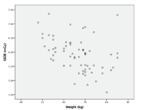
Figure 4. Linear regression between the SSDEs and the corresponding patient body weights. A moderate negative linear correlation is shown (r = - 0.41; P < 0.001)
EDs do not take into account patient body size; however, a linear regression test between EDs and patient body weights presents a moderate positive correlation between the two (Pearson correlation, r = 0.59; P < 0.001). The pattern can be seen in the scatter plot of the two data sets (Figure 5).
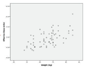
Figure 5.Linear regression between the EDs and the corresponding patient body weights. A moderate positive linear correlation is shown (r = 0.59; P < 0.001)
A statistical evaluation of the calculated ED and SSDE values
The mean dose values calculated for the set of the 85 patients was (5.9 ± 0.7) mGy through the SSDE estimation and (4.9 ± 0.3) mSv through the ED estimation. No linear correlation was seen between the SSDEs and EDs (Pearson correlation, r = - 0.13; P = 0.1). No evident pattern can be seen at the scatter plot of the two data sets, in figure 6.
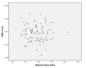
Figure 6. Linear regression between the EDs and the corresponding SSDEs. No evident pattern can be seen (r = - 0.13; P = 0.1)
Pairwise comparisons of the mean dose obtained with repeated-measures ANOVA showed that the dose values through the two methods differed by (0.92 ± 0.79) mSv, with a p value less than 0.001 (Table 1).
Table 1. Two dose estimation methods - Pairwise Comparison
Pairwise Comparison |
Mean Difference ± SD (mSv) |
P |
ED and SSDE |
0.92 ± 0.79 |
< 0.001 |
The respective Bland–Altman plot between SSDEs and EDs is shown in figure 7. It shows that the 95% Level of Agreement, LOA (mean difference ± 1.96 x SD) is 3.1 mSv for the dose estimate between the SSDEs and the EDs.
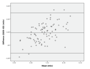
Figure 7. A Bland–Altman plot between SSDEs and EDs, showing the 95% Level of Agreement (LOA) boundaries (mean difference ± 1.96 x SD). A wide range of 3.1 mSv yields a relatively poor agreement between the two methods
Two methods to estimate patient dose from a CT scan, relying on the displayed on the CT console dose indices values, were evaluated. The first one is an ED estimation based on the displayed DLP [15] and the second one an SSDE evaluation based on the displayed CTDI [13].
These indices are based on different principles. The most important differences are the inclusion of tissue and radiation weighting factors in the ED estimation and the inclusion of the patient’s dimensions in the SSDE estimation. However, they can both offer an easy and prompt dose evaluation of a CT scan, through the two proposed methods.
In the current study, we found a weak linear correlation (r = - 0.13; P = 0.1) between EDs and SSDEs, evaluating the dose received by a set of 85 adult patients, for the torso body part of a low-dose whole body CT.
Pairwise comparisons showed that the dose values through the two methods differed by (0.92 ± 0.79) mSv, which is considered a significant difference (P < 0.001).
The Bland-Altman plot showed that the 95% LOA (mean difference ± 1.96 x SD) was 3.1 mSv wide for the dose estimation between ED and SSDE. These results show that the discrepancies between the two methods could be 47% at most, considering a mean dose of 5.9 mSv. These discrepancies seem highly significant and are clinically relevant. Therefore, these discrepancies indicate that dose estimation with these two methods should not be used interchangeably in a clinical setting.
The mean dose values calculated for the set of the 85 patients was
(5.9 ± 0.7) mGy by the SSDE estimation and
(4.9 ± 0.3) mSv by the ED estimation, for the torso body part.
Dose values referred to in other studies, range around 6.2 mSv (ICRP26) and 6.5 mSv (ICRP60) for 60 mAs, concerning a whole-body low-dose MDCT [2].
The two methods might not be expected to present identical values, since they are based on different principles, but it is of great interest to know if they can be used interchangeably in clinical routine. The difference between the two dose estimation methods can be attributed to a number of factors and possible sources of errors.
Firstly, both methods are strongly dependent on the displayed on the CT console dose values, either the CTDIvol or the DLP, and are therefore sensitive to the already stated weaknesses and possible inaccuracies relevant to the two indices [8,19].
When calculating the EDs, the displayed DLP values were used to account for the body part, omitting the head and legs fraction, because of the whole-body low dose protocol used for imaging. This may alter dose values considerably and it would be advised to repeat the calculations for patients examined following a low dose head protocol together with a low dose body protocol for scanning.
Moreover, the SSDE estimation strongly depends on defining the patient diameter, which can vary with the anatomic level and changes considerably over the examined area, especially when large areas are scanned. It also does not take into account the scan length but is based on typical abdominal scan lengths which can vary with patients’ height.
As also mentioned in other studies, further instructions on how to determine the patient diameter appear necessary for a general acceptance and right use of the SSDE concept [20].
Size-specific dose estimates (SSDE) can be used as an image-quality estimate [21]. The SSDE method of radiation dose optimization for torso CT should include SSDEs as a function of patient sizes, given an SSDE threshold curve based on experts’ valuation of image quality [21,22]. A low dose CT protocol used in MM imaging could produce doses that are slightly above the threshold SSDE curve.
Furthermore, SSDEs allow dose to be tailored to patient size in a straight way but how well SSDE works with automated tube current modulation was investigated recently [22]. A general relationship between CTDIvol-to-ED Conversion Coefficients and patient size was created and SSDE for exams acquired with both fixed and modulated tube currents could be estimated. Though in Whole Body Low dose Protocol, a fixed tube current is used, the modulated tube current use may be another positive option.
An incorporation of tissue weighting factors with the SSDE methodology would also provide a more refined estimate of risk. Moore et al used physical anthropomorphic phantoms to investigate the correlation of size-specific dose estimate (SSDE) with absorbed organ dose, for estimating patient organ dose in a pediatric population. Organ dose Correlation Factors (CF organ SSDE) and patient-specific SSDE estimate patient organ dose [23].
The two methods, based on CTDIvol and DLP, provide an easily applicable prompt dose estimation of a Whole-Body Low Dose CT scan in MM patient but are affected by different parameters and present notable differences in the resulting values. Thus, they are not to be used interchangeably in a clinical setting.
It is recommended that when patients with significant deviation from the reference person by ICRP (who weights around 70kg) are examined, dose could be estimated through the SSDE method.
Attention must also be given when scanning large anatomic areas, where both methods are sensitive to the mentioned parameters. Lastly, it is proposed that tissue weighting factors are incorporated with the SSDE methodology to provide a more refined estimate of risk.
- Delorme S, Baur-Melnyk A (2009) Imaging in multiple myeloma. Eur J Radiol.
- Horger M, Claussen C, Bross-Bach U, Vonthein R, Trabold T, et al. (2005) Whole-body low-dose multidetector row-CT in the diagnosis of multiple myeloma: an alternative to conventional radiography. Eur J Radiol 54: 289-97. [Crossref]
- Mahnken AH, Wildberger JE, Gehbauer G, Schmitz-Rode T, Blaum M, et al. (2002) Multidetector CT of the spine in multiple myeloma: comparison with MR imaging and radiography. AJR Am J Roentgenol 178: 1429-1436. [Crossref]
- Baur-Melnyk A, Buhmann S, Becker C, Schoenberg SO, Lang N, et al. (2008) Whole-body MRI versus whole-body MDCT for staging of multiple myeloma. AJR Am J Roentgenol 190: 1097-1104. [Crossref]
- Bannas P, Kröger N, Adam G, Derlin T (2013) Modern imaging techniques in patients with multiple myeloma. Rofo 185: 26-33. [Crossref]
- Pianko MJ, Terpos E, Roodman GD, Divgi CR, Zweegman S, et al. (2014) Whole-Body Low-Dose Computed Tomography and Advanced Imaging Techniques for Multiple Myeloma Bone Disease. Clin Cancer Res 20: 5888-97. [Crossref]
- Healy CF, Murray JG, Eustace SJ, Madewell J, O’Gorman PJ, et al. (2011) Multiple Myeloma: A Review of Imaging Features and Radiological Techniques. Bone Marrow Research 1-9.
- American Association of Physicists in Medicine. The measurement, reporting and management of radiation dose in CT. Report 96. AAPM Task Group 23 of the Diagnostic Imaging Council CT Committee. College Park, Md: American Association of Physicists in Medicine, 2008.
- Boone JM (2007) The trouble with CTD100. Med Phys 34: 1364-1371. [Crossref]
- Dixon RL (2006) Restructuring CT dosimetry-a realistic strategy for the future Requiem for the pencil chamber. Med Phys 33: 3973-3976. [Crossref]
- McCollough CH, Leng S, Yu L, Cody DD, Boone JM, et al. (2011) CT dose index and patient dose: they are not the same thing. Radiology 259: 311-316. [Crossref]
- Task Group on Control of Radiation Dose in Computed Tomography (2000) Managing patient dose in computed tomography: a report of the International Commission on Radiological Protection. Ann ICRP 30: 7-45. [Crossref]
- AAPM Report 204 (2011)Size-Specific Dose Estimates (SSDE) in Pediatric and Adult Body CT Examinations. American Assoc Physicists Med.
- McNitt-Gray MF1 (2002) AAPM/RSNA Physics Tutorial for Residents: Topics in CT. Radiation dose in CT. Radiographics 22: 1541-1553. [Crossref]
- Huda W (1997) Radiation dosimetry in diagnostic radiology. AJR Am J Roentgenol 169: 1487-1488. [Crossref]
- Huda W, Ogden KM, Khorasani MR (2008) Converting dose-length product to effective dose at CT. Radiology 248: 995-1003. [Crossref]
- Jones DG, Shrimpton PC (1993) Normalized organ doses for x-ray computed tomography calculated using Monte Carlo techniques. NRPB Report SR250. Didcot, Oxfordshire, England: National Radiological Protection Board.
- Pourjabbar S, Singh S, Padole A, Saini A (2014) Size-specific dose estimates: Localizer or transverse abdominal computed tomography images? World J Radiol 6: 210-217. [Crossref]
- Seibert JA, Boone JM, Wootton-Gorges SL, Lamba R (2014) Dose Is Not Always What It Seems: Where Very Misleading Values Can Result From Volume CT Dose Index and Dose Length Product. J Am Coll Radiol 11: 233-237. [Crossref]
- Kalender WA (2014) Dose in x-ray computed tomography. Phys Med Biol 59: R129-150. [Crossref]
- Larson DB (2014) Optimizing CT radiation dose based on patient size and image quality: the size-specific dose estimate method. Pediatr Radiol 44 Suppl 3: 501-505. [Crossref]
- McMillan K (2015) Estimating Radiation Dose Metrics for Patients Undergoing Tube Current Modulation CT Scans, UCLA Electronic Theses and Dissertations, Biomedical Physics 0119, UCLA.
- Moore BM, Brady SL, Mirro AE, Kaufman RA (2014) Size-specific dose estimate (SSDE) provides a simple method to calculate organ dose for pediatric CT examinations. Med Phys 41: 071917. [Crossref]