Background: Otological accidents resulting from mine explosions are polymorphous, mainly affecting deployed military personnel in our country. A delayed treatment, usually by lack of knowledge and / or association of injuries considered to be more serious, can worsen the auditory prognosis.
Methods: We have included patients treated for ear blast injury secondary to terrorist acts at the ENT department of the Military Hospital of Tunis between 2011 and 2017. All patients underwent a clinical evaluation and an audiometric exploration before and after treatment. A minimal follow-up of 12 months was required.
Results: Sixty patients were included in the study with an average age of 25 years. Thirty nine percent (39%) of patients had moderate hearing loss and tinnitus was present in 80% of patients. Treatment was based on corticosteroid therapy with or without hyperbaric oxygen therapy. At 12 months follow-up, hearing gain average was 20 dB and eight with perceptual hearing aggravated their hearing loss by 15 to 20 dB. Only 37.5% of patients reported a decrease in tinnitus. Evolution, however, was multifactorial.
Conclusion: Blast injury is responsible of cochlear injuries who are often final and a major source of persistent morbidity affecting the professional life of the victims. Because of possibility of secondary aggravation, extended follow -up is largely justified.
ear blast, explosion, therapeutic results
Blast injury is a pathological process that induces injuries in an organism exposed to a shock wave during an explosion.
According to the mechanism of injury, there are four types of blast injuries: primary cause by the wave itself, secondary caused by flying debris, tertiary caused by projection of the victim and quaternary (burns, inhalation of carbon monoxyde, ...) [1].
Organs with enclosed gas such as the middle ear, lungs and the digestive tract are the preferred targets of the primary blast and the severity of injuries is mainly due to the sudden variation of the pressure inside theses organs [2]. However, ear injuries are the most frequent because of a very low damage threshold (50% of the eardrums are perforated at 50 kPa) [2] and may interest either the middle or the inner ear. Initial symptomatology can involve; in various degrees, deafness, earache, tinnitus and vertigo [3,4].
In Tunisia, the incidence of these accidents increased considerably. However, this entity remains unclear, partially explaining the treatment delay.
Confirmation of the damage requires specialized clinical examination and audiometric investigations. Auditory restoration requires early management, but the prognosis remains multifactorial.
The aim of this study is to describe the different aspects of auricular blast injuries &as well as the post-therapeutic evolution in the medium and long term.
We conducted a longitudinal retrospective study including armed forces patients, hospitalized at the ENT unit of the Military Hospital of Tunis for the management of an ear primary blast injury.
Were excluded from our study, patients treated outside our structure, Fluid blasts and patients with missing data and / or having a post-treatment monitoring less than 12 months.
We noted the accident’s data (location, type of explosives) and patient’s data (age, sex, distance between the victim and the center of the explosion, the initial functional signs, therapeutic delay, the initial clinical examination, the results of audiometric explorations, extra-ENT injuries, treatment protocol, and the evolution of symptoms during a year).
Sixty patients were included in our study. They were all male with an average age of 25 years (20-42 years old). All patients were already exposed to extreme noises due to their professions and nine reported pre-existing hearing loss and/or tinnitus, but we had a basic audiometric assessment for only five of them, four of which had a 40 dB perception deafness. on average and one had isolated tinnitus treated medically.
The accident occurred as part of terrorist attacks in all cases. It was an explosion of a traditionally made device in an open space in 50 cases and in an enclosed space (by an explosive belt in a bus) in 10 cases. At the time, 35% of the patients were wearing their protective helmets. More than half (64%) of the patients were less than 10 meters from the center of the explosion and 30% were within five meters. The distance between the victims and explosion was roughly estimated by the patients themselves.
Delay to injury assessment and specialized exams ranged from few hours to more than three months. It often depended on the initial injury score and was longer for patients with extra-ENT injuries; especially vital ones (head trauma with coma in two cases, lower limb amputation supported by orthopedic surgeons in five cases, suffocating pneumothorax drained in one case, ocular injury with secondary blindness in two cases). In two cases, care was provided three months after the accident because of underestimation. Victims did not report initial hearing complaints.
Deafness and tinnitus were the most reported signs by patients (74% and 80% respectively). These signs were mostly bilateral (78%) and associated (67%). Less frequently, vertigo (10%), earache (27%) and otorrhagia (5 cases) were reported. Elsewhere, verbal discrimination disorders were observed after the accident in 65% of cases (39 patients) (Figure 1).
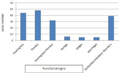
Figure 1. Functional signs reported by victims.
Initial otoscopic examination revealed tympanic perforation in 15 patients, eight of them were bilateral. All these perforations occurred in patients located within 10 meters of the explosion center or in an enclosed space. Elsewhere, there was tympanic hematoma or otorrhagia in six cases respectively.
The rest of the reports revealed secondary injuries due to patients falling on earth (tertiary blast) and/or screened by high speed projected debris (secondary blast). Displaced fracture of nasal bone was noted in two cases and head and neck wounds requiring surgical stitches were present in three cases.
A tonal audiometry was performed for all patients as soon as they were received at the department and complementary auditory evoked potentials assessment was claimed in the presence of pure perception deafness and/or unconformity between the clinical state and patient's tonal audiometry. In total, we reported pure hearing loss in 65% of cases (39 patients), pure transmission deafness in five cases (8%) and mixed hearing loss in 20% of cases. The attack was bilateral in 60% of cases and only four patients had normal hearing. By adopting the classification of the International office of Audiophonology (BIAP), majority of our patients had medium deafness (Table 1).
Table 1. Distribution according to initial average hearing loss. AHL: Average Hearing Loss.
hearing loss
(AHL) |
Slight
(AHL < 40 dB) |
Average
(AHL 40-60 dB) |
Deep
(AHL 61-80) |
Severe (AHL > 80 dB) |
% |
32% |
39% |
17% |
0% |
The most important hearing losses were observed in closed spaces blasts and when the distance between patients and the center of the explosion was less than five meters. Patients at more than 10 meters developed in most cases tinnitus, with a slight to moderate hearing losing audiometry. A CT scan of the rocks was performed in six cases with mixed hearing more than 40 dB, without improvement by treatment. It objectified a fractured anterior process of the right hammer in one case. Patients with vertigo were investigated magnetic resonance imaging (MRI) in all cases.
The therapeutic protocol was based on intravenous high dose of corticosteroid (300 milligrams per day of hydrocortisone hemisuccinate (HSHC), in three doses; for ten days followed by a regression over five days. This was associated with vasodilators (pentoxifylline; two tablets per day) and a preventive antibiotic (amoxicillin-clavulanic acid) in the presence of hematoma or tympanic perforation with or without otorrhagia.
After eliminating a possible contraindication (electrocardiogram, rhinological examination, chest x-ray, impedance measurement), a complementary treatment with hyperbaric oxygen therapy (HBOT) was performed in 35 patients in inner ear injuries regardless the hearing loss and / or in the presence of non-tolerated tinnitus. The mean onset time of HBOT was 12 days (four days in the case of isolated ENT involvement), but only 22 were able to complete the therapeutic protocol with a number of sessions between 8 and 22. The occurrence of Otitis directly related to barotrauma was the main cause of interruption of the sessions. Evolution under treatment was judged on subjective, clinical and audiometric data.
At the end of admission (10th day), a global sensation of satisfaction was reported by 40% of patients (24 cases). At six months of the accident, an improvement of the hearing was objectified by the tonal audiometry and /or by the study of the PEA in 18 cases with an average hearing gain of 20 dB and a total restoration of the hearing in eight patients (13%).
At 12 months, 12 patients (20%) still have painful hyperacusis and eight with perceptual hearing aggravated their hearing loss by 15 to 20 dB (Table 2).
Table 2. Mean hearing loss at the end of treatment. AHL: Average Hearing Loss.
Hearing |
Normal |
Mild hearing loss
(AHL < 40 dB) |
Average hearing loss (AHL 40-60dB) |
deep
hearing loss (AHL 61-80) |
severe hearing loss (AHL > 80 dB) |
% |
16% |
40% |
28% |
14% |
2% |
As for tinnitus, the evolution was judged on subjective criteria. Thus, at six months, only 37.5% (18 patients) reported a total disappearance or a significant decrease in the initial symptomatology. This improvement was noted at the end of the hospital stay in five cases. There was no significant difference between patients who received standard treatment and those who received the HBOT sessions.
In total, in our series, it appears that:
- The outcome was better in patients who were treated early (within seven days after the accident), those with medium initial deafness (average hearing gain of 25 dB) with a horizontal curve (improvement in 83% of cases). The combination with HBOT also appears to improve our therapeutic results
- On the other hand, a severe and/or bilateral pure sensorineural deafness, with a descending curve, the association with tinnitus, an intolerance to HBO hindering the finishing of the protocol as well as a late management and the proximity of the patient to the explosion core (<10 meters) were factors of poor prognosis in our series.
In Tunisia, despite the absence of official figures, the incidence of blast injuries has increased considerably since the revolution and the deployment of military personnel as part of the anti-terrorism war. Dougherty et al. [4] concluded that of 13226 US military personnel deployed in Iraq between 2004 and 2008, 1223 had ear blast injuries (30.7%). As for civilians, the prevalence of ear damage among all blasts was 21% in the Qureshi et al series [5].
The average age of our patients was 25 years, very similar to that of Dougherty's American series [4], which was 23.8 years old with a predominant male gender as in our study. This predominance of the young male population would have serious professional sociological consequences to taken into the account in case of poor auditory recovery. Thus, early diagnosis and adequate management are required to ensure a better functional prognosis and a successful professional reintegration.
As for damage; primary blast can interest all ear structures. Those of the outer ear are rare (burns, flag amputation, ...) and are the result of secondary blast resulting from metal splinters or other projections during the explosion. In a primary blast, symptoms result mainly from damage to the middle and/or inner ear and mainly result in deafness, tinnitus, otalgia and vertigo [5]. Hearing loss is the main complaint reported after an ear blast, but multiple signs are often associated (Table 3). In our series, it was reported by 74% of cases.
Table 3. Otological functional signs reported by different series.
|
Hearing loss
|
Tinnitus |
Otalgia |
Dizziness |
Verbal discrimination disorders |
Qureshi et al. [5] |
18% |
8% |
11% |
2% |
_ |
Ballivet et al. [6] |
61% |
88% |
34% |
20% |
56% |
Notre série |
74% |
80% |
28% |
18% |
76% |
At otoscopy, done as early as possible, the appearance of the tympanum differs according to the lesion, itself varying according to several data (intensity of the explosion, proximity of the victim to the explosion, place where it occurred, structures affected, ...). A tympanic perforation was objectified in 16% of the Dougherty series [4] and in 44% in the Ballivet series [6]. Bilateral perforations were noted in 10 to 66% of cases according to the authors and signed the severity of the explosion [6]. Otorrhagia, otorrhea and hemotympanum are often correlated with the presence of tympanic perforation. Ossicular lesions (dislocations and ossicular fractures) are even rarer (0.2 to 4% of lesions) [7] and the diagnosis can be difficult to make even with CT scans.
Among our patients, only 74% reported deafness but audiometry showed a loss of hearing in 94% of cases. The result of audiometry was probably altered by tinnitus. Perception deafness was the most common (78%) followed by mixed deafness (16%). Transmission or mixed hearing loss is due to tympanic perforation and ossicular involvement. The involvement of the inner ear, manifested by a pure perception deafness, results from a sensory cell tearing with displacement of the basilar membrane secondary to the shock wave. Achieving binaural hearing is particularly delicate because of a loss of the ability to locate sounds in space especially in the presence of complex sounds. This type of injury can seriously hamper the operational quality of military personnel on the ground [8]. The bilateral nature of the condition was found in 23% of the Nawaz series [9] and 38% in our series.
The prevalence of tinnitus varies from one series to another but remains the main complaints of the patients (Table 3). Its presence indicates an anterior labyrinth concussion [10]. The subjective nature of the complaint does not allow an objective and accurate assessment, but some authors suggest that their presence sign the viability of ciliary cells thus a possible good prognosis sign [11].
Rarely isolated, tinnitus is often associated with hearing impairment (56% in our series). However, their presence alone may be responsible of underestimation and delayed management [5]. Also, in our series, two patients with isolated tinnitus without initial hearing loss have been hospitalized after a period of 15 days and 3 months of the accident respectively. The evolution of tinnitus without treatment is capricious. Some studies have concluded that an almost complete disappearance can be seen within 24 hours and that treatment can be started only after in the absence of no improvement [12]. The type of tinnitus and the associated injuries are known to be important prognosis factors. Thus, wheezing and the presence of hearing impairment on high frequencies are more signs of bad prognosis than isolated low tones ringing. The discomfort described by patients is most often related to tinnitus than to the associated hearing loss [11,13].
Much less frequent, the mechanism of the vestibular system damage remains little known and several studies suggest that the imbalance results from central damage [14], especially since vertigo is often associated with head trauma. However, the symptomatology can be seen without any head trauma and can be secondary to the transmission of the shock wave to the peripheral vestibular system causing a concussion [10]. This is manifested either by the appearance of vertigo with the impression of rotation, or dizziness sensations such as imbalance, pitching or tread. These signs tend to move positively towards full recovery in a few days to several months even without treatment. Thus, several authors do not advocate any treatment of acute phase vertigo, especially if they are relatively tolerated [15]. It is their persistence that leads to further investigations (radiological and balance tests) looking for a labyrinth secondary residual fistula due to a labyrinth fracture caused by secondary or tertiary blast.
Little described in various series, other signs such as otalgia, otorrhagia and disorders of intelligibility can be caused by a blast attack. On the other hand, if the bleeding is often an alarming symptom for both the patient and the practitioner suggesting an urgent specialized examination, the otalgia and verbal discrimination disorders are often neglected in the acute phase of the accident, which can delay the treatment especially if the other signs are not that important. In our series, earache was described as the only sign of ear blast in two cases. The disorders of verbal discrimination were generally revealed by guided interrogations and rarely described spontaneously.
Factors influencing the severity:
Several authors have tried to identify the predictors of a severe blast. These were essentially explosive, space and/or patient related features.
In the study conducted by Qureshi et al. [5], an explosive weighing more than 80 kilograms was a highly significant factor in the occurrence of ear blast (p = 0.008), with symptomatology associating several signs (vertigo, tinnitus, severe deafness, ear pain and tympanic perforation).
A distance less than 10 meters from the center of the explosion appears, in several studies, as highly predictive of tympanic perforation with intense otalgia [4,5,16]. In our study as well, 83% of the perforations were objectified in patients located within a perimeter of less than 10 meters. This correlation was not, however, found in the studies of Jagade et al. [17] and Klamkam [2] despite an average distance of 5.6 meters from the explosion.
The occurrence of the accident in confined space was differently incriminated by authors. Thus, severe sensorineural deafness was reported in 73% of victims of explosions in an enclosed space and 62% of cases if the explosion occurred in an open space (p<0.05). This difference between locations did not influence the occurrence of functional or clinical signs in the Qureshi study [5]. Ten of our patients were victims of an explosion in a closed space and all had solid or bilateral tympanic perforation with a suffocating pneumothorax in one case.
According to Dougherty's study [4], it appears that wearing a helmet significantly reduces the risk of occurrence as well as the intensity of the damage. The use of earplugs or helmets reduces the intra-auricular pressure and therefore the risk of ear membranes rupture (both tympanic oval and round windows) [18]. Two of our patients developed a tympanic perforation despite wearing a helmet.
To increase the chances of recovery, treatment should be applied as early as possible, at best in the hours or days following the trauma. Some authors do not advocate any treatment before 24 hours in hope of a spontaneous disappearance, always possible, of the cochlear signs [15]. Similar to our attitude, in the absence of therapeutic consensus, the treatment of sensorineural deafness usually involves infusions of vasodilators, corticosteroids and drugs with hemorheological properties. In the presence of tympanic abnormalities, preventing local infections of a hemotympanum or tympanic perforation remains our first concern (antibiotics, local care, hygiene rules).
The spontaneous closure rate of a tympanic perforation reaches more than 83% after within few days to eight weeks for small ones (< 50% of the surface) [19,20]. In our series spontaneous healing was obtained in 2/3 of the cases. The subtotal perforations require, on the other hand, a surgical repair by myringoplasty. The prognosis remains more reserved for neurosensory deafness, and secondary aggravations, even at one year of the trauma, have been seen (10% in the Dougherty series [4] and 7% in the Fausti study [15]). In our series, a worsening of hearing was noted in six cases (12%) during surveillance despite well-conducted standard treatment.
Some authors suggest that the loss in bone conduction observed after a blast is the result of sudden deafness with the same at to the cochlear level including hypoxia, acidosis, cell swelling and a change of the cochlear microcirculation flow [21,22]. These lesions therefore justify the use of hyperbaric oxygen therapy (HBOT) ideally associated with general or intra-tympanic corticotherapy.
However, the results differ between the series and are rather encouraging. Pezzoli [23] reported good recovery in 70% (including 20% cure) of late-treated cases (30 days of evolution) by a standard 15-day HBOT protocol versus an unchanged aspect of the audiometric curve in 81 % of patients who did not receive HBOT. Recent phase I studies investigating the effect of low-pressure OHB (1.5 ATA) prescribed for 30 days for patients with chronic symptoms (deafness and / or tinnitus) after a blast accident appear promising with few side-effects [24].
The evolution of tinnitus is unpredictable. Although poorly validated, acute phase treatment is similar to that of deafness. The combination of oxygen therapy (OHBT) has only rarely been described as adjunctive therapy for tinnitus. In the absence of improvement with persistence of significant discomfort, rehabilitation and cognitive-behavioral therapies will be proposed.
Balance disorders management differs from one patient to another and according to the mechanism. Besides the specific treatment of a particular injury (perilymphatic fistula by trauma of the rock, vertigo secondary to head trauma, ...), vestibular rehabilitation remains essential [15]. However, blast induced vertigo rarely persists for more than a few days. In our series, the spontaneous regression was observed in six cases (67%) and only one patient kept unilateral compensated vestibular deficit.
Ear blast injuries are polymorphic, affecting several components of the acoustic-vestibular system. It remains a relatively common condition among military personnel deployed in combat zones, but its incidence seems to be increasing in civilians. However, the lack of parallelism between important functional complaints and poor clinical examination often leads to an under estimation of the diagnosis.
Despite taking prompt and adequate care, cochlear injuries are often final and a major source of persistent morbidity affecting the professional life of the victims (often young workers) with a significant psychological impact. In addition to these therapeutic difficulties, a lack of proven effectiveness of the traditional means of protection remains an issue. In any case, given the risk of secondary aggravation, regular monitoring and extended psychological support in these patients is largely justified.
- DePalma RG, Burris DG, Champion HR, Hodgson MJ (2005) Blast injuries. N Engl J Med 352: 1335-1342. [Crossref]
- Pahor AL (1981) The ENT problems following the Birmingham bombings. J Laryngol Otol 95: 399-406. [Crossref]
- Klamkam P, Jaruchinda P, Nivatwongs S, Muninnobpamasa T, Harnchumpol P, et al. (2013) Otologic manifestations from blast injuries among military personnel in Thailand. Am J Otolaryngol 34: 287-291. [Crossref]
- Dougherty AL, MacGregor AJ, Han PP, Viirre E, Heltemes KJ, et al. (2013) Blast-related ear injuries among U.S. military personnel. J Rehabil Res Dev 50: 893-904. [Crossref]
- Qureshi TA, Awan MS, Hassan NH, Aftab AH, Ali SA (2017) Effects of bomb blast injury on the ears: The Aga Khan University Hospital experience. J Pak Med Assoc 67: 1313-1317. [Crossref]
- Ballivet de Régloix S, Crambert A2, Maurin O3, Lisan Q1, Marty S1, et al. (2017) Blast injury of the ear by massive explosion: a review of 41 cases. J R Army Med Corps 163: 333-338. [Crossref]
- Brismar B, Bergenwald L (1982) The terrorist bombing explosion in Bologna, Italy 1980: an analysis of the effects and injuries sustained. J Trauma 22: 216-220. [Crossref]
- Kubli L, Brungart D, Northern J (2018) Effect of blast injury on auditory localization in military service members. Ear and Hearing 39: 457-469. [Crossref]
- Nawaz G, Ulhaq N, Khan AR (2014) Bomb blast injuries to the ear: The Peshawar experience. J Med Sci 22: 193-196.
- Garth RJ (1994) Blast injury of the auditory system: a review of the mechanisms and pathology. J Laryngol Otol 108: 925-929. [Crossref]
- Lin YT, Young YH (2008) Retrocochlear mass lesion in mid-frequency sudden deafness. Otolaryngol Head Neck Surg 138: 13-17. [Crossref]
- Shah A, Ayala M, Capra G, Fox D, Hoffer M (2014) Otologic assessment of blast and non blast injury in returning Middle East-deployed service members. Laryngoscope 124: 272-277. [Crossref]
- Breeze J, Cooper H, Pearson CR, Henney S, Reid A (2011) Ear injuries sustained by British service personnel subjected to blast trauma. J Laryngol Otol 125: 13-17. [Crossref]
- Akin FW, Murnane OD (2011) Head injury and blast exposure: vestibular consequences. Otolaryngol Clin Nord Am 44: 323-334. [Crossref]
- Fausti SA, Wilmington DJ, Gallun FJ, Myers PJ, Henry JA (2009) Auditory and vestibular dysfunction associated with blast-related traumatic brain injury. J Rehabil Res Dev 46: 797-810. [Crossref]
- Remenschneider AK, Lookabaugh S, Aliphas A, Brodsky JR, Devaiah AK, et al. (2014) Otologic outcomes after blast injury: the Boston Marathon experience. Otol Neurotol 35: 1825-1834. [Crossref]
- Jagade MV, Patil RA, Suhail IS, Kelkar P, Nemane S, et al. (2008) Bomb blast injury: effect on middle and inner ear. Indian J Otolaryngol Head Neck Surg 60: 324-330. [Crossref]
- Ritenour AE, Baskin TW (2008) Primary blast injury: update on diagnosis and treatment. Crit Care Med 36: S311-S317. [Crossref]
- Harrison CD, Bebarta VS, Grant GA (2009) Tympanic membrane perforation after combat blast exposure in Iraq: A poor biomarker of primary blast injury. J Trauma 67: 210-211. [Crossref]
- Rijswijk JB, Dubach P (2017) Binaural tympanic-membrane perforations after blast injury. N Engl J Med 376: e41. [Crossref]
- Chen W, Wang J, Chen J, Chen Z (2013) Relationship between changes in the cochlear blood flow and disorder of hearing function induced by blast injury in guinea pigs. Int J Clin Exp Pathol 6: 375-384. [Crossref]
- Sun H, Qiu X, Hu J, Ma Z (2018) Comparison of intratympanic dexamethasone therapy and hyperbaric oxygen therapy for the salvage treatment of refractory high-frequency sudden sensorineural hearing loss. Am J Otolaryngol 39: 531-535. [Crossref]
- Pezzoli M, Magnano M, Maffi L, Pezzoli L, Marcato P, et al. (2015) Hyperbaric oxygen therapy as salvage treatment for sudden sensorineural hearing loss: a prospective controlled study. Eur Arch Otorhinolaryngol 272: 1659-1666. [Crossref]
- Harch PG, Andrews SR, Fogarty EF, Amen D, Pezzullo JC, et al. (2012) A phase I study of low-pressure hyperbaric oxygen therapy for blast-induced post-concussion syndrome and post-traumatic stress disorder. J Neurotrauma 29: 168-185. [Crossref]