We present a method for visualizing tongue motion in the oral stage of swallowing. Conventional computed tomography clearly distinguishes air, swallowing contrast agent, and bone. However, because the tongue contacts the palate, it is difficult to clearly display contours of the tongue. We devised a double-contrast tongue surface imaging technique using sodium alginate, barium (positive contrast), and golden flaxseed oil (negative contrast). This facilitates safe visualization of tongue and soft palate motion during swallowing.
swallowing CT, double-contrast, tongue, soft palate, 320-ADCT
Swallowing contrast imaging and swallowing endoscopy are commonly performed to assess clinical status in dysphagia rehabilitation and to evaluate treatment effects. We have successfully used computed tomography (CT) for dynamic analysis during swallowing. For contrast imaging evaluation of swallowing (swallowing CT) using a 320-row area detector computed tomography (320-ADCT) scanner, we typically set a special chair in the semisitting position, and perform serial radiography to coincide with the timing of swallowing of the contrast agent We then create sequential three-dimensional computed tomographic (3DCT) images. This new technique makes it possible to carry out detailed analysis of the morphological changes of the swallowing organs and the stages that contrast agent passes through.
In conventional CT imaging, air, swallowing contrast agent, and bone are clearly distinguished. The tongue plays an important role during swallowing motion but displaying a clear outline of the tongue is difficult because it contacts the palate (Figure 1). Therefore, we devised a double-contrast technique to image the tongue surface with a liquid mixture of sodium alginate and barium as the positive contrast agent and golden flaxseed oil as the negative contrast agent.
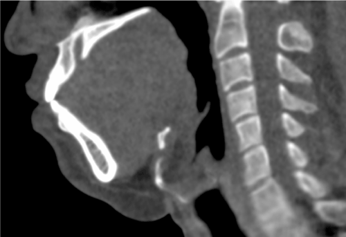
Figure 1. Conventional computed tomography image during swallowing (sagittal plane). The tongue and palate are in contact, so displaying a clear outline of the tongue is difficult
Double-contrast technique
We used 90 mL of 5% sodium alginate solution (Alloid G, Kaigen Pharma Co., Ltd., Hiroshima, Japan) mixed with 10 mL of 100% w/v barium as contrast agent for the mucosal surface (1500 HU). To ensure uniform consistency of the mixture, it was agitated for 10 min in a barium shaker (BR-6, Thermal Chemical Industry, Co., Ltd., Tokyo, Japan; Figure 2). First, subjects took a sip of 10 mL of the contrast agent for the mucosal surface, rolled it in the mouth for 10 s so that it sticks to the entire surface of the tongue, and then swallowed it. A representative oral photograph of the contrast agent attached to the tongue and palate is shown in Figure 3. Second, subjects swallowed 10 mL of golden flaxseed oil (Amaniyu, Nippon Flour Mills Co., Ltd., Tokyo, Japan) as a negative contrast agent (-50 HU). CT imaging was performed during swallowing.
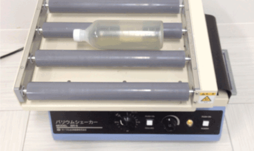
Figure 2. Mixture of the double-contrast agent for the mucosal surface. Barium shaker containing 9 mL of sodium alginate solution and 1 mL of 100% w/v barium mixed by stirring for 10 min
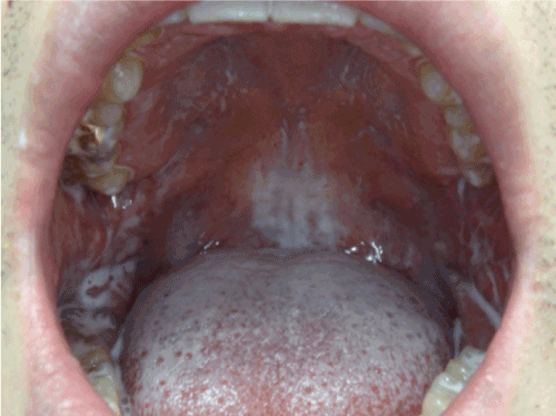
Figure 3. Oral photograph. The contrast agent for the mucosal surface is attached to the tongue and palate
Equipment
We used a 320-row area detector computed tomography scanner (320-ADCT, Aquilon ONE, Toshiba Medical Systems Corporation, Otawara, Japan). A special reclining chair for swallowing CT was inclined at 45 degrees in the semisitting position and placed at the side of the bed opposite to the CT equipment. CT images were taken with the gantry inclined 22 degrees (Figure 4). This chair (co-produced by Tomei Brace Co., Ltd., Seto, Japan, and Aska Corporation, Kariya, Japan ) adjusts the angle of the back side and the position of the seat surface in a back and forth direction, making it possible to insert the face into the scan plane.
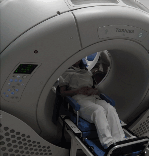
Figure 4. Positioning for 320-row area detector computed tomography imaging. A special chair for swallowing computed tomography (CT) is placed at the side of the bed opposite to the CT equipment and is reclined in a semisitting position inclined at 45 degrees. CT images are taken with the gantry inclined at 22 degrees
Scanning conditions and image production
Scanning conditions and positioning for swallowing CT are shown in Table 1. Scanning conditions were 120 kV, 40 mA, and 3.3 s of serial radiography. After scanning, images of 10 frames/s were produced from the data using a reconstruction function (Adaptive Iterative Dose Reduction 3D, Toshiba Medical Systems Corporation, Otawara, Japan) to reduce radiation exposure. Using a workstation for 3D images (Ziostation2, Ziosoft Inc., Tokyo, Japan), the images of air, golden flaxseed oil, bone, and skin surface were reconstructed by the volume rendering (VR) method, and the images of the tongue and soft palate were reconstructed by the surface rendering (SR) method. Following overlaying of each part of the images, we observed the 3D images during swallowing motion along the time sequence.
Table 1. Exposure parameters for 320-ADCT
| |
Location selected for CT scan |
CT scan |
Scan mode |
Volume |
Dynamic Volume |
kV |
120 |
120 |
mA |
10 |
40 |
Gantry revolution time [sec/rot] |
0.275s/1rot |
3.3s /12rot |
Detector configuration [mm] |
160 |
160 |
mAs |
2.75 |
11 |
FOV [mm] |
240(S) |
240(S) |
CTDIvol [mGy] |
0.6 |
26.2 |
DLP [mGy・cm] |
9.6 |
419.3 |
This study was approved by the ethics committee of Fujita Health University (HM16-135).
We successfully observed the images obtained from 320-ADCT in the axial, coronal, and sagittal planes of multiplanar reconstruction for each organ in the oral cavity and pharynx that is involved in swallowing (Figure 5). In the sagittal plane, the contrast agent attached to the surface of the tongue was detected and the border between the tongue and palate became clear. In the median sagittal plane, the preparatory, oral, pharyngeal, and esophageal stages and the end of swallowing were all observed along the time sequence. The transfer of the food bolus and the motion of the soft palate and tongue were also confirmed (Figure 6).
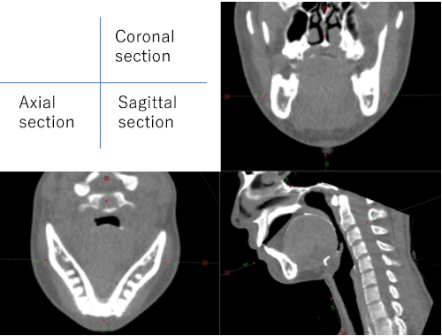
Figure 5. Multiplanar reconstruction images. Contrast agent stuck to the tongue surface, with clear depiction of the border between the tongue and palate
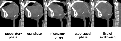
Figure 6. Computed tomography images of tongue median (sagittal plane). Transfer of the food bolus and the motion of the soft palate and tongue are confirmed in the preparatory, oral, pharyngeal, and esophageal stages, and at the end of swallowing
In the oral stage, the subject scoops up the food bolus with the tongue and touches the tip of the tongue to the posterior aspect of the upper incisors. Next, the subject pushes down the tongue median and transfers the bolus of food to the pharynx. The pharyngeal stage involves the closure of the upper pharynx by elevation of the soft palate and the closing of the pharynx by elevation of the larynx. In the esophageal stage, the esophageal entrance is dilated, the upper esophageal sphincter is closed after the bolus of food passes through, and the swallowing process ends. The 3D overlay images of VR and SR are shown in Figure 7. The swallowing motion shown in Figure 6 was displayed in 3D. Particularly, the depression that appears on the tongue median during the phase of holding the food bolus on the tongue and the depression that appears on the elevated soft palate and the tongue base during propulsion became distinct.
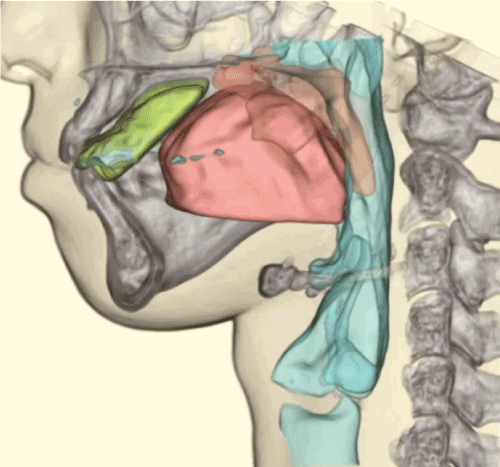
Figure 7. Three-dimensional overlay images of volume rendering and surface rendering. Bone (white), food bolus (yellow), tongue (pink), palate (orange), and respiratory tract (aqua) are seen. The depression seen on the median of the tongue during the holding of the food bolus on the tongue, and the depression that appears on the elevated soft palate and the tongue base during propulsion of the food bolus are clearly depicted. (CT, computed tomography)
This technical note describes a method for visualizing tongue motion during the oral stage of swallowing. This newly revised swallowing CT imaging technique is carried out using a mixture of sodium alginate and barium as a positive contrast agent, which sticks to the tongue and palate. Golden flaxseed oil is used as a negative contrast agent, as the bolus of food. This double-contrast technique enabled us to depict the motion of the tongue and palate during swallowing.
Other radiographic tests involving a double-contrast technique include double-contrast upper gastrointestinal examination, double-contrast barium enema, and double-contrast arthrography. CT is used for double-contrast arthrography. In these tests, air is used as a negative contrast agent in addition to barium, a positive contrast agent. We used sodium alginate to facilitate the temporary attachment of barium to the surface of the tongue. Then, the subject swallows the oil as a negative contrast agent. In patients with residual teeth, the border of the buccal mucosa and teeth is clear. Even in edentulous patients, using the double-contrast technique allows the border of the buccal mucosa, alveolar ridge, and tongue to be observed.
In terms of the formulation, sodium alginate solution is a green, viscous aqueous solution, which is used for mucosal protective and hemostatic effects. This formulation has high adhesiveness. In Japan, it is commonly used as a drug and in the United States, it is used for various purposes in food, drugs, and dental impression materials. In recent years, it has been used as a base for drug delivery systems and film preparation [1]. Extracted from brown seaweed, it has high safety and is used in various industries. It has few side effects including diarrhea and constipation. It mixes readily with barium, and by swirling the mixture in the mouth for 10 s, it can easily coat the entire oral mucosa. Golden flaxseed oil is an edible oil reported to be rich in “α-linolenic acid and is effective for preventing heart disease [2]. When consumed in large amounts, it is known to often cause loose stool and diarrhea. It is easy to consume and does not elute barium, hence it has utility as a negative contrast agent during CT imaging.
Numerous specialized studies exist as the tests for dysphagia. These include modified barium studies, ultrasonography, radionuclide imaging or scintigraphy, and endoscopy. Others are magnetic resonance imaging and ultrafast CT [3]. For swallowing tests, we used the 320-ADCT, which is an ultrafast CT device that has a detector of 0.5 mm×320 rows with a length of 16 cm. The 16-cm length enables dynamic analysis of swallowing from the pharynx to the cervical esophagus. Swallowing CT has been used not only in research but also in clinical practice, and various studies with the use of cross-sectional and 3D imaging have been reported [4-6].
Reducing radiation exposure is important in performing swallowing CT due to the need for serial radiography. In the first CT imaging protocol reported in 2011 when the swallowing CT test was first introduced, the dose of radiation exposure was a maximum of 3.9 mSv [7,8]. Introducing a reconstruction method for reducing radiation exposure and optimizing the protocol reduced the dose of exposure to 1.9 mSv in the latest protocol [9,10]. Studies using low-radiation-exposure protocols have reported the time and timing [11-15], the square measure and volume of the swallowing organ [16-21], and travel distance of the hyoid bone [14,15,20].
Image assessment during dysphagia rehabilitation is important. Dynamic images are most useful to capture the features and extent of disorders, clarifying the utility of treatments and compensatory techniques.3 This novel imaging technique for tongue motion will enable observation of propulsion of the food bolus by the tongue and the dynamic state during mastication, and will contribute to further development of the use of kinematics and oral devices in rehabilitation.
DK designed the study and wrote the initial draft of the manuscript. NF contributed to analysis and interpretation of data and assisted in the preparation of the manuscript. All other authors have contributed to data collection and interpretation, and critically reviewed the manuscript. All authors approved the final version of the manuscript and agree to be accountable for all aspects of the work in ensuring that questions related to the accuracy or integrity of any part of the work are appropriately investigated and resolved.
We would like to thank the staff of our hospital’s radiology department and rehabilitation department, and Ziosoft, Inc., for assistance in our study.
Conflicts of Interest and Source of Funding: Daisuke Kanamori received a Grant-in-Aid for Young Scientists (B), 2016-2018 (#16K21459) from the Japan Society for the Promotion of Science.
The authors have no conflicts of interest directly relevant to the content of this article.
- Agricultural Marketing Service, US Department of Agriculture (2015) Technical Evaluation Report: Alginates: Handling/Processing. Available at: https://www.ams.usda.gov/sites/default/files/media/Alginates%20TR%202015.
- National Center for Complementary and Integrative Health. Flaxseed and Flaxseed Oil. November 30, 2016. Available at: http://nccih.nih.gov/health/flaxseed/ataglance.htm.
- Jones B, Donner M, (2003) Normal and Abnormal Swallowing: Imaging in Diagnosis and Therapy. 2nd edn. New York: Springer-Verlag.
- Fujii N, Inamoto Y, Saitoh E, Baba M, Okada S, et al. (2011) Evaluation of swallowing using 320-detector-row multislice CT. Part I: Single- and multi-phase volume scanning for three-dimensional morphological and kinematic analysis. Dysphagia 26: 99-107. [Crossref]
- Inamoto Y, Fujii N, Saitoh E, Baba M, Okada S, et al. (2011) Evaluation of swallowing using 320-detector-row multislice CT. Part II: Kinematic analysis of laryngeal closure during normal swallowing. Dysphagia 26: 209-217. [Crossref]
- Inamoto Y, Kagaya H, Saitoh E, Kanamori D, Shibata S, et al. (2012) Inter-rater and intra-subject reliability for the evaluation of swallowing kinematics using 320-row area detector computed tomography. Jpn J Compr Rehabil Sci 3: 59-65.
- Kanamori D, Kagaya H, Fujii N, Inamoto Y, Nakayama E, et al. (2011) Examination of the distance measurement error and exposed dose when using a 320-row area detector CT: a comparison with video uoroscopic examination of swallowing. Jpn J Compr Rehabil Sci 2: 18-23.
- Kobayashi M, Koshida K, Suzuki S, Katada K (2012) Evaluation of patient dose and operator dose in swallowing CT studies performed with a 320-detector-row multislice CT scanner. Radiol Phys Technol 2: 148-155. [Crossref]
- Kobayashi M, Asada Y, Matsubara K, Minami K, Suzuki S, et al. (2018) Swallowing computed tomography: dose estimation in a phantom study conducted at various patient reclining angles. Radiat Prot Dosimetry 178: 87-94. [Crossref]
- Kobayashi M, Asada Y, Haba T, Matsunaga Y, Matsubara K, et al. (2019) Optimisation of Swallowing CT Examination: Dose Reduction and Image Quality. Radiat Prot Dosimetry pii: ncz029. [Crossref]
- Inamoto Y, Saitoh E, Okada S, Kagaya H, Shibata S, et al. (2013) The effect of bolus viscosity on laryngeal closure in swallowing: kinematic analysis using 320-row area detector CT. Dysphagia 28: 33-42. [Crossref]
- Shibata S, Inamoto Y, Saitoh E, Kagaya H, Aoyagi Y, et al. (2017) The effect of bolus volume on laryngeal closure and UES opening in swallowing: Kinematic analysis using 320-row area detector CT study. J Oral Rehabil 44: 974-981. [Crossref]
- Pongpipatpaiboon K, Inamoto Y, Saitoh E, Kagaya H, Shibata S, et al. (2018) Pharyngeal swallowing in older adults: Kinematic analysis using three-dimensional dynamic computed tomography. J Oral Rehabil 45: 959-966. [Crossref]
- Shibata S, Kagaya H, Inamoto Y, Saitoh E, Okada S, et al. (2011) Swallowing maneuver analysis using 320-row area detector computed tomography (320-ADCT). Jpn J Compr Rehabil Sci 2: 54-62.
- Inamoto Y, Saitoh E, Ito Y, Kagaya H, Aoyagi Y, et al. (2018) The Mendelsohn maneuver and its effects on swallowing: Kinematic analysis in three dimensions using dynamic area detector CT. Dysphagia 33: 419-430. [Crossref]
- Inamoto Y, Saitoh E, Okada S, Kagaya H, Shibata S, et al. (2015) Anatomy of the larynx and pharynx: effects of age, gender and height revealed by multidetector computed tomography. J Oral Rehabil 42: 670-677. [Crossref]
- Nakayama E, Kagaya H, Saitoh E, Inamoto Y, Hashimoto S, et al. (2013) Changes in pyriform sinus morphology in the head rotated position as assessed by 320-row area detector CT. Dysphagia 28: 199-204. [Crossref]
- Iida T, Kagaya H, Inamoto Y, Shibata S, Saitoh E, et al. (2017) Measurement of pharyngo-laryngeal volume during swallowing using 320-row area detector computed tomography. Dysphagia 32: 749-758. [Crossref]
- Wattanapan P, Kagaya H, Inamoto Y, Saitoh E, Shibata S, et al. (2018) Evaluation of pharyngoesophageal segment using 320-row area detector computed tomography. Ann Otol Rhinol Laryngol 127: 888-894. [Crossref]
- Okada T, Aoyagi Y, Inamoto Y, Saitoh E, Kagaya H, et al. (2013) Dynamic change in hyoid muscle length associated with trajectory of hyoid bone during swallowing: analysis using 320-row area detector computed tomography. J Appl Physiol 115: 1138-1145. [Crossref]
- Mulheren RW, Inamoto Y, Odonkor CA, Ito Y, Shibata S, et al. (2019) The association of 3-D volume and 2-D area of post-swallow pharyngeal residue on CT imaging. Dysphagia 34: 665-672. [Crossref]