Abstract
We report the observation of a 57-year-old woman who was investigated for ecchymotic purpura of the folds and periorbita evolving in the context of diffuse bone pain, asthenia and a deterioration of the general condition. Macroglossia, hypotension, proteinuria and rhythm disturbances were associated with this clinical history. A bone marrow aspiration performed in the presence of hypogammaglobulinemia allowed the diagnosis of Myeloma and the cutaneous signs were linked to amyloidosis which was confirmed by performed rectal biopsy.
Keywords
diffuse ecchymotic purpura, periorbital ecchymotic purpura, macroglossia, hypogammaglobulinemia, amyloidosis, myeloma
Introduction
The combination of diffuse purpura, periorbital ecchymotic purpura, macroglossia, arterial hypotension and hypoglobulinemia is highly suggestive of amyloidosis and should lead to a priority search for dysglobulinemia.
Case presentation
HT, 57 years old woman, with a history of stable hypertension under monotherapy (ARA2), was investigated for petechial purpura. Physical examination consisted ecchymosis after vomiting, associated with diffuse petechial purpura (elbow, fingers, inframammary folds, oral mucosa….). (Figure 1 and Figure 2). Macroglossia was also noted with ecchymotic spots on the oral mucosa (Figure 3 and Figure 2D) and the concomitant onset of a tendency to hypotension, which justified the discontinuation of antihypertensives by the patient treating physician.

Figure 1. Periorbital ecchymosis

Figure 2. Diffuse petechial purpura. (2A) Elbow crease; (2B) Dorsum of the fingers; (2C) Inframammary folds; (2D) Oral mucosa
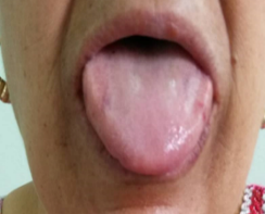
Figure 3. Macroglossia
The patient also reported chest pain, which led to a stress echocardiogram before hospitalization, which was then negative. A complete blood count revealed a normochromic, non-regenerative microcytic anemia. Thyroid function tests, electrolytes, and hemostasis assessment were unremarkable. Proteinuria of 2.2 g/L was observed, with preserved renal function, as was hyperglobulinemia of 5.7 g/L, (Figure 4) with no evidence of a monoclonal component on immunotyping, albumin of 36.4 g/L, an ESR of 51 mmHg, and a ferritin level of 299 ng/ml. The 8-hour serum cortisol level was low at 50.5 nmol/L. Standard X-rays of the skull, the cervical, dorsal and lumbar spine, the pelvis and the femurs revealed lytic lesions known as "punch-out geodes," appearing as multiple, well-circumscribed, round, oval lacunae with no peripheral reaction (Figure 5).

Figure 4. Blood protein electrophoresis showing Hypoglobulinemia

Figure 5. X-ray of the skull profile highlighting geodes
No deep tumor syndrome was found in abdominal tomodensitometry scan, and the kidneys were of normal size. Given this presentation, amyloidosis was suspected and confirmed by rectal biopsy which revealed issue amyloid deposits demonstrate apple-green birefringence when stained with Congo red and viewed under polarizing microscopy. The second step consisted of finding the etiology of this amyloidosis, the context very suggestive of a malignant hemopathy. So, the diagnosis of myeloma was made based in bone marrow biopsy revealing the presence of 34% plasma cells on the myelogram.
In summary the diagnosis of multiple myeloma with secondary AL amyloidosis was held. A lesion assessment was initiated to evaluate organ damage attributable to amyloidosis (neurological, cardiac, etc.) as well as a pre-therapeutic assessment in anticipation of chemotherapy. The course was punctuated by worsening dyspnea related to nosocomial pneumonia, but a glossy appearance of the myocardium suggested cardiac amyloidosis, the cause of the patient's sudden death (rhythmias due to myocardial amyloid infiltration?).
Results and discussion
Diffuse ecchymotic purpura, particularly periorbital [1,2], macroglossia, a tendency toward arterial hypotension (due to adrenal infiltration), renal involvement (with preserved kidney size), and myocardial infiltration, which compromises life prognosis due to excitability disorders (as reported in our case) [3,4], are well established manifestations of amyloidosis in the literature and should be recognized by all practitioners. The ecchymosis purpura, especially in the periorbital area, develop with minor trauma (pinch purpura) because of fragility from amyloid deposition around cutaneous vessels is usual and constitutes a classical mode of revelation but is observed in only 20% of the patients. Multiple myeloma is often the cause of these skin‘s manifestations and should be screened for even in the absence of a monoclonal peak, particularly in elderly patients.
Conclusion
While purpura can be a sign of various conditions, its presence, especially periorbital purpura is more characterized for amyloidosis, particularly the light chain (AL) type. So, for long time periorbital ecchymoses are classically considered as pathognomonic but a late sign of AL amyloidosis [1]. Nevertheless many authors have recently described this sign of transthyretin familial amyloid polyneuropathy that refuting its pathognomonic character in AL amyloidosis [5]. Amyloidosis has a poor prognosis and can expose, as reported in our case, to sudden death attributable to amyloid infiltration of the myocardium. The literature review estimates the association of myeloma and amyloidosis at approximately 20%. This is AL amyloidosis (Light-chain Amyloidosis) which is characterized by the extracellular deposition of a protein material composed of monoclonal light chains (most often lambda) and other proteins (amyloid component P), organized in β-pleated sheets forming fibrils. The deposit is often multi-organ and only the brain is spared. The progressive increase in deposits will modify the structure and function of the affected organs, giving, depending on the case, restrictive heart disease, nephrotic syndrome, digestive, hepatic, peripheral neurological, joint, muscular and cutaneous involvement.
Conflicts of interest
The author declares no conflict of interest.
References
- Visconti L, Cernaro V, Ferrara D, Lacava V, Ricciardi CA, et al. (2015) Periorbital purpura: A pathognomonic but late sign of AL amyloidosis. G Ital Nefrol 32: gin-32.5.8. [Crossref]
- Sapkota S, Kuehl S, Pulluri B (2022) Do not ignore those raccoon eyes; they may indicate lethal AL Amyloidosis. Case Rep Oncol 15: 1039-1048. [Crossref]
- Wechalekar AD, Fontana M, Quarta CC, Liedtke M (2022) AL amyloidosis for cardiologists: Awareness, diagnosis, and future prospects: JACC: CardioOncology state-of-the-art review. Cardio Oncol 4: 427-441. [Crossref]
- Tuñón J, Oliva-Encabo R, Cortés M (2014) Diagnosis of cardiac amyloidosis by skin lesions. Rev Esp Cardiol (Engl Ed) 67: 666. [Crossref]
- Jhawar N, Reynolds J, Nakhleh R, Lyle M (2023) Hereditary transthyretin amyloidosis presenting with spontaneous periorbital purpura: A case report. Eur Heart J Case Rep 7: ytad108. [Crossref]