A 36-year-old female farmer presented with dry cough for 3 months and dyspnea for 20 days. On admission to our hospital, physical examination showed bilateral pulmonary crepitus. Analysis of arterial gas on room air revealed hypoxemia. Pulmonary function testing showed a forced vital capacity of 39% and a diffusion capacity of 54% of the predicted values. Chest radiography (Figure 1) showed bilateral diffuse miliary nodules; chest computed tomography (Figure 2) showed diffuse disseminated small nodules and partially confluent to form larger nodules or consolidations. Pathological examination of transbronchial biopsy (Figure 3) showed lepidic growth of nonmucinous cuboidal cells along intact and mildly thickened alveolar septa, which was diagnosed as nonmucinous bronchoalveolar carcinoma. The mutation of EGFR gene was negative. Unfortunately, the patient died from respiratory failure 2 months later.
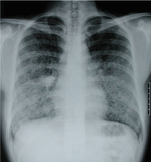
Figure 1. Chest radiography.
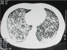
Figure 2. Chest computed tomography.
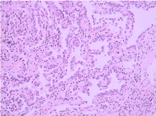
Figure 3. Pathological examination of transbronchial biopsy
Article Type
Short Communication
Publication history
Received date: June 28, 2017
2021 Copyright OAT. All rights reserv
Accepted date: July 18, 2017
Published date: July 20, 2017
Copyright
© 2017 Xiang-Dong MU. This is an open-access article distributed under the terms of the Creative Commons Attribution License, which permits unrestricted use, distribution, and reproduction in any medium, provided the original author and source are credited.
Citation
Xiang-Dong MU (2017) Diffuse bronchoalveolar carcinoma. Gen Med Open 1: DOI: 10.15761/GMO.1000105
Corresponding author
Xiang-dong MU
Department of Respiratory and Critical Care Medicine, Peking University First Hospital, Beijing 100034, China
E-mail : bhuvaneswari.bibleraaj@uhsm.nhs.uk