A patient with coronary artery ectasia and 2 vessel disease was treated by PCI using DES in the RCA and a magnesium-based bioresorbable vascular scaffold (BVS) in the LAD. Follow- up examination after one year using optical coherence tomography revealed late acquired stent malapposition in the DES-treated artery due to the progression of coronary artery ectasia whereas the lesion treated by the BVS showed an excellent result.
coronary artery ectasia, bioresorbable vascular scaffold, drug eluting stent
In January 2020, a 69 years old female patient with a history of hypertension and smoking was admitted to our department due to stable angina CCS 3. Echocardiography showed normal left ventricular function and elective coronary angiography revealed 2 vessel disease with chronic total occlusion of the mid right coronary artery (RCA) and high degree stenosis of the left artery descending (LAD). Additionally, mid LAD showed an ectatic vessel segment directly adjacent to the stenotic lesions (Figure 1). As the patient refused bypass surgery, a staged PCI was planned starting 14 days later with the CTO-PCI of the RCA where 3 DES were implanted from the distal to proximal (DES Promus Premier Select, Boston Scientific, USA; sizes: 3.0 x 38mm; 3.0 x 38mm; 3.5 x 24mm. (Figure 2), 3 months later (22.04.2020), the patient was scheduled for elective PCI of the mid LAD. At this time, angiography showed a patent RCA (Figure 3). Due to the ectatic vessel segments of the LAD directly neighboring the stenotic lesions, it was planned to perform the PCI using a magnesium-based bioresorbable vascular scaffold as it could be expected that any part of the scaffold that did not reach the ectatic vessel wall would be resorbed rapidly thereby avoiding persisting malapposition of stent struts which otherwise would harbor the potential risk of late stent thrombosis. After an uneventful procedure with implantation of one BVS (Magmaris, Biotronik, Germany; size: 3.5 x 25mm; post-dilatation with 3.5mm NC balloon; Figures 3,4), the patient was sent home the next day and kept on dual antiplatelet therapy (DAPT). Before the planned cessation of DAPT, an elective repeat angiography was performed on 24.03.2021. As seen from (Figures 5,6), the LAD was found without signs of restenosis or progression of the ectasia and the implanted magnesium BVS was completely resorbed. On the other hand, OCT of the RCA showed several sites with late malapposition of the implanted DES probably due to the new development of coronary artery ectasia (Figure 7). The decision was made to implant another 3 DES in order to achieve complete stent apposition (Orsiro, Biotronik, Germany; sizes: 4.0 x 13mm; 4.0 x 15mm; 4.0 x 13mm; post-dilatation with 4.5mm NC balloon) (Figure 8). The patient was advised to stay on DAPT for another 6 months and to undergo repeat intracoronary imaging before stopping clopidogrel.
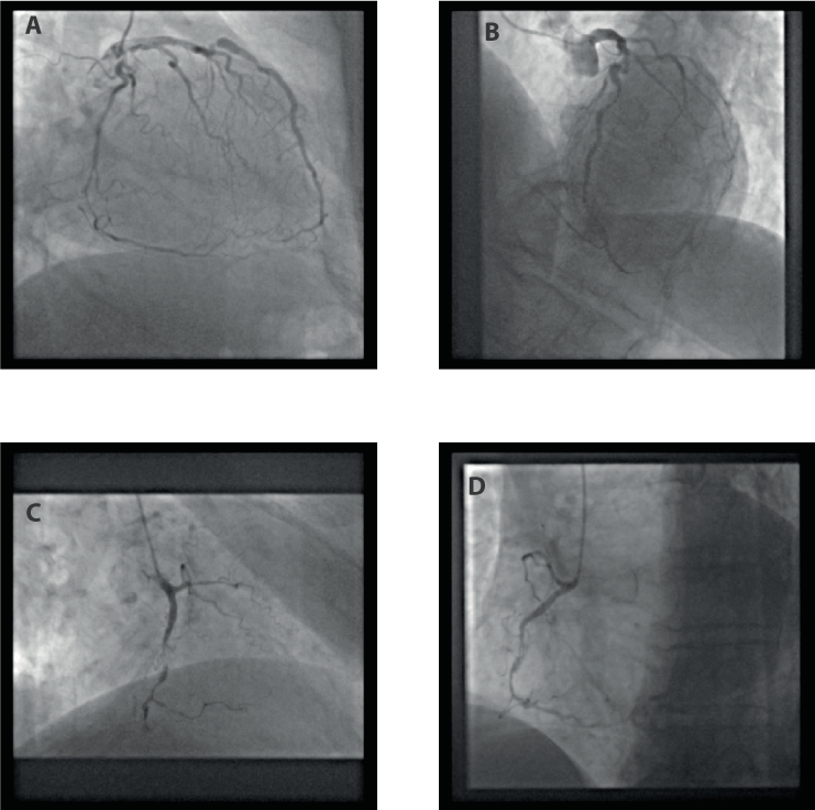
Figure 1. Coronary angiography (16.01.2020). A + B: left coronary artery. Note the retrograde filling of the distal right coronary artery. C + D: right coronary artery.
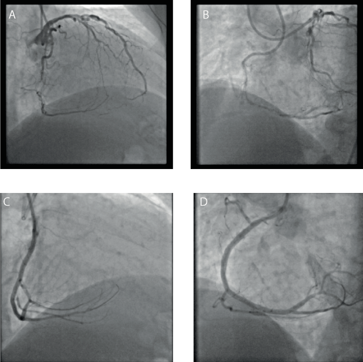
Figure 2. Percutaneous coronary intervention of right coronary artery (RCA) with implantation of 3 drug-eluting stents (31.01.2020). A + B: before PCI (simultaneous injection into left and right coronary artery). C + D: RCA after PCI.
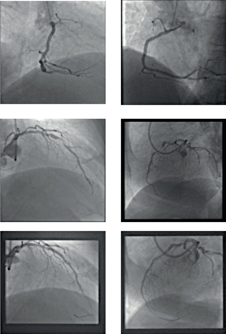
Figure 3. Percutaneous coronary intervention of left artery descending (LAD) using a Magmaris bioresorbable scaffold (22.04.2020). A+ B: angiography of right coronary artery (no further intervention). C + D: LAD before PCI. E + F: LAD after PCI with Magmaris.
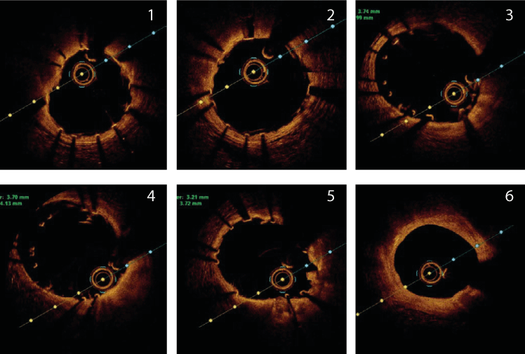
Figure 4. Optical coherence tomography of left artery descending from distal (1) to proximal (6) after implantation of Magmaris-BVS (22.04.2020). The „malapposition“ of the Magmaris- BVS in (2), (3) and (4) was taken into account without further intervention due to the anticipated resorption of the struts.
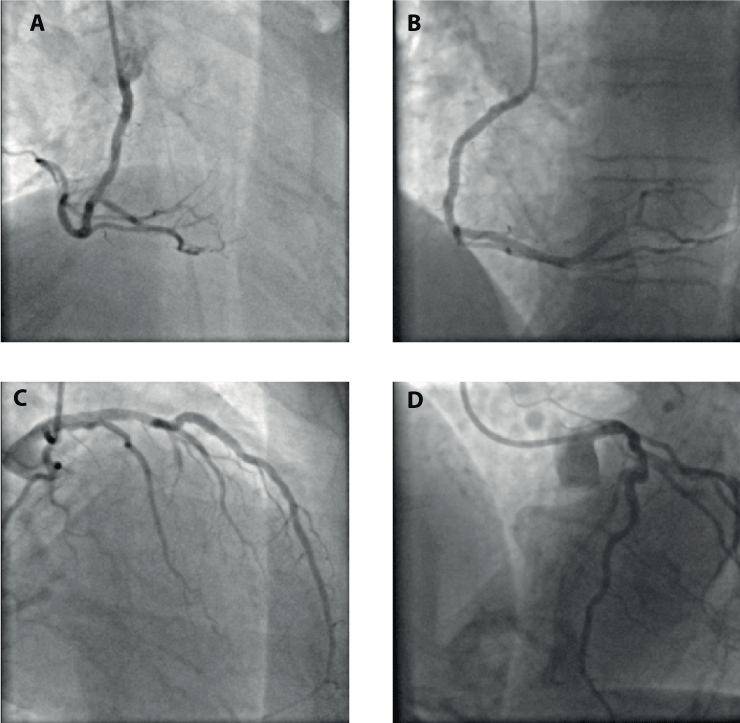
Figure 5.Coronary angiography (follow-up, 24.03.2021). A + B: right coronary artery. C + D: left coronary artery.
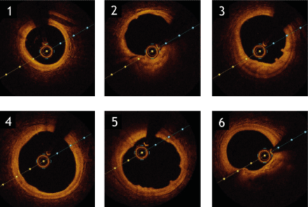
Figure 6. Optical coherence tomography of left artery descending from LAD from distal (1) to proximal (6); Magmaris-BVS has been resorbed completely with some typical residual humps“ seen at the endoluminal vessel wall (24.03.2021).
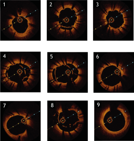
Figure 7. Optical coherence tomography of right coronary artery from distal (1) to proximal (9). Due to the developing ectasia of the RCA, the formerly embedded DES struts now are malapposed (24.03.2021).
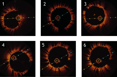
Figure 8. Optical coherence tomography of right coronary artery from distal distal (1) to proximal (6) after additional DES implantation (24.03.2021).
Coronary artery ectasia is defined as segmental dilatation with a diameter of 1.5 times compared with an adjacent normal coronary artery [1]. It has been described with varying frequencies (0.85% per total angiograms in [2]; 4.5% per total angiograms in [3]) and is often found in combination with atherosclerotic heart disease (ASHD, 75% in 2; 85% in [3]). In a cohort of patients presenting with acute myocardial infarction (MI), CAE was present in 3.0% of cases [4]. Until today, there are no clearly established special treatment strategies for these patients.
In our patient suffering from angina CCS 3 arising from 2 vessel disease, a strategy of staged PCI was pursued. We started with the PCI of the RCA which was successfully completed with the implantation of 3 DES. Although intracoronary imaging of the acute result of this PCI was not performed, angiography indicated proper stent sizing and no signs of procedural complications. 3 months later, PCI of the LAD was scheduled which a priori was a complex situation because the target stenosis was in direct neighborhood with a dilated vessel segment so that the risk of acute stent malapposition had to be considered. Therefore, after individual risk- benefit evaluation the decision was made to use a bioresorbable Magmaris-BVS to treat this lesion. The decision was based on the assumption that the stenosis would be sufficiently covered by the BVS whereas in segments where the scaffold would not reach the ectatic vessel wall an accelerated resorption of the “malapposed” struts would take place. The magnesium-based Magmaris-BVS was preferred over other BVS as it had been shown previously that PLLA-based scaffolds are linked to unfavorable longterm outcomes [5] which has not been described for Magmaris [6,7]. 11 months later, follow-up examination indeed showed an excellent morphological result in the LAD without restenosis and complete resorption of the scaffold as intended. However, OCT analysis of the previously implanted
DES in the RCA indicated significant late acquired stent malapposition. In general, it has been described that after CTO-PCI malapposed stent struts are a frequent finding due to several reasons [8]. Nevertheless, in our patient we consider the development of coronary ectasia as the main reason for the extensive late malapposition according to the morphological appearance (tissue bridges between the struts) although this can’t be proven definitely.
We decided to treat the late malapposition of the DES by implantation of further stents of a larger diameter which finally corrected the finding as seen from OCT. This strategy was based on data from previous studies that have emphasized the role of stent malapposition as a major factor of late stent thrombosis and myocardial infarction [9-11] although so far there are no prospective studies which have shown a benefit of any specific therapy in this situation.
Taken together, we present the case of a patient with coexisting CAE and symptomatic 2 vessel ASHD that was treated by PCI using DES in one vessel (RCA) and a magnesium based BVS in the other (LAD). Follow-up examination revealed late acquired stent malapposition in the DES-treated artery due to progression of the CAE whereas the lesion treated by BVS showed an excellent result. We believe that this finding warrants further investigation in clinical trials.
- Swaye PS, Fisher LD, Litwin P, Vignola PA, Judkins MP, et al. (1983) Aneurysmal coronary artery disease. Circulation 67: 134-138.
- Willner NA, Ehrenberg S, Musallam A, Roguin A (2020) Coronary artery ectasia: prevalence, angiographic characteristics and clinical outcome. Open Heart 7: e001096.
- Harikrishnan S, Sunder KR, Tharakan J, Titus T, Bhat A, et al. (2000) Coronary artery ectasia: angiographic, clinical profile and follow-up. Indian Heart J 52: 547-553. [Crossref]
- Doi T, Kataoka Y, Noguchi T, Shibata T, Nakashima T, et al. (2017) Coronary Artery Ectasia Predicts Future Cardiac Events in Patients With Acute Myocardial Infarction. Arterioscler Thromb Vasc Biol 37: 2350-2355. [Crossref]
- Kereiakes DJ, Ellis SG, Metzger C, Caputo RP, Rizik DG, et al. (2017) 3-Year Clinical Outcomes With Everolimus-Eluting Bioresorbable Coronary Scaffolds: The ABSORB III Trial. J Am Coll Cardiol 70: 2852-2862. [Crossref]
- Abellas-Sequeiros RA, Ocaranza-Sanchez R, Bayon-Lorenzo J, Santas-Alvarez M, Gonzalez-Juanatey C (2020) 12-month clinical outcomes after Magmaris percutaneous coronary intervention in a real-world cohort of patients: Results from the CardioHULA registry. Rev Port Cardiol 39: 421-425. [Crossref]
- Verheye S, Wlodarczak A, Montorsi P, Torzewski J, Bennett J, et al. (2020) BIOSOLVE-IV-registry: Safety and performance of the Magmaris scaffold: 12-month outcomes of the first cohort of 1,075 patients. Catheter Cardiovasc Interv 98: E1-E8. [Crossref]
- Sherbet DP, Christopoulos G, Karatasakis A, Danek BA, Kotsia A, et al. (2016) Optical coherence tomography findings after chronic total occlusion interventions: Insights from the "AngiographiC evaluation of the everolimus-eluting stent in chronic Total occlusions" (ACE- CTO) study (NCT01012869). Cardiovasc Revasc Med 17: 444-449. [Crossref]
- Cook S, Eshtehardi P, Kalesan B, Raber L, Wenaweser P, et al. (2012) Impact of incomplete stent apposition on long-term clinical outcome after drug-eluting stent implantation. Eur Heart J 33: 1334-1343. [Crossref]
- Souteyrand G, Amabile N, Mangin L, Chabin X, Meneveau N, et al. (2016) Mechanisms of stent thrombosis analysed by optical coherence tomography: insights from the national PESTO French registry. Eur Heart J 37: 1208-1216. [Crossref]
- Adriaenssens T, Joner M, Godschalk TC, Malik N, Alfonso F, et al. (2017) Prevention of Late Stent Thrombosis by an Interdisciplinary Global European Effort I. Optical Coherence Tomography Findings in Patients With Coronary Stent Thrombosis: A Report of the PRESTIGE Consortium (Prevention of Late Stent Thrombosis by an Interdisciplinary Global European Effort). Circulation 136: 1007-1021. [Crossref]


