Abstract
Introduction: Children with craniosynostosis account for numerous referrals to contemporary pediatric neurosurgeons. While many of these referrals do not result in surgery, meticulous diagnosis and planning is essential for proper care and relieving parent anxiety.
Methods: We retrospectively reviewed pediatric patients ages 0-18 months who underwent corrective surgery for craniosynostosis. Imaging sent by the referring physician was compared to those obtained prior to surgery.
Results: Fifty-seven patient charts were reviewed. Average age at time of presentation was 3.7 months. Sixteen presented with no imaging, 4 with an ultrasound, 25 with X-rays, 7 with a computed tomography (CT) scan, and 5 with magnetic resonance imaging (MRI).
Discussion: Our surgical team favors CT with 3D reconstructed imaging prior to intervention for planning as well as diagnostic confirmation. Of the 34 patients who presented without a CT, 15 (44%) were found to have an incorrect diagnosis on the initial imaging reports. Most commonly this included diagnoses of positional plagiocephaly, a normal scan, or a suture fusion pattern different than what was found surgically or on CT.
Conclusion: When compared to X-rays, the CT has a higher probability of allowing for proper visualization of the sutures. In addition, by using low-dose pediatric protocols, the difference in radiation exposure between the CT and an X-ray is negligible.
Keywords
computed tomography, craniosynostosis, cranial vault reconstruction, magnetic resonance imaging, ultrasound, x-ray
Introduction
Intriguing clinicians and scientists for centuries, irregularities in skull shape can manifest from various etiologies [1,2]. Although the focus of modern surgeons is aimed towards correcting prematurely fused sutures (e.g., syndromic and non-syndromic craniosynostoses), positional plagiocephaly resultant from sleep position is a more common presentation and can often be addressed via conservative measures [2,3]. Ethnic variations in head shape must also be considered when evaluating patients, and several genetic loci have been implicated [2,4-6]. Since the early 1970’s where the surgical management of these complicated disorders was first described [7], the specialties of craniofacial surgery and pediatric neurosurgery have witnessed a rapid evolution of corrective operations [1,4].
Recognizable patterns of craniosynostosis include trigonocephaly (metopic), scaphocephaly (sagittal), plagiocephaly (unilateral coronal or lambdoid), brachycephaly (bilateral coronal or lambdoid), turricephaly (coronal and sagittal), and kleeblattschädel or cloverleaf skull (coronal, lambdoid, and sagittal), amongst others [4,5,8-10]. In addition, minor facial sutures and secondary craniosynostosis (usually due to prolonged supine positioning during infancy) may occur.
Children with craniofacial anomalies represent a large portion of referrals to contemporary pediatric neurosurgeons. Beyond the medical, developmental, and cosmetic considerations of a missed anatomical deformity, abnormal skull shape prompts a great degree of parental anxiety [10,11]. While many referrals do not necessitate surgical correction, success in management is dependent on meticulous diagnosis and planning [8,12].
Early diagnosis is essential for optimizing results, especially with the growing recognition impeded calvarial growth can alter neuronal development [13]. There are several considerations when deciding on imaging for this young population. We retrospectively reviewed surgically confirmed cases of craniosynostosis (i.e., patients who underwent operative intervention) to gain a better sense of the imaging practice in our encatchment area and provide insight into imaging patterns by referring providers.
Methods
Our hospital is a tertiary care center for pediatric and craniofacial surgery. We reviewed charts of pediatric patients aged 0-18 months (at their first appointment) who presented between January 2008 and March 2018. Those with a diagnosis of craniosynostosis who subsequently underwent corrective surgery were included. Institutional review board approval was obtained for this study.
Imaging obtained prior to neurosurgical evaluation (given by the referring physician) and imaging needed – both for diagnostic confirmation and in some cases, operative planning – prior to surgical intervention were recorded. Cases with a false negative finding (initially referred as plagiocephaly but ultimately required surgical correction for craniosynostosis based on imaging and clinical presentation) and those with an incorrect diagnosis on initial imaging (based on surgical findings) were noted.
Results
Fifty-seven pediatric patients 0-18 months of age who underwent operative intervention for craniosynostosis were identified. Eight additional patients (aged 19 months to 44 months) were outside of the age range specified and were not reviewed. Average age at time of presentation was 3.7 months old.
Upon referral, 16 presented with no imaging (28%), 4 with a cranial ultrasound (7%), 25 with X-rays (44%), 7 with a computed tomography (CT) scan (12%), and 5 with magnetic resonance imaging (MRI; 9%). For those who presented with no imaging but referral diagnosis of irregular head shape, 4 (25%) were referred for suspicion of positional plagiocephaly and not craniosynostosis, highlighting the difficulty in diagnosing craniosynostosis clinically.
At our institution, patients generally undergo a pediatric protocol CT scan with three-dimensional (3D) reconstructions as part of surgical planning [14-16]. Thus, it was unsurprising that the 7 patients who had CT imaging by the referring provider did not require additional imaging prior to surgery, and none showed a craniosynostosis pattern during surgery that differed from the imaging findings on initial CT. Amongst the 34 children who presented with a cranial ultrasound, skull X-ray, or MRI, 23 (68%) were referred for CT prior to surgery.
From the group of 25 patients who presented with an X-ray (the most common imaging modality on presentation), 7 (28%) went for surgery without additional imaging due to a classical clinical and radiographic presentation, while 17 (68%) underwent CT scans. Of note, 4 of these 25 children (16%) had X-rays reported as having no evidence of craniosynostosis but were found to have prematurely fused sutures on CT scan. Another 4 (16%) were reported as having craniosynostosis but had incorrect sutures identified on plain radiographs.
Of the four patients who presented with an ultrasound, one was diagnosed correctly as having sagittal synostosis. Of the remaining three, two were found to have craniosynostosis on CT requiring reconstruction that was not seen on ultrasound. The other was thought to have sagittal synostosis alone, but found to have metopic synostosis as well intraoperatively.
Five (9%) of patients who underwent surgery presented with an MRI to the initial consultation. Of these, one (20%) had radiographic and clinical findings that matched the operative findings. However, the other 4 (80%) elicited strong clinical suspicion (despite report of normal sutures) warranting an CT with 3D reconstructions, and all subsequently had both radiologic and intraoperative confirmation of craniosynostosis.
Discussion
Our data presents a unique view into the imaging practices of referring providers within the cohort of surgically confirmed cases of craniosynostosis referred to one surgeon. The imaging available at the time of referral is correlated with intraoperative findings.
As mentioned, our surgical team (involving pediatric neurosurgery and pediatric craniofacial plastic surgery specialists) favors CT with 3D reconstructed imaging for planning as well as diagnostic confirmation prior to intervention, unless the presentation imaging and clinical picture are unequivocal. It is unsurprising that patients who presented with a CT upon initial consultation were all confirmed intraoperatively. In addition, roughly two-thirds of those who presented without a CT underwent one prior to surgery. Of the 34 patients who presented without a CT, 15 (44%) were found to have an incorrect diagnosis when CT was performed and/or intraoperatively.
The armamentarium of imaging modalities available to referring pediatricians has grown significantly. Differentiating between a congenitally absent, closed (apposed), and fused sutures requires a discerning eye from the radiologist’s perspective and careful amalgamation of the clinical presentation by clinicians [4,8]. Radiologists and surgeons alike must consider the primary and secondary etiologies of calvarial asymmetry when reviewing images [4-6,9]. Historically, variations in interpretation and emerging radiologic criteria have led to inter- and intra-observer disagreements, though modern techniques have reduced this concern [17,18].
Bony craniofacial growth is achieved by systematic widening of the sutures and gradual ossification of these enlarging membranous plates. The conduit for this widening is applied force from the growing infant brain. Once found, early surgical correction in infancy generally results in a better cosmetic result than if undertaken later in life, as roughly 80% of brain growth occurs in the first two years [17].
Primary signs of craniosynostosis seen on plain film X-ray are changes in the sutures themselves, including sclerosis or bony bridging, heaping up of bone (beaking), or indistinctness of the suture. Secondary signs are the result of redirecting the normal vector forces of brain growth away from the immobile fused suture, which can change based on the age of the patient at the time the suture closed, the status of the other sutures, and other factors [9,17]. In some cases, X-rays may only reveal secondary signs.
MRI can be a useful adjunct in the investigation of more complicated cases of craniosynostosis, especially if there is suspicion for a syndromic craniosynostosis or a secondary cause of calvarial asymmetry (e.g., defects in the hindbrain or posterior fossa, underlying tumors causing raised intracranial pressure, structural parenchymal defects, etc.) [5,19,20]. Magnetic resonance venography (MRV) is a non-invasive option for evaluating for potentially aberrant venous sinuses prior to surgery, while avoiding the additional ionizing radiation and contrast administration of a CT venogram [1,5]. Most of the patients in our cohort who presented with an MRI had the study to evaluate for other intracranial abnormalities and not necessarily craniosynostosis. None of these studies included black bone or other dedicated sequences to evaluate bony structures, which are available in some centers.
Sonographic evaluation provides an inexpensive alternative that is devoid of ionizing radiation [4,17,21]. Most infants will tolerate an ultrasound without sedation furthering its utility, though this requires someone skilled with the technique to obtain and interpret the images [22,23]. Given the near-perfect sensitivity and specificity of ultrasound in diagnosing premature suture fusion [24], ultrasound is an attractive first-line screening tool in infants, though there is an upper limit of about 8-12 months for its usefulness [21]. However, the information gathered from an ultrasound is generally insufficient to guide surgical management and additional imaging is recommended before operating [2].
CT has emerged as the imaging of choice for diagnostic confirmation as well as surgical planning [1,3,25]. While initially reliant on plain film radiographs with multiple views for evaluation [9,17], the detail afforded by thin-slice (<1mm) CT has allowed for vastly increased levels of detail, particularly in evaluating the orbit and skull base [2,4,5,8]. Similarly, windowing images to evaluate the parenchyma grants simultaneous insight into underlying abnormalities such as ventricular dilatation or asymmetry, encephaloceles, or porencephalic cysts, amongst others [8,17]. In addition, signs of raised intracranial pressure – which can occur in up to 55% of patients with single suture and 90% of those with multi-suture craniosynostosis – can be evaluated with more advanced imaging techniques [4,9].
3D reconstructed models based on CT imaging has been another area of rapid advancement in the craniofacial field. Reconstructed images act as powerful adjuncts in arriving at the diagnosis, especially with regards to surgical planning (Figure 1). Given the uniquely artistic and multi-dimensional nature of these operative endeavors, visualizing the calvarium in a more tangible manner allows for greater pre-operative decision-making [17].
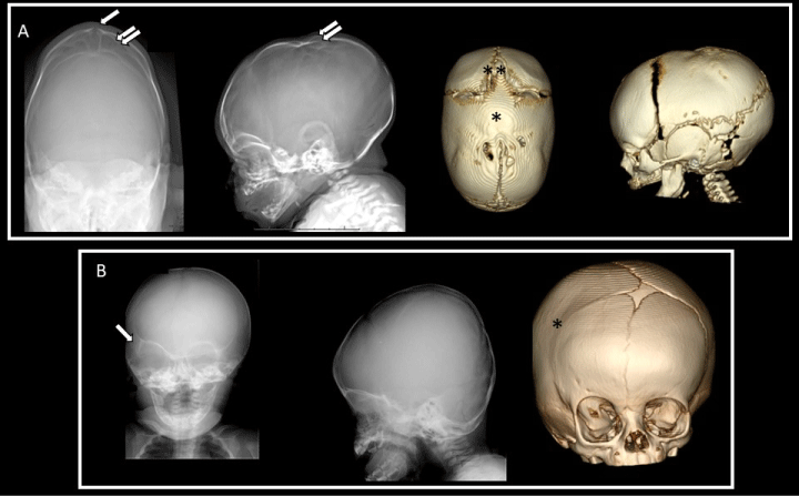
Figure 1: Comparison between plain X-ray and CT scan findings in patients with craniosynostosis. (A) 2 month old patient, plain X-rays on left interpreted as revealing patency of the sagittal suture (arrow), with bony changes suggestive of cephalohematoma (double arrow). 3D CT scan on right revealing synostosis of the anterior ½ of the sagittal suture (asterisk), with extension of the bony fusion across the anterior fontanelle (double asterisk). (B) 4 month old patient, X-rays interpreted as revealing patency of all major sutures, despite right harlequin eye (arrow), with CT scan on right revealing right coronal suture craniosynostosis (asterisk).
Much like X-rays which can miss a fused suture if the beam is not in a plane tangential to the fused suture, volume averaging when reformatting models from CT can result in missed deformities [9,10]. Thus, reconstructions should be derived from appropriately protocoled images. 3D images can result in a much higher accuracy and inter-observer agreement than other modalities [4]. This must be weighed against the risks of higher radiation doses required for higher resolution images due to the increased lifetime risk of radiation exposure in children [4,25-27]. Fortunately, improvements in CT imaging with rapid low-dose protocols have reduced radiation exposure by up to 89% using these protocols [11,28].
Badve et al. eloquently summarized the different imaging options in evaluating children with irregular head shapes. They note that X-rays provide a “rapid, cost-effective screening tool in children with low pretest probability,” while CT scans are the modality of choice in children of higher concern [1]. Ultrasound is a radiation-free option for children in whom there is low suspicion, and MRI is reserved for those with concern for intracranial abnormalities. However, different authors have diverging preferences, and there is currently no one best test [4].
In reviewing our data, we found a large variety in the number of presentations and imaging findings. Ultimately, it remains up to the surgeons to determine if additional imaging is needed as there are benefits to each modality. Pediatric neurosurgeons and craniofacial surgeons must remain vigilant to ensure that all patients are assessed clinically and that proper imaging is obtained in patients with a high index of suspicion despite negative imaging, and similarly that those who are undergoing surgery have appropriate pre-operative investigations.
Some limitations to this study exist, primarily based around its retrospective nature. We wanted to limit our analysis to surgically confirmed cases of craniosynostosis, and thus our data does not take into account referrals that were not made based on normal imaging reports or children who did not undergo surgical correction for imaging confirmed diagnoses of craniosynostosis. The familiarity of the radiology team in evaluating pediatric craniosynostosis will alter these findings between institutions.
Conclusions
In patients suspected of having craniosynostosis, a CT scan is the study of choice to evaluate the cranial sutures. When compared to X-rays, the CT has a higher probability of allowing for proper visualization of the sutures. In addition, by using low-dose pediatric protocols, the difference in radiation exposure between the CT and an X-ray is reduced. Ultimately, the clinical evaluation of children with abnormal head shapes by an experienced provider continues to play an indispensable role in determining the appropriate imaging and interventions required.
Disclosures
The authors report no conflict of interest concerning the materials or methods used in this study or the findings specified in this paper.
REFERNCES
- 1. Badve CA, K MM, Iyer RS, Ishak GE, Khanna PC (2013) Craniosynostosis: imaging review and primer on computed tomography. Pediatr Radiol 43: 728-742. [Crossref]
- 2. Kirmi O, Lo SJ, Johnson D, Anslow P (2009) Craniosynostosis: a radiological and surgical perspective. Semin Ultrasound CT MR 30: 492-512. [Crossref]
- 3. Kajdic N, Spazzapan P, Velnar T (2018) Craniosynostosis - Recognition, clinical characteristics, and treatment. Bosn J Basic Med Sci 18: 110-116. [Crossref]
- 4. Kotrikova B, Krempien R, Freier K, Muhling J (2007) Diagnostic imaging in the management of craniosynostoses. Eur Radiol 17: 1968-1978. [Crossref]
- 5. Nagaraja S, Anslow P, Winter B (2013) Craniosynostosis. Clin Radiol 68: 284-292. [Crossref]
- 6. Ketwaroo PD, Robson CD, Estroff JA (2015) Prenatal Imaging of Craniosynostosis Syndromes. Semin Ultrasound CT MR 36: 453-464. [Crossref]
- 7. Tessier P (1971) The definitive plastic surgical treatment of the severe facial deformities of craniofacial dysostosis. Crouzon's and Apert's diseases. Plast Reconstr Surg 48: 419-442. [Crossref]
- 8. Thompson D, Jones B, Hayward R, Harkness W (1994) Assessment and treatment of craniosynostosis. Br J Hosp Med 52: 17-24. [Crossref]
- 9. Harshbarger R, Kelley P, Leake D, George T (2010) Low dose craniofacial CT/rapid access MRI protocol in craniosynostosis patients: decreased radiation exposure and cost savings. Plastic Reconstructive Surgery 126: 4-5.
- 10. Mafee MF, Valvassori GE (1981) Radiology of the craniofacial anomalies. Otolaryngol Clin North Am 14: 939-988. [Crossref]
- 11. McAlister WH (1998) Invited commentary: posterior deformational plagiocephaly. Pediatr Radiol 28: 727-728. [Crossref]
- 12. Fernbach SK, Feinstein KA (1991) Radiologic evaluation of the child with craniosynostosis. Neurosurg Clin N Am 2: 569-585. [Crossref]
- 13. Brooks ED, Beckett JS, Yang J, Timberlake AT, Sun AH, et al. (2018) The Etiology of Neuronal Development in Craniosynostosis: A Working Hypothesis. J Craniofac Surg 29: 49-55. [Crossref]
- 14. Nagayama Y, Oda S, Nakaura T, Tsuji A, Urata J, et al. (2018) Radiation Dose Reduction at Pediatric CT: Use of Low Tube Voltage and Iterative Reconstruction. Radiographics 38: 1421-1440. [Crossref]
- 15. Jonczyk-Potoczna K, Frankiewicz M, Warzywoda M, Strzyzewski K, Pawlak B (2012) Low-dose protocol for head CT in evaluation of hydrocephalus in children. Pol J Radiol 77: 7-11. [Crossref]
- 16. Morton RP, Reynolds RM, Ramakrishna R, Levitt MR, Hopper RA, et al. (2013) Low-dose head computed tomography in children: a single institutional experience in pediatric radiation risk reduction: clinical article. J Neurosurg Pediatr 12: 406-410. [Crossref]
- 17. Fernbach SK (1998) Craniosynostosis 1998: concepts and controversies. Pediatr Radiol 28: 722-728. [Crossref]
- 18. Alderman BW, Fernbach SK, Greene C, Mangione EJ, Ferguson SW (1997) Diagnostic practice and the estimated prevalence of craniosynostosis in Colorado. Arch Pediatr Adolesc Med 151: 159-164. [Crossref]
- 19. Vargo JD, Hasan A, Andrews BT (2018) Identification and Management of Cranial Anomalies in Perinatology. Clin Perinatol 45: 699-715. [Crossref]
- 20. Sawh-Martinez R, Steinbacher DM (2019) Syndromic Craniosynostosis. Clin Plast Surg 46: 141-155. [Crossref]
- 21. Proisy M, Bruneau B, Riffaud L (2019) How ultrasonography can contribute to diagnosis of craniosynostosis. Neurochirurgie 65: 228-231. [Crossref]
- 22. Soboleski D, McCloskey D, Mussari B, Sauerbrei E, Clarke M, et al. (1997) Sonography of normal cranial sutures. AJR Am J Roentgenol 168: 819-821. [Crossref]
- 23. Sze RW, Parisi MT, Sidhu M, Paladin AM, Ngo AV, et al. (2003) Ultrasound screening of the lambdoid suture in the child with posterior plagiocephaly. Pediatr Radiol 33: 630-636. [Crossref]
- 24. Safran T, Viezel-Mathieu A, Beland B, Azzi AJ, Galli R, et al. (2018) The State of Technology in Craniosynostosis. J Craniofac Surg 29: 904-907. [Crossref]
- 25. Kim HJ, Roh HG, Lee IW (2016) Craniosynostosis : Updates in Radiologic Diagnosis. J Korean Neurosurg Soc 59: 219-226. [Crossref]
- 26. Brenner D, Elliston C, Hall E, Berdon W (2001) Estimated risks of radiation-induced fatal cancer from pediatric CT. AJR Am J Roentgenol 176: 289-296. [Crossref]
- 27. Ginat DT, Lam D, Kuhn AS, Reid R (2018) CT Imaging Findings after Craniosynostosis Reconstructive Surgery. Pediatr Neurosurg 53: 215-221. [Crossref]
- 28. Dempsey RF, Monson LA, Maricevich RS, Truong TA, Olarunnipa S, et al. (2019) Nonsyndromic Craniosynostosis. Clin Plast Surg 46: 123-139. [Crossref]