Objective: The objective of this research is to investigate the validity and applicability of ultrasonography as a diagnostic test for difficulties in intubation through direct laryngoscopy and/or ventilation under facial mask, comparing it with the traditional clinical method of screening.
Method: A double blind accuracy study was performed at Ophir Loyola Hospital, Belém-PA, Brazil. Patients of both genders, between 18 and 80 years of age, with body mass index between 18 and 40 kg/m2, submitted to general anesthesia were included. Traditional clinical inspection, ultrasound scanning of the airways and after anesthetic induction were performed, easy or difficult ventilation as well as difficulty during direct laryngoscopy based on Cormack-Lehane classification were observed. For inspection related to ventilation and intubation, the traditional clinical measurements established in the literature were used. For sonographic inspection, we analyzed the distances: chin-hyoid, skin-hyoid, skin-epiglottis midpoint, skin-thyroid cartilage, skin-anterior commissure of the vocal cords and skin distance-cricoid cartilage. The data were analyzed by the T-student and Mann-Whitney tests, and their validity was verified by the ROC curve.
Results: As for the prediction of direct laryngoscopy, the ultrasonography obtained greater validity as a diagnostic test, surpassing the clinical method. Presenting sensitivity of 71.4%, specificity of 60.3% and high negative predictive value of 92.1%, better predicting the real cases of difficult intubations by the proposed cervical distance measurements. When evaluated in isolation, mask ventilation did not present statistical relevance by ultrasonography. A formula to estimate the difficulty during laryngoscopy based on ultrasound measurements was listed and will serve as a basis for the creation of software for the development of new technologies to assist in diagnostic screening.
Conclusion: It was concluded that ultrasonography is a valid and superior diagnostic test to the clinician to anticipate difficulties with direct laryngoscopy, being an effective test at the bedside.
Airway, ultrasonography, direct laryngoscopy, intubation, ventilation
The clinical evaluation of the airways is essential for the planning of anesthetic-surgical acts that require obtaining and maintaining a definitive means as the primary way to ensure the oxygenation and life of the patient during the peri-operative act [1].
Improper airway management is the most frequent cause of complications related only to anesthesia, being responsible for 30% of deaths, having as main sequelae: permanent brain injury, cardiorespiratory arrest, unnecessary tracheostomies, airway trauma and death [2].
Prior airway evaluation is mandatory, according to the norms of the Federal Council of Medicine and the Brazilian Society of Anesthesiology and should be performed in all patients who will undergo elective surgical procedures [2].
With the recent introduction of new technologies, the use of ultrasonography (USG) is increasingly present in the reality of anesthesiology, and this method can be used to evaluate and anticipate a risk airway, guiding behaviors, creating protocols and markedly reducing the comorbidities associated with the anesthetic-surgical act [3].
The USG is a practical, fast, non-invasive and painless method, becoming a promising instrument to be used in the near future as a routine approach not only to the airway of critically ill patients, but mainly in those electives, in which the attending physician has the obligation to reduce as much as possible the risks inherent to the execution of the procedure [4].
In this context, the use of the USG should emerge as an effective method to adapt to the reality and technical requirements of procedures performed in most surgical blocks. It is an efficient method, when we compare it to other non-invasive means of imaging, because it presents low costs and can even be incorporated into the budget of public health services with low capital [5].
The purpose of this study was to investigate the validity and applicability of ultrasonography as a diagnostic test to anticipate difficulties regarding oral tracheal intubation and/or ventilation under facemask.
This is a study of the diagnostic accuracy test type, developed in the surgical block of the Ophir Loyola Hospital (HOL), in Belém-PA, Brazil, approved by the research ethics committee of the Center for Tropical Medicine of the Federal University of Pará and the Ophir Loyola Hospital.
Sample: Patients who met the following inclusion criteria were included in the study: patients of both genders; age range between 18 and 85 years; with body mass index (BMI) between 18 and 40 kg/m2; submitted to general anesthesia with neuromuscular blockade, with the need to obtain an advanced and definitive airway by oral tracheal intubation through direct laryngoscopy. Patients with anatomical deformities of any kind, which could generate a deviation of the median axis of the airway and therefore incur a measurement bias (clinical data such as dysphonia, hoarseness, dysphagia, chronic cough or proof of deformity by imaging method were considered excluding factors) were also excluded, patients who had their procedures cancelled for any reason (clinical complications
Data collection: An individual questionnaire on clinical inspection of the airways was initially applied, investigating the following variables: presence of beard, BMI, age; lack of teeth; history of snoring and easy or difficult ventilation.
For clinical inspection of the oral tracheal Intubation, the following variables were studied: Mallampati classification, Comarck-Lehane classification, cervical mobility, inter-incisive distance (greater or less than 3 cm), sternum-chin distance (greater or less than 12.5 cm), presence of prominent central incisors, history of difficult intubation.
Then, ultrasound scanning of the airways was performed painlessly and non-invasively, with the patient awake, in dorsal decubitus position and with the head in hyperextension at the same level of the body (earlobe at sternum height). Six different ultrasound windows were performed in each patient to measure the following distances: hyoid-chin (convex probe, with longitudinal windowing- Figure 1), skin-hyoid (linear probe, with transversal windowing-Figure 2), skin-epiglottis midpoint (linear probe, with transversal windowing-Figure 3), skin-thyroid cartilage (linear probe, with transversal windowing-Figure 4), skin-anterior commissure of the vocal cords (linear probe, with transversal windowing-Figure 4) and skin-cricoid cartilage (linear probe, with transversal windowing-Figure 5).
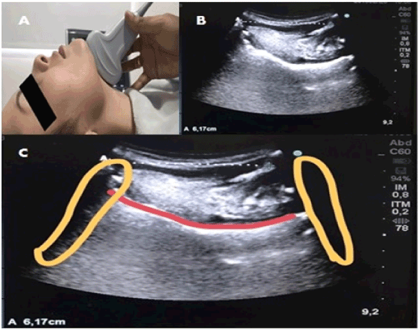
Figure 1. Sonoanatomy (Hyoid Distance - Mind). A. Probe positioning for windowing; B. Sonoanatomy; C: Subtitled sonoanatomy: yellow (left: acoustic shadow of the chin/ right: acoustic shadow of the hyoid bone); in red (surface of the tongue).
Source: Research Protocol.
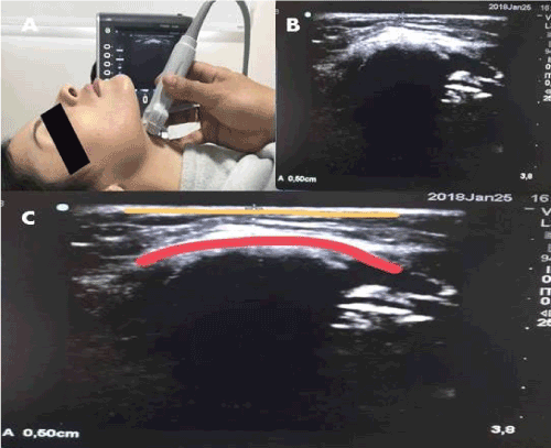
Figure 2. Sonoanatomy (Skin Distance - Hyoid). A. Probe positioning for windowing; B: Sonoanatomy; C. Subtitled Sonoanatomy: yellow (skin surface)/ red (hypere - hyoid bone chica line with posterior acoustic shadow).
Source: Research Protocol.
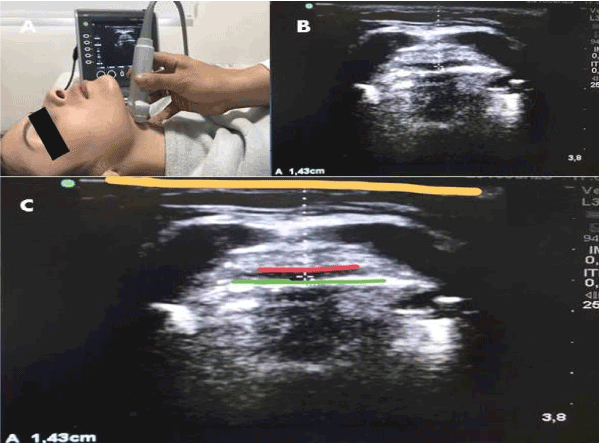
Figure 3. Sonoanatomy (Skin Distance - Epiglottis). A: Window probe positioning/ B: Sonoanatomy. C: Sonoanatomy: yellow (skin surface) / red (epiglottis cartilage -hypoechoic image) / green (hyperechoic line: air interface - epiglottis mucosa).
Source: Research Protocol.

Figure 4. Sonoanatomy (Skin Distance - Thyroid Cartilage). A. Probe positioning for windowing. B: Sonoanatomy. C. Sonoanatomy: Yellow (Skin Surface)/ Blue (Thyroid Cartilage); Red (anterior commissure of vocal cords)/ Green (vocal cords).
Source: Research Protocol.
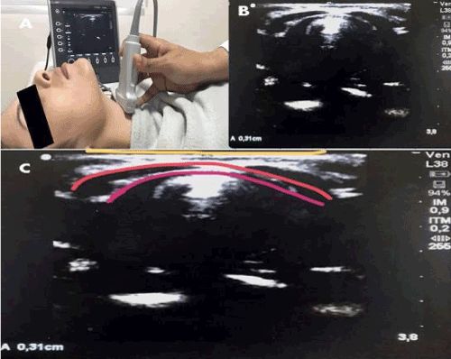
Figure 5. Sonoanatomy (Skin Distance - Crichoid Cartilage). A: Probe positioning for windowing; B. Sonoanatomy; C. Sonoanatomy: Yellow (Surface of the skin)/ Red (Cricoid cartilage - hypoechoic)/ Pink (hyperechoic line of the air - mucosa interface).
Source: Research Protocol.
For sonographic inspection (which evaluated the prediction of both direct laryngoscopy intubation and mask ventilation), the following variables were analyzed: distance from the chin to the hyoid bone, distance from the skin to the hyoid bone, distance from the skin to the midpoint of the epiglottis, distance from the skin to the thyroid cartilage, distance from the skin to the anterior commissure of the vocal cords and distance from the skin to the cricoid cartilage.
For comparison between sonographic measurements and clinical inspection of the airways regarding ventilation under face mask, Mann-Whitney and Student T tests were used according to the normality of the variables, adopting as gold standard the report of the operator during manual ventilation with positive pressure (if easy or difficult).
For comparison between sonographic measurements and clinical inspection of the airways in relation to oral tracheal intubation by direct laryngoscopy, we adopted as the gold standard the visualization of the vocal cords according to the Cormack - Lehane classification (Grade I or II - Easy and Grade III or IV - Difficult) for this, we used the Student's T test and the Mann-Whitney test, according to the normality of the variables.
Multiple logistic regression analysis was another method used to identify the model that best describes the sonographic measurements in relation to the classification of the degree of intubation difficulty found by direct laryngoscopy. This model was also adopted to exclude the influence of any possible confounding bias present in the collection. After inserting the variables with p<0.20 in the model, from the bivariate analyses, the next step was to rotate again a multiple logistic model with the variables that presented p<0.20. Later, p<0.05 was adopted as the significance level for identifying the final model and the really relevant measures.
After the identification of the final model, a simulation was performed to evaluate how many patients the model correctly classified. It was adopted that if the model indicated a chance higher than 30%, it would be considered as a difficult intubation and easy intubation otherwise.
Cormack-Lehane's classification was considered the gold standard for confirmation of results. The results were then compared between the clinical method and the ultrasound method, using the ROC curve to identify the most valid diagnostic test for airway screening.
Data analyses were performed by SPSS, version 20.0. In all work, the significance level of 5% was used.
The study included 144 patients, containing the clinical and ultra-sonographic variables specific to evaluate the prediction of difficulty in ventilation under mask and direct laryngoscopy. Regarding ventilation under facemask, 16 patients (11.11%) presented difficult ventilation. Correlating the estimated difficult ventilation (considering the presence of two or more clinical risk factors, according to the literature) with the actual ventilation, obtained in the work, a specificity (42.2%) and sensitivity (93.8%) of the clinical screening were evidenced. It had a positive predictive value of 16.8% and a negative predictive value of 98.1% (Table 1).
Table 1. Estimated ventilation by clinical factors x real ventilation. Source: Research protocol.
Estimated Ventilation |
Real Ventilation |
Total |
Easy |
% |
Hard |
% |
Easy |
54 |
42.2 |
1 |
6.3 |
55 |
Difficult |
74 |
57.8 |
15 |
93.8 |
89 |
Total |
128 |
100 |
16 |
100 |
144 |
Ultrasound measurements were correlated to actual ventilation (easy or difficult). It was evidenced that none of the sonographic measurements presented significant differences between the groups that had easy ventilation and difficult ventilation (p>0.05) (Table 2).
Table 2. Ultrasonographic measurements (both genders) x mask ventilation. Source: Research protocol. Mann - Whitney test.
Measures |
Freq. |
Average |
Standard deviation |
Average |
IIQ |
p* Value |
Skin - Hyoid |
Difficult Ventilation |
16 |
9.9 |
3.3 |
9.1 |
12.2 |
0.4764 |
Easy Ventilation |
128 |
9.2 |
3.1 |
8.7 |
3.7 |
|
Skin - Cyanoid |
Difficult Ventilation |
16 |
5.7 |
2.3 |
5.5 |
7.1 |
0.8189 |
Easy Ventilation |
128 |
5.6 |
1.8 |
5.4 |
2.2 |
|
Skin - Epiglottis |
Difficult Ventilation |
16 |
19.8 |
3.6 |
19.1 |
21.5 |
0.3387 |
Easy Ventilation |
128 |
18.7 |
3.3 |
18.7 |
4.9 |
|
Skin - Thyroid |
Difficult Ventilation |
16 |
5.1 |
2.4 |
4.5 |
6.3 |
0.7723 |
Easy Ventilation |
128 |
5.2 |
2.3 |
4.7 |
3.2 |
|
Hyoid - Mento |
Difficult Ventilation |
16 |
46.8 |
5 |
48 |
50.1 |
0.1163 |
Easy Ventilation |
128 |
44.5 |
5.6 |
44 |
6.6 |
|
Vocal Chord Skin |
Difficult Ventilation |
16 |
9.8 |
2.4 |
9 |
11.4 |
0.9949 |
Easy Ventilation |
128 |
9.7 |
2.4 |
9.2 |
2.5 |
|
Regarding oral tracheal intubation by direct laryngoscopy, it was considered easy when Cormack-Lehane grade I or II was found and difficult if grade III or IV, out of a total of 144 patients, 29 (20.14%) presented difficult intubation. Regarding the frequency of Mallampati test, it can be observed that most of the patients included were classified as grade II (45.80%) and regarding the risk factors for difficult intubation the most prevalent was Mallampati grade III or IV present in 29.2% of the sample (Table 3).
Table 3. Frequency of Mallampati Test and clinical risk factors for difficult intubation.
Variables |
Frequency |
% |
Mallampati |
Grade I |
36 |
25 |
Grade II |
66 |
45.8 |
Grade III |
24 |
16.6 |
Grade IV |
18 |
12.5 |
Risk Factors |
Mallampati grade 3 or 4 |
42 |
29.2 |
Sternum distance <12,5 |
7 |
4.9 |
Inter-incisive distance <3 |
1 |
0.7 |
Prominent central incisors |
1 |
0.7 |
Limited cervical mobility |
0 |
0 |
History of difficult intubation |
0 |
0 |
The risk factors exposed in Table 3 were accounted and grouped in (Table 4) according to the general incidence being related to difficult intubation.
Table 4. Frequency of the number of risk factors X Real Intubation (Easy or Difficult). Source: Research protocol.
Number of Risk Factors |
Easy |
% |
Difficult |
% |
Total |
% |
0 |
87 |
75.65% |
12 |
41.38% |
99 |
68.75% |
1 |
26 |
22.61% |
14 |
48.28% |
40 |
27.78% |
2 |
1 |
0.87% |
3 |
10.34% |
4 |
2.78% |
3 |
1 |
0.87% |
0 |
0.00% |
1 |
0.69% |
General Total |
115 |
100.00% |
29 |
100.00% |
144 |
100.00% |
Table 5 groups the clinical risk factors for difficult intubation exposed in Table 3 and Table 4 (0 to 1 factor-easy intubation and 2 or more factors-difficult intubation). This allowed evidencing a specificity of 98.3% and sensitivity of 10.3%, with a positive predictive value of 60% and a negative predictive value of 81.4%.
Table 5. Risk Factors (aggregates) X Real Intubation (Easy or Difficult). Source: Research protocol.
Estimated Intubation |
Real intubation |
Total |
% |
|
Easy |
% |
Difficult |
% |
|
|
0 a 1(Easy) |
113 |
98.3 |
26 |
89.7 |
139 |
96.5 |
2 ou + (Difficult) |
2 |
1.7 |
3 |
10.3 |
5 |
3.5 |
Total |
115 |
100 |
29 |
100 |
144 |
100 |
Regarding the sonographic measurements and the correlation between oral tracheal intubation (easy or difficult), the distances: skin-cricoid, skin-epiglottis and hyoid-ment were statistically significant with p<0.05 (Table 6).
Table 6. Sonographic measurements (both genders) X Intubation (easy or difficult). Source: Research protocol. *Test of Mann - Whitney/* Test t-student.
Measures |
Freq |
Avarage |
Standard deviation |
Avarage |
IIQ |
P value |
Skin - Hyoid |
Difficult Intubation |
29 |
10.48 |
4.088 |
10.3 |
6.95 |
0.0757* |
Easy Intubation |
115 |
8.964 |
2.721 |
8.7 |
3.6 |
|
Skin - Cyanoid |
Difficult Intubation |
29 |
6.662 |
2.409 |
6.5 |
3.85 |
0.0041* |
Easy Intubation |
115 |
5.311 |
1.522 |
5.2 |
2.3 |
|
Skin - Epiglottis |
Difficult Intubation |
29 |
20.36 |
3.691 |
20.4 |
9.35 |
0.0062** |
Easy Intubation |
115 |
18.46 |
3.165 |
18.6 |
4.8 |
|
Skin - Tireoid |
Difficult Intubation |
29 |
6.2 |
3.335 |
5.5 |
4.8 |
0.2318* |
Easy Intubation |
115 |
4.957 |
1.839 |
4.5 |
3.1 |
|
Hyoid - Mento |
Difficult Intubation |
29 |
41.63 |
6.168 |
41.8 |
9.05 |
0.0006** |
Easy Intubation |
115 |
45.58 |
5.159 |
44.7 |
6.1 |
|
Skin - Vocal Cords |
Difficult Intubation |
29 |
10.88 |
3.329 |
9.9 |
4.4 |
0.056* |
Easy Intubation |
115 |
9.457 |
1.999 |
9.1 |
2.7 |
|
The correlation of sonographic measurements of only male patients with intubation (easy or difficult) showed that no measurement was statistically significant (p>0.05). (Table 7) correlates the sonographic measurements of only male patients with intubation (easy or difficult), showing that all measurements were statistically significant (p<0.05).
Table 7. Sonographic measurements (female gender) X Intubation (easy or difficult). Source: Research protocol. *Test of Mann - Whitney/* Test t-student.
Measures |
Freq |
Avarage |
Standard deviation |
Avarage |
IIQ |
P value |
Hioid Skin |
Difficult Intubation |
14 |
12.9 |
3.6 |
13.1 |
5.8 |
0.0001** |
Easy Intubation |
78 |
9.5 |
2.8 |
9 |
4.1 |
|
Skin - Cyanoid |
Difficult Intubation |
14 |
7.6 |
2.4 |
7.6 |
4.6 |
0.0032* |
Easy Intubation |
78 |
5.5 |
1.6 |
5.4 |
2.1 |
|
Skin - Epiglottis |
Difficult Intubation |
14 |
20.2 |
4.2 |
19.5 |
5.9 |
0.0088** |
Easy Intubation |
78 |
18 |
2.9 |
18.6 |
4.6 |
|
Skin - Thyroid |
Difficult Intubation |
14 |
8.2 |
3.5 |
7.55 |
5.35 |
0.0132* |
Easy Intubation |
78 |
5.5 |
1.7567 |
5.6 |
3.025 |
|
Hyoid - Mento |
Difficult Intubation |
14 |
40.8143 |
5.0253 |
40.55 |
7.325 |
0.0044* |
Easy Intubation |
78 |
45.3782 |
4.7838 |
44.4 |
5.325 |
|
Skin - Vocal Cords |
Difficult Intubation |
14 |
13.0429 |
3.3768 |
13 |
5.25 |
0.0012* |
Easy Intubation |
78 |
10.05 |
1.9256 |
9.6 |
2.3 |
|
From the quartile analysis, a reference value was inferred to serve as a cut-off for each sonographic measurement (in females), correlating them to the probability of predicting an easy or difficult intubation (Chart 1).
Chart 1. Borderline reference values of ultrasound measurements by quartiles. Source: Research protocol.
Measures |
Reference Value |
Quartil |
Skin - Hyoid |
> 10.3 mm |
1 |
Skin - Cyanoid |
> 5.4 mm |
1 |
Skin - Epiglottis |
> 17.4 mm |
1 |
Skin - C. Tireoid |
> 5.7 mm |
1 |
Hyoid - Mento |
< 43.9 mm |
3 |
Skin - Vocal Cords |
> 10.1 mm |
1 |
It was observed that the greater the distance of the skin-hyoid, skin-cricoid, skin-epiglottis, skin- thyroid cartilage and skin-anterior commissure of the vocal cords, the greater the association with the difficulty of intubation. While the shorter the hyoid distance, the greater the associated intubation difficulty.
From the data obtained from Chart 1, the sonographic measurements were associated between themselves (Table 8), considering as a prediction of difficult intubation if present in the patient 4 or more measurements which exceed their reference values established by the quartile method. Obtaining a test specificity of 60.3% and sensitivity of 71.4%. With a positive predictive value of 24.4% and a negative predictive value of 92.1%.
Table 8. Association of sonographic measurements (by quartiles) X Type of Intubation. Source: Research protocol.
|
Easy Intubation |
% |
Difficult Intubation |
% |
Easy <4 measures |
47 |
60.3 |
4 |
28.6 |
Difficult > = 4 measures |
31 |
39.7 |
10 |
71.4 |
Total |
78 |
100 |
14 |
100 |
When plotting the sonographic measurements versus the clinical measurements on the ROC (Receiver Operating Characteristic) curve as predictors of difficult intubation, it was noted that the area obtained by the sonographic tests considering the presence of 4 or more measurements that exceed the reference values was 66% of the area under the curve, against 56.5% of the area obtained by the clinical variables. The validity of the diagnostic test is greater when performed by ultrasonography (Figure 6).
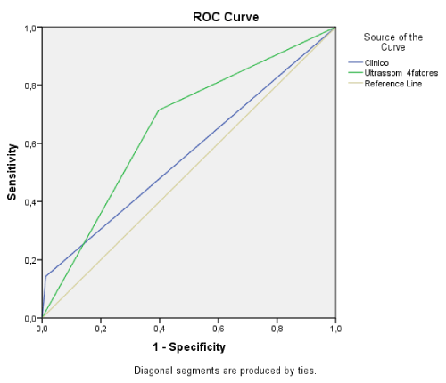
Figure 6. Validity of ultrasonography X Clinical inspection. Clinical Measurements: 56.5 % of the area/ Ultrasonography Measurements: 66% of the area.
Source: Research protocol.
Multiple logistic regression analysis was also performed (Table 9) to identify the model that best describes the measures regarding the classification of intubation difficulty found in both genders. After the identification of the final model with p<0.05 a simulation was performed to evaluate how many patients the model correctly classified. It was adopted that if the model indicated a chance higher than 30% it would be considered as a difficult intubation and an easy intubation otherwise. The method presented specificity of 87.8% and sensitivity of 48.3%, with a negative predictive value of 87% and a positive predictive value of 50%.
Table 9. Logistic Regression of Ultra sonographic Measurements X Real Intubation. Chance > 30 % = difficult intubation. Source: Research protocol.
Estimated Intubation |
Real intubation |
Total |
% |
Easy |
% |
Difficult |
% |
Easy |
101 |
87.8 |
15 |
51.7 |
116 |
80.6 |
Difficult |
14 |
12.2 |
14 |
48.3 |
28 |
19.4 |
Total |
115 |
100 |
29 |
100 |
144 |
100 |
In the multiple logistic regression analysis, the variables that were statistically significant (p<0.05) were gender and sonographic measurements of hyoid distances-chin and skin-anterior commissure of the vocal cords. The greater the distance between the skin-vocal cords and the shorter the hyoid-chin distance, the greater the chance of difficult intubation. By plotting the logistic regression results and confronting them with the clinical variables in the ROC curve, in order to analyze the validity of the tests to anticipate difficulties regarding intubation. It was obtained that the area of the sonographic test corresponded to 68.1% below the curve, against 54.3% of the area occupied by clinical inspection tests. Again, the ultrasonography presents a greater validity as a diagnostic test when compared to traditional clinical methods (Figure 7).
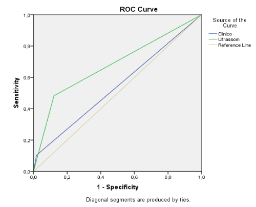
Figure 7. Validity of ultrasonography (logistic regression) X Clinical Inspection. Clinical Measurements: 54.3% of the area/ Ultrasound Measurements: 68.1% of the area.
Source: Research protocol.
Facemask ventilation is the most basic procedure among airway management skills. Patients who are identified as having difficult ventilation are potentially at higher risk in their driving [6]. Of the 144 patients studied, 16 (11.11%) were identified as having difficult ventilation, resembling the prevalence of difficult ventilation in the study by Bradley et al. [7] who described a 0.08% to 15% incidence of difficult ventilation depending on the population studied, unlike the studies by Nekhendzy and Tanaka [8] and Langeron et al. [9] with incidences of 1.4% and 5%, respectively.
An important differential of this study is that the sample was obtained in an oncologic hospital, with many of these patients in a consumptive stage of the disease, which can alter the anatomy of the upper airways at cervical level. It is known that in the consumptive syndromes a high consumption of lean mass and fat mass occurs in the topography of the extremities and at central level. The first affected areas are: hair, eyes, teeth, oral cavity and cervical tissues, due to the high energy-protein consumption of many neoplasms [10,11].
Analyzing the 144 patients, a sensitivity of 93.8% and specificity of 42.2% was obtained by clinical screening to identify difficult ventilation under mask, surpassing the findings of Langeron et al. [9] who anticipated only 17% of his sample.
When performing sonographic screening to predict possible difficult mask ventilation, no statistical relevance was obtained in any of the USG variables, and it was not possible to isolate any specific image measurement that could influence ventilation prediction. Therefore, the present study cannot confirm the usefulness of USG as a test to predict the degree of difficulty of ventilation under a facemask.
This result is largely due to the fact that the measurements studied by the USG present a lower topography in relation to most anatomical structures above the floor of the oral cavity, and which are directly related to ventilation under mask. Moreover, the method is unable to take into account factors external to the patient's face, such as clinical criteria, which are essential for a good sealing and coupling of the mask over the patient's face and consequently good ventilation [12].
Regarding direct laryngoscopy and oral tracheal intubation, 29 (20%) of the 144 patients studied had direct laryngoscopy with Cormack-Lehane grade 3 or 4, being considered difficult intubation. For Kandemir et al. [13] the distribution of Cormack-Lehane grade I and II and grade III and IV in a sample of 290 patients was 92.1% and 7.9%, respectively.
In order to increase the chance of correct clinical method in relation to oral tracheal intubation, the variables were grouped in easy and difficult prediction. Based on this premise, the research obtained a sensitivity of 10.3% and a specificity of 98.3% of the traditional clinical method. It was above the study made by Huitink and Bowman [14] that obtained a sensitivity of only 7% when evaluating 3391 patients with difficult intubation. The study also obtained better results than Norskov et al. [15], who when analyzing 188,064 patients through clinical predictors, observed that 93% of difficult intubations were not anticipated, and among those anticipated, only 25% were really difficult.
The positive predictive value of the current clinical trials was 60%, while the negative predictive value was 81.4%, meaning that in 18.6% of the cases in which the clinical examination attested easy airway, in fact were cases of difficult airway not anticipated, incurring a considerable risk to the patient.
In a study by Kandemir et al. [13], correlating the Mallampati test with sternomental distance (greater or less than 12.45 cm) to predict difficult intubation, found sensitivity of 18.75%, specificity of 90.48%, positive predictive value of 60% and negative predictive value of 59.38%, showing a higher margin of error (40.62%) related to the diagnostic test in indicating an easy airway, being in fact a difficult intubation.
The validity of ultrasound screening as a diagnostic test to anticipate difficult direct laryngoscopy in this study was observed through the statistical significance for both genders in three measurements (distance skin - cricoid; skin - epiglottis and hyoid-chin).
By separating the sonographic analysis between the genders, it was observed that all six measurements presented statistical significance in females, while none presented relevance in males. This discrepancy is probably due to two factors. First, the number of women in the sample was almost double the number of men. Added to this, the profile of the sample studied was of oncologic patients, many of them with consumptive syndrome (deproteinized, with loss of fat mass and lean mass).
Women, because they have different fat distribution and constitution, despite losing cervical mass, use more intensively the consumption of peripheral areas of the body rich in fat, such as buttocks, breasts and extremities, metabolizing more lentifiably and at a lower rate the tissues at the cervical level [11].
Three sonographic measurements showed statistical relevance (when analyzed in both genders, it was preferred however to stipulate the cut of reference values evaluating the female population, thus avoiding a possible measurement bias. The data generate values that may serve as reference for the adult population in general but does not exclude the need for other studies in larger and better equalized samples with healthy patients of both genders.
The values found from ultrasound measurements to predict possible difficult direct laryngoscopy were: hyoid-mind distance of 43.9 mm (the shorter the distance, the greater the prediction of difficulty); skin-hyoid distance of 10.3 mm (the greater the distance, the greater the chance of difficulty); skin-cricoid distance of 5.4 mm (the greater the distance, the greater the chance of difficulty); skin distance-epiglottis of 17.4 mm (the greater the distance, the greater the difficulty); skin distance-thyroid cartilage of 5.7 mm (the greater the measure, the greater the difficulty) and skin distance-anterior commissure of the vocal cords of 10.1 mm (the greater the distance, the greater the predicted difficulty).
By associating the six sonographic measurements between themselves, in order to increase the hit rate, it was considered difficult to predict intubation in patients who present 4 or more measurements that exceed their reference values established by the quartile method. A test specificity of 60.3%, sensitivity of 71.4%, positive predictive value of 24.4%, and negative predictive value of 92.1% was then obtained. Thus, it can be affirmed that the ultrasound is able to predict more accurately those patients who really have difficult intubation.
While clinical screening, despite having a higher specificity, has a low sensitivity and a low negative predictive value, it becomes more susceptible to errors in order to predict an easy laryngoscopy in real situations of difficult laryngoscopy, greatly increasing the risk of the procedure. Thus, the diagnostic test based solely on clinical measurements exposes the patient to risk situations such as airway trauma [2].
To better evaluate the relationship of the ultrasound diagnostic test, together with the clinical screening tests, regarding the prediction of difficult direct laryngoscopy, the data and reference values obtained were plotted on the ROC curve. The ultrasound test showed a larger area under the curve of 66% compared to 56.5% of the area obtained by the clinical tests in the quartile method, meaning, therefore, that in the prediction of direct laryngoscopy and difficult intubation, the ultrasound test is more valid than the traditional clinical tests for airway evaluation.
Ultrasonography provides high resolution images, identifying anatomical structures of the upper airways comparable to those evidenced by computed tomography and magnetic resonance imaging [16].
In a study by Adhikari et al. [17], assessing the relationship between difficult laryngoscopy and ultra-sonographic variables (thickness of fat tissue in the neck at the level of the hyoid bone and thyroid membrane), comparing them with the evaluation of preoperative clinical criteria, the authors found that among the 51 patients analyzed, 12% (6) presented with difficult laryngoscopy.
Probably the highest value of the hyoid skin distance identified by the author (16.9 mm), compared to that found by this research (10.3 mm), is due to the fact that in the study of Adhikari et al. [17] it was performed in the American population, healthy, with unspecified BMI indices, with different adipose composition from the sample of oncologic patients of the research, who also had their BMI stipulated and limited.
A work performed with 50 obese patients, correlating the ultra-sonographic screening by measuring the cervical fat index at the level of the anterior commissure of the vocal cords, comparing their ability to predict difficult intubation with that of traditional methods, resulted in 9 cases of difficult intubation, and the skin distance-anterior commissure of the vocal cords had a cut-off of 28 mm. When associated to neck circumference above 45 cm, they showed a strong correlation with the incidence of difficult laryngoscopy, the imaging method being the predictor with the highest level of influence on the outcome. The authors concluded that sonographic measurements are a good diagnostic screening test, agreeing with the findings of this study regarding the validity of the test [18].
Komatsu et al. [19], when studying 62 obese people, 20 of them with difficult intubation, evaluated the adipose skin tissue index-anterior commissure of the vocal cords, through the ultrasound, relating it to the mentioned clinical parameters, concluded that none of these factors had statistical relevance to predict a difficult laryngoscopy. Disagreeing with the findings of this research.
A study of 74 adults, comparing the skin distance-epiglottis measured by ultrasonography with the findings of difficult direct laryngoscopy by Cormack-Lehane classification, found that the greater this distance, the more likely the occurrence of difficult oral tracheal intubation. Estimating a 27.5 mm cut-off to predict difficulty with sensitivity of 64.7% and specificity of 77.1% [20].
Another cross-sectional study with 203 patients evaluating the correlation of the ultrasound method and the traditional clinical methods with the findings of direct laryngoscopy showed that the skin distance-hyoid and the skin distance-epiglottis had a good correlation with predicting difficult intubation (74%) and moderate correlation between the skin distance - anterior commissure of the vocal cords and skin - epiglottis (60%). With the area under the ROC curve being greater in the sonographic measurements over the other analyzed variables. Thus, we conclude that sonographic measurements are individual laryngoscopy and difficult intubation predictors [21].
The study by WU et al. [21], agrees with this study, ratifying that the sonographic method as a diagnostic test has greater validity than the traditional clinical method for predicting difficulty during direct laryngoscopy.
In the analysis of multiple logistic regression, the variables which were statistically significant were gender and the sonographic measurements of hyoid distances-chin and skin-anterior commissure of the vocal cords. The greater the skin distance-anterior commissure of the vocal cords and the lower the hyoid distance-chin, the greater the chance of difficult intubation (ratifying the one found by the quartile method) and agreeing with all the other studies cited.
This method presented specificity of 87.8% and sensitivity of 48.3%, with negative predictive value of 87% and positive predictive value of 50%. Still, in this new multiple logistic regression analysis (covering both genders), a higher sensitivity and negative predictive value were obtained when compared to the traditional clinical method (sensitivity of 10.3% and negative predictive value of 81.4%) in relation to the ability to anticipate difficult direct laryngoscopy situations.
The regression method does not allow establishing a cut-off to stipulate reference values for the statistically significant variables, but its results (chance higher or lower than 30%) are governed by the logarithmic formula: Logit Pi = - 0.6741 + (1.9135 x Value of variable 1) - (0.123 x Value of variable 2) + (0.3858 x Value of variable 3), shown in Fig. 24 of the results.
By plotting the sonographic data obtained by logistic regression and the clinical screening data in a ROC curve, it was obtained that the ultrasonography showed a larger area under the curve (68.1%) in relation to the clinical variables (54.3%). Ratifying then that the diagnostic ultrasonography test is more valid than the traditional clinical method to anticipate difficult situations of direct laryngoscopy and oral tracheal intubation.
The research concluded that only in the question of mask ventilation, the clinical tests have greater validity and applicability than the ultrasound test for predicting difficult ventilation, because there was no statistical relevance on this topic (ventilation).
Regarding direct laryngoscopy and oral tracheal intubation, it can be stated that the ultrasound test has greater validity when compared to clinical tests as a screening method. Ultrasonography can better predict laryngoscopy and difficult intubation situations when they really occur, as well as to rule out the possibility of difficulty during laryngoscopy and intubation when those are really easy. Because it has greater sensitivity and a higher negative predictive value, the ultrasound test gains greater validity and applicability in predicting difficult laryngoscopy.
- Cangiani LM, Carmona MJC, Torres MLA, Bastos CO, Ferez D, et al. (2011) Tratado de anestesiologia SAESP. São Paulo: Atheneu.
- Canga JC (2007) Métodos diagnósticos para via aérea difícil Análise crítica. Dissertations. Hospital Heliopolis.
- Kristensen MS, Theo WHL (2013) The ultrasound probe in the hands of anesthesiologist: A powerful new tool for airway management. Anesthesiology News.
- Sutagatti JG, Kurdi MS ((2016) Upper airway imaging and its role in preoperative airway evaluation. Med J 9: 300-306.
- Kundra P, Mishra SK, Ramesh A (2011) Ultrasound of the airway. Ind J Anaesth 55: 456-462. [Crossref]
- Crawley SM, Dalton AJ (2014) Predicting the difficult airway. Crit Care Pain 15: 253-257.
- Bradley P, Chapman G, Crooke B, Greenland K (2016) Airway assessment. Australian and New Zealand college of anaesthetists.
- Nekhendzy V, Tanaka P (2009) Análise do algoritmo da ASA para a via aérea difícil. In: Martins MP, Moraes JMS, Pires OC. Controle da Via Aérea (SBA vida). Rio de Janeiro: Sociedade Brasileira de Anestesiologia.
- Langeron O, Masso E, Huraux C, Guggiari M, Bianchi A, et al. (2000) Prediction of difficult mask ventilation. Anesthesiolgy 92: 1229-1236. [Crossref]
- Macedo AVD, Rocha MOC (2010) Avaliação e tratamento da perda de peso involuntária e significativa. Revista Médica de Minas Gerais 20: 115-123.
- Pinheiro KMK. Massaia IFDS, Gorzoni ML, Marrochi LC, Fabbri RMA (2011) Investigação de síndrome consumptiva. Arq Med Hosp Fac Cienc Med Santa Casa São Paulo 56: 87-95.
- Fleisher AL, Gaiser G (2018) Medical-Procedures, mask ventilation. Procedures Consult.
- Kandemir T, Şavlı S, Ünver S, Kandemir E (2015) sensitivity of the combination of mallampati scores with anthropometric measurements and the presence of malignancy to predict difficult intubation. Turk J Anaesthesiol Reanim 43: 7-12. [Crossref]
- Huitink JM, Bouwman RA (2015) The myth of the difficult airway: airway management revisited. Anaesthesia 70: 141-257. [Crossref]
- Nørskov AK, Rosenstock CV, Wetterslev J, Astrup G, Afshari A, et al. (2015) Diagnostic accuracy of anaesthesiologists’ prediction of difficult airway management in daily clinical practice: a cohort study of 188064 patients registered in the Danish Anaesthesia Database. Anaesthesia 70: 272-281. [Crossref]
- Bajracharya GR, Truong AT, Truong DT, Cata JP (2015) Ultrasound-assisted evaluation of the airway in clinical anesthesia practice: past, present and future. Int J Anesthesiol Pain Med 1: 1-10.
- Adhikari S, Zeger W, Schmier C, Crum T, Craven A, et al. (2011) Pilot study to determine the utility of point-of-care ultrasound in the assessment of difficult laryngoscopy. Acad Emerg Med 18: 754-758. [Crossref]
- Ezri T, Gewürtz G, Sessler DI, Medalion B, Szmuk P, et al. (2005) Prediction of difficult laryngoscopy in obese patients by ultrasound quantification of anterior neck soft tissue. Anaesthesia 58: 1111-1114. [Crossref]
- Komatsu R, Sengupta P, Wadhwa A, Akça O, Sessler DI, et al. (2007) Ultrasound quantification of anterior soft tissue thickness fails to predict difficult laryngoscopy in obese patients. Anaesth Intensive Care 35: 32-37. [Crossref]
- Pinto J, Cordeiro L, Pereira C, Gama R, Fernandes HL, et al. (2016) Predicting difficult laryngoscopy using ultrasound measurement of distance from skin to epiglottis. J Crit Care 33: 26-31. [Crossref]
- Wu J, Dong J, Ding Y, Zheng J (2014) Role of anterior neck soft tissue quantifications by ultrasound in predicting difficult laryngoscopy. Med Sci Monit 20: 2343-2350. [Crossref]