Abstract
In the present paper, cutaneous viral papillomatosis determined in one cattle was evaluated with concern to clinical and histopathological findings. The study material was a six years old Jersey cow that she had three deliveries. In clinical examination, diffuse papillomatous growth was detected in all the skin surface of the mammary glands. Samples collected with an excisional biopsy from mammary skin tissue sent for histopathological examination. In the histopathological examination of the specimens revealed the cutaneous viral papillomatosis of the skin from the mammary glands region. As a result, in cutaneous viral papillomatosis case age, breed, environment and applied treatment reported to be important.
Key words
cow, cutaneous papillomatosis, teat
Introduction
Papilloma viruses are connected to papovaviridae family comprising affiliated, tiny and double chain DNA [1-5].
Skin papillomatosis is a viral disease constituted by papillomavirus characterized by epithelial proliferation [6]. Cutaneous papillomas are benign type of tumor of stratified squamous epithelium [7,8]. This tumor is named as papillomatosis when it emerges from more than one and is usually seen on the skin and mucosas [3].
Direct contact is an important infection reason in the emergence of skin papillomatosis and contaminated materials like cord, earrings, tuberculin injections, heredity, hormonal disorders, vitamin deficiencies and mutation factors play also important role [1,5-7,9-13].
It is usually occurs in mammals and avians [14,15]. It is generally reported in cows, horses, and dogs. However, it occurs mostly in cows [10,13]. Although skin papillomas emerge in almost every part of the body in cows, it is detected mostly around the head, neck, mammary glands, back, stomach and extremities [3,7].
12 different species-specific serotypes of the virus have been reported. BPV 1, 2 and 5 causes fibropapillomatosis, whereas BPV 3, 4 and 6 are the reasons for the epithelial papillomatosis [8,10,17]. Type 1, 5 and 6 may cause papillomas in the skin of the mammary glands in cows [11,17]. Infection made by BPV 6 in the mammary glands may affect excretory ducts and is a reason for economic loss [18].
The aim of this paper to share the papillomatous phenomenon with a different appearance than the known cutaneous papillomas.
Case history
Disease of this study is a Jersey race cow having 3 births aged 6.5 years. In clinical examination, papillomatosis was seen in all the epidermal surface of the skin particularly from teats (Figure 1,2). For the treatment purpose Theranecron was applied with no sign of recovery.
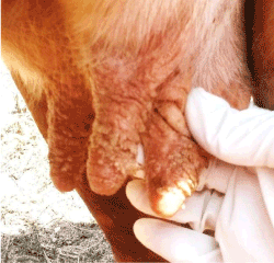
Figure 1. Clinical appearance of the case.
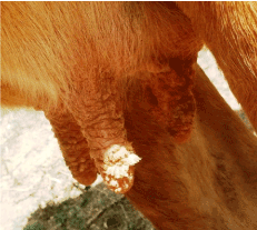
Figure 2. Skin of the all teats are covered with multiple papillomatous proliferation.
Histopathology
Epidermis revealed marked ortho keratotic and para keratotic hyperkeratosis. The dermal papillae were elongated with irregular rete ridge formations covered by acanthotic epidermis (Figure 3). In the granular layer numbers and sizes of keratohyalin granules were elevated (Figure 4). In the stratum spinosum layer numerous koilocytes characterized by eccentric, slightly enlarged nuclei that were displaced by large perinuclear vacuole were also common finding (Figure 4).
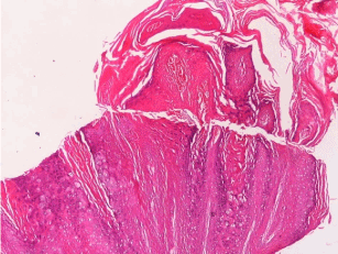
Figure 3. Epidermis is showing marked hyperkeratosis with irregular rete ridge formations. H&Ex4.
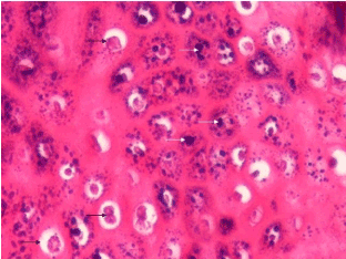
Figure 4. In the granular layer numerous various sized keratohyalin granules (white arrows) with presence of numerous koilocytes (black arrows). H&Ex40.
Discussion
Cutaneous papillomatosis causing widespread lesions is more frequently detected in domestic and wild animals. Cows are the most affected animals. The disease is more widely known for the cows under 2 years, but it may be seen any age group [7,17,19]. The tumors in cows’ 5-years or above are usually ocular squamous cell carcinomas but in our case cutaneous papilloma is detected.
Papillomaviruses are resistant to environment, detergent and high temperatures [5]. Congenital and acquired deficiencies in cellular immunity pave the way for papilloma emergence [5]. Villiers and his colleagues, in a research they conducted on human papilloma, proved that cutaneous tumor developments in allograft receptors occurred 6-7 years after explosion to sun and transplantation [20]. They also showed that following the immune suppressive treatments, incidence of cutaneous malignancies significantly increased.
Profilactic and therapeutic vaccination can be applied against papilloma viruses in cows [18].
Theranecron (Tarantula cubensis) is reported to be effective in treatments of cutaneous papillomatosis in cows and in the growth of mammary gland tumors [21]. However, in our case it was not effective since it was not used regularly.
Lesions caused by Papillomavirus have an important effect on animal health and is the reason for huge economic loss in farm animals. In our case, following Papillomavirus infection on the skin of the mammary glands and teats it was reported a significant decrease in milk production of the animal.
In conclusion, ages, environment of animal and treatment type applied are important in cows’ cutaneous papillomatosis along with early diagnosis and adequate treatments are the significant factors that elevate success rate.
Acknowledgement
I would like to thank Mr. Mahmut SÖZMEN (Ondokuz Mayis University Veterinary Faculty Pathology Department) for his advice during the preparation of this report.
References
- Gül Y (2012) Gevis getiren hayvanlarin iç hastaliklari (Sigir, Koyun, Keçi) 456-457. Medipres Matbaacilik Ltd. Sti. Malatya.
- Özsoy SY, Özyildiz Z, Güzel M (2011) Clinical, pathological and immunohistochemical findings of bovine cutaneous papillomatosis. Ankara Üniv Vet Fak Derg, 58, 161-165.
- Atasever A (2005) Bir sigir sürüsünde deri papillomatozis olgulari. Ankara Üniv Vet Fak Derg, 52: 197-200.
- Rector A, Van Ranst M (2013) Animal papillomaviruses. Virology 445: 213-223. [Crossref]
- de Villiers EM, Fauquet C, Broker TR, Bernard HU, zur Hausen H (2004) Classification of papillomaviruses. Virology 324: 17-27. [Crossref]
- Munday JS (2014) Papillomaviruses in felids. Vet J 199: 340-347. [Crossref]
- Freitas AC, Silva MAR, Jesus ALS, Mariz FC, Cordeiro MN (2011) Albuquerque BMF and Batista MVA: Recent insights into Bovine Papillomavirus. Afr J Microbiol Res 5: 6004-6012.
- Campo MS (1997) Bovine papillomavirus and cancer. Vet J 154: 175-188. [Crossref]
- Risvanli A, Kalkan C (2001) Ineklerde meme papillomatozisi ile mastitis arasindaki iliski. Vet Bil Derg 17: 143-147.
- Alpay G, Yesilbag K (2009) Mastitis Olgularinda Viruslarin Rolü. Uludag Univ J Fac Vet Med 28: 39-46.
- Öcal H, Yildiz A, Apaydin AM, Kaygusuzoglu E (2001) Inek ve düvelerde meme papillomatozisinin tedavisi ve etiyolojisi üzerine çalisma. F Ü Saglik Bil Derg 15: 23-26.
2021 Copyright OAT. All rights reserv
- Van Doorslaer K (2013) Evolution of the papillomaviridae. Virology 445: 11-20. [Crossref]
- Nicholls PK, Stanley MA (2000) The immunology of animal papillomaviruses. Vet Immunol Immunopathol 73: 101-127. [Crossref]
- Bossart GD, Ewing RY, Lowe M, Sweat M, Decker SJ, et al. (2002) Viral papillomatosis in Florida manatees (Trichechus manatus latirostris). Exp Mol Pathol 72: 37-48. [Crossref]
- Börkü MK, Atalay O, Kibar M, Çam Y, Atasever A (2007) Ivermectin is an effective treatment for bovine cutaneous papillomatosis. Res Vet Sci 83: 360-363.
- Kirmizigül AH, Gökçe E, Sözmen M, Yildirim Y, Beytut E (2010) Ivermektin’in sigir deri papillomatozisinde tedavi etkinligi. Kafkas Univ Vet Fak Derg 16: 627-631.
- de Villiers EM (1998) Human papillomavirus infections in skin cancers. Biomed Pharmacother 52: 26-33. [Crossref]
- Tas A, Karasu A, Aslan L, Atasoy N, Ilhan F (2009) Iki sigirda oküler yassi hücreli karsinom olgusu. Y Y U Vet Fak Derg 20: 69-71.
- Campo MS (1997) Vaccination against papillomavirus in cattle. Clin Dermatol 15: 275-283. [Crossref]
- Sykes JE, Luff JA (2013) Viral papillomatosis. In, Sykes JE (Ed): Canine and Feline Infectious Disease. e-book. Chapter: 26:261-262.
- Albay MK, Sahinduran S, Kale M, Karakurum MÇ, Sezer K: Influence of Tarantula cubensis Extract on the treatment of the oral lesions in cattle with Bluetongue disease. Kafkas Univ Vet Fak Derg 16: 593-596.