Abstract
The variability in clinical presentation and clinical course of patients with myocardial bridges (MB) hinders their management. Therefore, we assessed the correlation between the clinical profile of three patients and their coronarography data, coronary computerized tomography and the hemodynamic analysis of intracoronary pressure indexes measured with pressure wire. We studied validated indexes of coronary physiology; the resting distal coronary to aortic pressure ratio (Pd/Pa), Fractional Flow Reserve (FFR) and Diastolic Pressure Ratio (DPR). In addition, we propose specific systolic dynamic coronary compression (DCC) severity indexes in patients with MB; maximum percentage fall in systolic pressure at the end of systole (Δp/Pa%) and FFR/DPR ratio, among others. Cases 1 and 2 were mild (both with or without betablocker treatment) and similar among them (no anatomic or angiographic differences). However, they do manifest normal overall or mildly impacted translesional pressure ratios without a clear diastolic compromise. Case 3 had severe MB, which suggested that severe clinical presentation is associated with longer and deeper MB and more tortuosity, as well as greater systolic and diastolic angiographic DCC with intracoronary nitroglycerine (NTGic). Case 3 also showed lower basal values of conventional coronary physiology indexes that further worsened with NTGic and hyperemia with adenosine, consistent with greater DCC. Whilst treatment with betablockers is beneficial in all cases, there may be a greater response in patients with more dynamic systolic compromise (greater Δp/Pa%, lower FFR/DPR ratio in hyperemia) in the intracoronary hemodynamic study. We believe the proposed ratios could help to identify patients who will present more severe angina, a higher number of events, and who will benefit most from intense beta-blocker treatment. So, in patients with MB, a complete anatomical and functional evaluation, including specific physiological indexes, would probably be recommended to predict their clinical course and guide treatment strategies.
Keywords:
case report, fractional flow reserve (FFR), diastolic pressure ratio (DPR), diastolic and systolic compression, myocardial bridge.
Introduction
Myocardial bridging (MB) is a frequently congenital variant in which a portion of an epicardial coronary artery (most commonly the middle segment of the left anterior descending [LAD] artery) adopts an intramuscular course [1]. Its clinical relevance varies from frequent asymptomatic cases, coronariography and coronary computerized tomography findings [2,3] to patients with refractory angina, acute myocardial infarction or sudden death [4]. The variability in clinical course and treatment decisions are related to differences in anatomy (depth, length or tortuosity), degree and duration of milking effect (constriction of the artery during systole) and abnormalities in the remaining vessels (proximal associated atherosclerosis, vasospasm, etc).
Current treatment depends on the degree of ischemia according to the classification proposed by Schwarz et al [5]: Type A are symptomatic patients with incidental findings on angiography, who require no treatment while there are no signs of ischemia; Type B are those with objective signs, with angina and positive ischemia test, in these cases, pharmacological treatment is warranted; and Type C patients are those with or without signs and who present altered intracoronary hemodynamics and may require revascularization if symptoms become refractory. However, variability continues to cause controversy over its management. The strategy in asymptomatic patients or those with class 1-2 angina is conservative. Nevertheless, treatment decision is harder in symptomatic patients when outcomes are not always favorable, especially in those who are candidates for stent revascularization [6] or surgery [7]. In this regard, stent implantation is related to perforation during the stent deployment [8], fracture [9] or thrombosis [10,11] and also with repetitive stenosis [12]. In these patients the correlation between anatomic and hemodynamic parameters has not been analyzed, though it would help in management decision.
Coronary invasive functional assessment by fractional flow reserve (FFR) analysis is useful in fixed atherosclerotic lesions [13]. However, systolic compression can generate a normal or negative systolic pressure gradient across the MB, with higher distal pressure causing flow reversal observed by intracoronary Doppler. This phenomenon can be exacerbated with intracoronary nitroglycerine (NTGic). This negative systolic pressure gradient would underestimate the functional impact of MB stenosis on FFR [14]. To avoid this systolic artifact, some authors have analyzed diastolic flow reserve indexes such as diastolic FFR or instantaneous wave-free ratio (iFR) [15-17].
The ideal provocative stimuli to confirm the hemodynamic relevance of MB have not been clearly defined either. Coronary vasodilators may also unmask or exacerbate the severity of the milking effect [18,19]. FFR in MB has been performed by dobutamine stimulation or even an association with adenosine to cause maximum vasodilatation to analyze diastolic FFR (less artifact due to ischemia over-estimation in MB patients) [16]. However, these studies were performed in patients to whom ischemia was revealed, and not in asymptomatic or negative test patients (majority of MB patients), in which dobutamine may over-estimate the MB-related ischemia.
To avoid estimation errors of full-cycle coronary pressure indexes that analyze averaged values, we propose adding a morphological analysis of the pressure curves when dealing with functional stenosis that change during the cardiac cycle. For this reason, a deep intracoronary hemodynamic analysis specifically aimed at patients with MB was performed. In addition, to complete cardiac cycle indexes (distal coronary pressure to aortic pressure ratio [Pd/Pa] and FFR), we analyzed the instantaneous distal/proximal pressure ratios at the end of systole and systole-diastole ratios aimed at assessing systolic compression by MB.
Our main objective was to better understand the mechanisms that explain the clinical situation and evolution in patients with MB. To this end, we studied the correlation between angiographic, functional and coronary computerized tomography (cCT) data in three patients with MB in LAD artery and their different clinical profiles. Finally, since no pressure indexes that can analyze dynamic MB stenosis have been studied so far, we propose specific indexes.
Methods
Patient Inclusion: Three patients were selected. These had suspected or confirmed myocardial ischemia with different clinical profiles and were referred for coronarography. The existence of MB was considered evident when a dynamic systolic milking effect (≥50% systolic diameter stenosis not detected or less evident in diastole) was present at coronary angiography after intracoronary nitrates administration. Angina severity was assessed using the Canadian Cardiovascular Society (CCS) angina classification system [20].
Angiographic Assessment and Quantitative Coronary Angiography: The presence of the MB in the anterior descending artery was confirmed in no less than two angiographic projections separated by at least 25 degrees. The diagnosis and angiographic analysis were separately performed by two expert interventional cardiologists. Assessment of coronary lesions by quantitative coronary angiography analysis (QCA) was automatically performed with dedicated software (Coronary Quantification Package, Philips Medical System) with a temporal resolution of 25 images per second, including MB length, depth and distance to the ventricle cavity, and temporal analyses (time and percentage duration of the systolic compression).
Pressure wire - Physiological Assessment: Physiological Assessment was performed after angiographic diagnosis, using the intracoronary pressure wire (PW) OptoWire (Opsens Medical) with a second-generation fiber optic sensor FidelaTM.
Whole cycle indexes were calculated by performing ratios on coronary pressure measurement; from the mean coronary pressure measured distal to the stenosis (Pd) to aortic pressure (Pa) [21], during resting (Pd/Pa) [22], and hyperemic FFR [21] states. The diastolic pressure ratio (DPR) index was also calculated from each individual waveform as an average Pd/Pa over the entire period of diastole over five consecutive cardiac cycles as previously described [23-25], using software provided by OpSens.
Pressure curves were recorded and indexes calculated basally (resulting indexes: Pd/Pa resting and DPR resting), after infusion of 200 µg NTGic (resulting indexes: Pd/Pa 90 seconds after NTGic and DPR 90 seconds after NTGic), and after maximum hyperemia caused by intracoronary adenosine 300 µg + 200 µg NTGic (resulting indexes: FFR and DPR during hyperemia [DPRh]) to analyze the hemodynamic effect of vasodilator drugs.
To date, no pressure indicators have been studied that can analyze dynamic stenosis due to MB. We propose specific indexes in patients with MB: maximum percentage fall in systolic pressure at the end of systole (Δp/Pa%, where Δp is the difference between Pa minus Pd), calculated to assess the dynamic systolic compromise by milking effect; FFR/DPR, since FFR is a complete cycle index that encompasses the dynamic and static components, while DPR is mainly influenced by fixed stenosis (a lower ratio is indicative of a greater systolic drop in coronary pressure, suggesting a greater systolic dynamic coronary compression [DCC]) and [Δp/Pa]/FFR and [Δp/Pa]/DPR during hyperemia as global indexes of maximum coronary pressure drop caused by dynamic and static coronary stenosis per MB.
The fixed stenosis component was assessed with indexes of complete cycle (Pd/Pa and FFR) or complete diastole (diastolic pressure ratio, DPR) validated in this type of stenosis, with known cut-off points [26-29].
Coronary CT (cCT): The anatomical features were analyzed by means of cCT angiogram with helicoidal 80 multidetector CT (MDCT) technology PUREViSION Aquilion PRIME (Toshiba Medical Systems). The MB length, maximum depth, and proximity to the right ventricle cavity were measured.
Case studies and Results
The baseline characteristics have been described in (Table 1). For all cases, coronarography, pressure wire and computed tomography results are shown (Table 2, 3, 4). Coronary angiographies, aortic and coronary CT images are also shown (Figure 1, 2, 3) respectively.
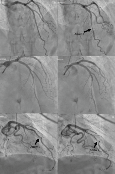
Figure 1. Left coronary angiography. Angiographies performed to CASE 1, (upper row, right oblique projection 10º /cranial projection 30º), CASE 2 (medium row, right oblique projection 15º /cranial projection 30º) and CASE 3 (lower row, right oblique projection 25º /cranial projection 10º) for diastole (left) and systole (right). The MB from case 3 presented milking effect even at the end of diastole (arrow 1) and kinking in the distal segment during systole (arrow 2). Partial coating of MB in case 1 (arrow 3).
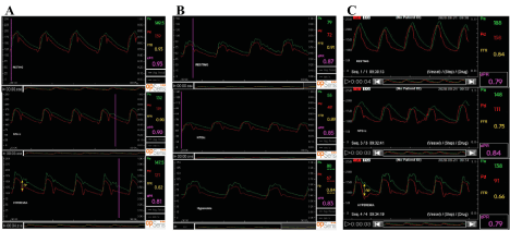
Figure 2. Aortic and distal pressure curves in the basal anterior descending artery after NTGic and hyperemia with with PW OptoWire and Opsens pressure monitor. CASE 1 (A), CASE 2 (B) and CASE 3 (C). Arrow indicates maximum fall (Δp) in systolic pressure (Figures 2A and 2C). In all cases, the figure shows from top to bottom the resting state, the effect of intracoronary nitroglycerin and the data during hyperemia. Aortic pressure (Pa) values are marked in green, pressure distal to the stenosis (Pd) in red, fractional flow reserve (FFR) in yellow and diastolic
pressure ratio (DPR) in pink.
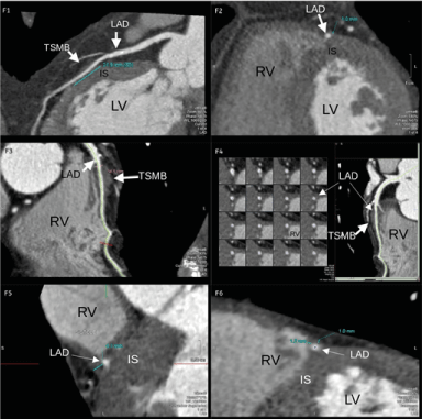
Figure 3. Coronary CT images. CASE 1 (F1-F2) and CASE 3 (F3-F6). F1. Longitudinal section of the tunneled segment in the interventricular septum of the anterior descending artery where the radiologist measured the MB length (21.5 mm). F2. Cross section of the tunneled segment where the depth (1 mm) and distance to ventricular cavities are measured. F3. Longitudinal section of the MB with tunneled segment 56.5 mm deeper proximally and closer to RV distally. F4. Transversal and longitudinal sections showing the location of the tunneled segment of the MB in the interventricular septum. F5 and F6. Extended cross sections of the MB where the maximum depth of 4.1 mm was measured (F5) and the minimum distance to the right ventricle of 1.2 mm at distal level where the depth was only 1 mm (F6). LV: Left Ventricle, IS: Interventricular septum, RV: Right Ventricle, TSMB: Tunneled Segment Myocardial Bridge, LAD: Left Arterial Descending.
CASE 1 (Type A): An asymptomatic 47-year-old man was admitted after an isolated episode of typical chest pain, without electrocardiogram changes or raised troponin I (TrI). The stress test was negative both clinically and electrically (ST segment was -0.6 mm). Given the study findings regarding pressure wire, discharge was decided without treatment. The patient remained symptom-free at six months.
CASE 2 (Type B): A 62-year-old man with typical angina of grade 2 CCS on moderate effort for the last six months and positive stress test (with ST segment -1.3 mm). Treatment was commenced with low dose bisoprolol (2.5 mg/day) and the patient became asymptomatic. After the study, discharge was decided continuing the same treatment. The patient was asymptomatic at six months.
CASE 3 (Type C): A 78-year-old man treated with a stent in the right coronary artery due to unstable angina with positive stress test in 2011. MB was observed in the long segment of the proximal LAD artery. The patient was admitted several times because of unstable angina in 2017 and 2018. There were no coronarography changes (MB remained) and vasospasm in the right coronary artery was observed. The patient continued treatment with nitrates and diltiacem. Due to mixed angina episodes, the man visited the emergency service several times. Treatment was switched to low dose bisoprolol (2.5 mg/day). The patient was admitted after two months with multiple episodes of grade 3-4 typical mixed angina (rest/effort) with temporary ST depression, normal levels of TrI (1.2 ng/dL) and positive stress test. Another coronarography was recommended because of refractory angina, which revealed similar MB in the LAD. Despite clinical refractoriness the patient was deemed a poor candidate for angioplasty given the cCT findings (risk of rupture at the right ventricle). A coronary bypass to the LAD artery was finally performed. The patient is currently asymptomatic.
Summary of Results
Coronary Angiography and cCT Findings: Oligosymptomatic patients without treatment (case 1) or with betablocker treatment (case 2) revealed shorter lengths of MB on angiography than case 3 with refractory angina (Figure 1). Angiography also showed an inferior degree of systolic dynamic compression in cases 1 and 2 (34-39% basally compared to 72% in case 3). However, after NTGic, systolic dynamic compression was similar in all three cases (Figure 2).
In cases 1 and 2, the duration of absolute and relative DCC was shorter, mainly systolic. Nevertheless, for case 3, with a longer MB length (61 mm), the milking effect remained until the end of diastole (whole cycle) producing a compressive stenosis of greater severity (which resembled a fixed stenosis) (Table 2). In these severe cases, intracoronary imaging was important to identify other factors such as coronary spasm, endothelial dysfunction, and atherosclerotic disease that could explain the greater symptomatology of patient 3. In addition, case 3 presented marked distal kinking as it abruptly penetrated the interventricular sulcus, further increasing the resistance to flow. cCT confirmed greater length and depth of the MB in case 3 with a course very close to the right ventricle (Figure 3).
Intracoronary Pressure Indexes: Measurement of non-hyperemic Pd/Pa and DPR indicator (cut-off points <0.91 and <0.89, respectively) with PW presented normal parameters (0.93) for case 1 and values in the limits of severity (0.91 and 0.87 respectively) for case 2, possibly explaining their paucisymptomatic presentation. Indicators measured for case 3 were lower (Pd/Pa 0.84, DPR 0.79), possibly justifying the patient´s severe angina grade 3-4 CCS. Logically, after NTGic, DCC of the MB increased and coronary pressure ratios worsened, with greater impact on the Pd/Pa index value (with systolic component) compared to DPR. Although, cases 1 and 2 were not markedly modified with vasodilator stimuli. After maximal vasodilation by NTGic administration and hyperemia after intracoronary adenosine, the FFR and DPRh indexes fell in all patients to a similar extent. Although the FFR value only reached hemodynamic relevance in case 3 (FFR 0.66), being slightly higher than 0.80 in cases 1 and 2 (Table 3).
Regarding dependent Δp/Pa%, there were no significant differences at rest among the three cases (17 to 21%) with clear worsening after NTGic (with or without intracoronary adenosine). A greater drop was evident in case 3 (Δp/Pa% NTGic 37 % and Δp/Pa% hyperemia 40%) (Table 3) who presented greater severity of angina and dynamic stenosis in angiography. Likewise, the ratio [Pd/Pa]/DPR was similar in the three patients at baseline (1.00-1.06), decreasing more with NTGic in case 3 (17%). The FFR/DPRh was lower in case 3 (0.84) compared to cases 1 and 2 (1.00 and 1.01, respectively). Finally, the [Δp/Pa]/FFR and [Δp/Pa]/DPRh ratios were less than 0.4 in cases 1 and 2 compared to 0.61 and 0.51 in case 3, respectively (Table 3).
Discussion of Results
The clinical impact of MB is highly variable for two reasons: differences in terms of the anatomic characteristics and the dynamic nature of the systolic (and at times diastolic) compromise of coronary flow. The detailed analysis of our patients enabled us to understand this variability when correlating the anatomic and functional information.
Anatomically and angiographically, patient 3 with the longer MB was the one to present the worst clinical severity. Additionally, case 3 presented higher maximum epicardial depth and more tortuosity. Studies have directly related length and especially depth with a higher degree of DCC and severity of angina [26-28,30-34], which would explain the greater symptoms of patient 3. However, no relationship with major adverse events has been demonstrated [35].
Angiographic assessment with QCA reveals case 3 has a more severe DCC, which behaved like a severe fixed lesion by presenting flow limitation throughout the entire cardiac cycle. It can be seen as an angiographic stenosis throughout the cardiac cycle, with residual stenosis of 57% at the end of diastole and marked separation of systolic and diastolic Pa and Pd pressure curves. Meanwhile, in cases 1 and 2 the DCC occupied only 32-41% of the cardiac cycle. The duration of DCC both in systole and diastole is a main factor causing ischemia. Thus, as expected, the longer DCC in case 3 caused a greater deterioration of the coronary hemodynamic indexes.
In more refractory patients like case 3, the functional study revealed abnormality in the resting non-hyperemic parameters (Pd/Pa 0.84 and DPR 0.79), which were not so clear in milder patients, like cases 1 and 2 with values close to the proposed cut-off points for fixed coronary stenosis (0.91 and 0.89). The same is true when NTGic and maximum hyperemia (NTGic + intracoronary adenosine) were used. Cases 1 and 2 resulted in Pd/Pa and DPR values around or slightly lower than the limit of normality (0.89), same for FFR (0.82-0.84), whereas results were clearly altered in case 3 (Pd/Pa 0.75 and FFR 0.66). Therefore, all the indicators of coronary physiology (complete cycle Pd/Pa and FFR or diastolic DPR) differentiated both patient profiles. In the most symptomatic cases, MB behaved as a mixed type fixed-dynamic stenosis in contrast to the dynamic-only behavior of the mildest ones. In contrast, the indexes that assessed systolic DCC at rest [Δp/Pa% and (Pd/Pa)/DPR] would not differentiate the two profiles (both around 20% and 1.03, respectively).
The proposed global dynamic coronary stenosis indexes ([Δp/Pa]/FFR and [Δp/Pa]/DPRh ratios) would be excellent markers of the maximum severity of resistance to coronary flow caused by the MB, as they encompass the maximum dynamic and fixed components. They were clearly superior in patient 3 ([Δp/Pa]/FFR 0.61 and [Δp/Pa]/DPRh 0.51) compared to patients 1 (0.38 and 0.38, respectively) and 2 (0.27 and 0.28), mainly due to lower values of FFR and DPRh in patient 3. The value is maximal in hyperemia, when systolic DCC (Δp/Pa) is higher; and is longer in diastole, with lower FFR or DPR during hyperemia. We believe these ratios could help to identify patients who will present more severe angina, a higher number of events, and who will benefit most from intense beta-blocker treatment.
Other authors in the literature performed the functional assessment with PW in symptomatic MB patients with stimuli such as dobutamine. Most of these studies defend the use of dobutamine over adenosine, as it causes more frequently ischemia due to MB, as it provokes, in addition to coronary hyperemia, a specific increase in the severity of DCC. Dobutamine increases both the dynamic (inotropic effect) and fixed components of coronary stenosis (longer duration of DCC and shorter duration of diastole) caused by MB [15-16,36-37]. These studies also suggest that adenosine increases the rates of systolic and global DCC by causing hyperemia, but it does not allow to differentiate patients according to their clinical profile. We measured DPRh with intracoronary adenosine as an equivalent to the diastolic FFR evaluated with intravenous dobutamine as proposed by other authors [16-17,36]. However, DPRh with intracoronary adenosine did not reach in our patients (case 3 DPRh 0.79) the cut-off point of <0.75 proposed for hemodynamic relevance in the literature.
Although dobutamine together with NTGic would be the ideal provocation test in patients with MB, it does not seem necessary in asymptomatic patients as it can give a false impression of severity. In these mild cases, the alteration caused by MB was only systolic DCC and specifically exacerbated by intracoronary vasodilator stimuli (NTGic), assessed by angiography or with systolic pressure drop ratios (Δp/Pa). On the other side, for patients with severe angina, the results gathered in this case series showed that the assessment of MB with NTGic alone or with adenosine may be enough to analyze its hemodynamic relevance. However, coronary pressure indexes had intermediate values with NTGic, hiding the real compromise of the DCC caused by MB in case 2. This is the case for patients with benign functional and anatomical data (but with significant symptoms or discrepancy in the anatomical/angiographic/functional information) or in those with beta-blocker treatment (as in patient 2). In these particular cases, use of dobutamine could be useful to avoid discrepancies.
There remain many unknowns about the long-term effects of COVID-19 and who is most vulnerable. Our case demonstrates that patients suffering from cardiac arrest from COVID-19 can benefit from eCPR. Patient selection for eCPR should consider scarcity of resources and complex constellations of symptoms in COVID-19 patients. This case demonstrates one successful outcome, but it should be noted that more data for this patient population is crucial. ECMO programs should collaborate with public health and hospital officials to devise a plan for this therapy so it may be offered to patients who can benefit from it.
Conclusion
In patients with MB the symptoms were related to the anatomical and angiographic characteristics, as well as to the functional alterations in indexes of coronary physiology, assessed with pressure guide wire measurements (especially if coronary vasodilator treatment was associated). In this case series, the patient with worse clinical severity had greater length and depth of MB, and longer duration of milking effect, as well as a greater drop in coronary pressure values Pd/Pa, FFR and diastolic DPR. Hemodynamic indexes specifically proposed to describe DCC varied according to clinical and angiographic severity. In patients with MB, a complete anatomical and functional evaluation, including specific physiological parameters, would probably be recommended to predict their clinical course and guide treatment strategies. It would be advisable to extend these data with larger clinical trials.
Acknowledgements
We would like to thank the entire Interventional Cardiology Unit members of the H. Universitario de Guadalajara for their selfless help and support in the management of this publication.
Declarations of interest
None
Author Contributions
;All the authors have contributed to the conception, design, coordination of the study, interpretation of results, critically revised the draft, and finally approved the version to be published.
Funding
This research did not receive any specific grant from funding agencies in the public, commercial, or not-for-profit sectors.
References
- Mohlenkamp S, Hort W, Ge J, Erbel R (2002) Update on myocardial bridging. Circulation 106: 2616-2622. [Crossref]
- Konen E, Goitein O, Sternik L, Eshet Y, Shemesh J, et al. (2007) The prevalence and anatomical patterns of intramuscular coronary arteries: a coronary computed tomography angiographic study. J Am Coll Cardiol 49:587-593. [Crossref]
- Kramer JR, Kitazume H, Proudfit WL, Sones FM Jr (1982) Clinical significance of isolated coronary bridges: benign and frequent condition involving the left anterior descending artery. Am Heart J 103: 283-288. [Crossref]
- Desseigne P, Tabib A, Loire R (1991) Myocardial bridging on the left anterior descending coronary artery and sudden death. Apropos of 19 cases with autopsy. Arch Mal Coeur Vaiss 84:511-516. [Crossref]
- Schwarz ER, Gupta R, Haager PK, Vom Dahl J, Klues HG, et al. (2009) Myocardial bridging in absence of coronary artery disease: proposal of a new classification based on clinical-angiographic data and long-term follow-up. Cardiology 112:13-21. [Crossref]
- Haager PK, Schwarz ER, Vom Dahl J, Klues HG, Reffelmann T, et al. (2000) Long term angiographic and clinical follow up in patients with stent implantation for symptomatic myocardial bridging. Heart 84: 403-408. [Crossref]
- Sun X, Chen H, Xia L, Zhao D, Ding W, et al. (2012) Coronary artery bypass grafting for myocardial bridges of the left anterior descending artery. J Card Surg 27: 405-407. [Crossref]
- Ernst A, Bulum J, Šeparović Hanževački J, Lovrić Benčić M, Strozzi M, et al. (2013) Five-year angiographic and clinical follow-up of patients with drug-eluting stent implantation for symptomatic myocardial bridging in absence of coronary atherosclerotic disease. J Invasive Cardiol 25: 586-592. [Crossref]
- Tandar A, Whisenant BK, Michaels AD (2008) Stent fracture following stenting of a myocardial bridge: report of two cases. Catheter Cardiovasc Interv 71:191-196. [Crossref]
- Agirbasli M, Hillegass WB Jr, Chapman GD, Brott BC (1998) Stent procedure complicated by thrombus formation distal to the lesion within a muscle bridge. Cathet Cardiovasc Diagn 43:73-76. [Crossref]
- Jiang Q, Liang C, Wu Z (2012) Myocardial bridging is a potential risk factor of very late stent thrombosis of drug eluting stent. Med Sci Monit 18: HY9-HY12. [Crossref]
- Tsujita K, Maehara A, Mintz GS, Poon M, Maiolino G, et al. (2009) Cross-sectional and longitudinal positive remodeling after subintimal drug-eluting stent implantation: multiple late coronary aneurysms, stent fractures, and a newly formed stent gap between previously overlapped stents. J Am Coll Cardiol Intv 2:156-158. [Crossref]
- Singh IM, Subbarao RA, Sadanandan S (2008) Limitation of fractional flow reserve in evaluating coronary artery myocardial bridge. J Invasive Cardiol 20: E161-166. [Crossref]
- Murtaza G, Mukherjee D, Gharacholou SM, Nanjundappa A, Lavie CJ, et al. (2020) An updated review on myocardial bridging. Cardiovasc Revasc Med 21:1169-1179. [Crossref]
- Hakeem A, Cilingiroglu M, Leesar MA (2010) Hemodynamic and intravascular ultrasound assessment of myocardial bridging: fractional flow reserve paradox with dobutamine versus adenosine. Catheter Cardiovasc Interv 75: 229-236. [Crossref]
- Escaned J, Cortés J, Flores A, Goicolea J, Alfonso F, et al. (2003) Importance of diastolic fractional flow reserve and dobutamine challenge in physiologic assessment of myocardial bridging. J Am Coll Cardiol 42: 226-233. [Crossref]
- Tarantini G, Barioli A, Nai Fovino L, Fraccaro C, Masiero G, et al. (2018) Unmasking myocardial bridge–related ischemia by intracoronary functional evaluation. Circ Cardiovasc Interv 11: e006247. [Crossref]
- Shimori T, Raizner AE, Chahine RA, Awdeh M, Luchi RJ (1977) Myocardial bridges in man: clinical correlations and angiographic accentuation with nitroglycerin. Cathet Cardiovasc Diagn 3: 59-65. [Crossref]
- Hongo Y, Tada H, Ito K, Yasumura Y, Miyatake K, et al. (1999) Augmentation of vessel squeezing at coronary-myocardial bridge by nitroglycerin: study by quantitative coronary angiography and intravascular ultrasound. Am Heart J 138: 345-350. [Crossref]
- Kaul P, Naylor CD, Armstrong PW, Mark DB, Theroux P, et al. (2009) Assessment of activity status and survival according to the Canadian Cardiovascular Society angina classification. Angina severity be assessed using grading measures such as the Canadian Cardiovascular Society (CCS) angina classification system. Can J Cardiol 25: e225-e231. [Crossref]
- Pijls NH, De Bruyne B, Peels K, Van Der Voort PH, et al. (1996) Measurement of fractional flow reserve to assess the functional severity of coronary-artery stenoses. N Engl J Med 334: 1703-1708. [Crossref]
- Hwang D, Jeon KH, Lee JM, Park J, Kim CH, et al. (2017) Diagnostic performance of resting and hyperemic invasive physiological indices to define myocardial ischemia: validation with 13N-ammonia positron emission tomography. JACC Cardiovasc Interv 10: 751-760[Crossref]
- Lee JM, Park J, Hwang D, Kim CH, Choi KH, et al. (2017) Similarity and difference of resting distal to aortic coronary pressure and instantaneous wave-free ratio. J Am Coll Cardiol 70: 2114-2123. [Crossref]
- Van’t Veer M, Pijls NHJ, Hennigan B, Watkins S, Ali ZA, et al. (2017) Comparison of different diastolic resting indexes to iFR: Are they all equal? J Am Coll Cardiol 70: 3088-3096. [Crossref]
- Johnson NP, Li W, Xi Chen, Hennigan B, Watkins S, et al. (2019) Diastolic pressure ratio: new approach and validation vs. the instantaneous wave-free ratio. Eur Heart J 40: 2585-2594. [Crossref]
- Kim SS, Min Ko S, Song MG, Hwang HG (2011). Systolic luminal narrowing and morphologic characteristics of myocardial bridging of the mid-left anterior descending coronary artery by dual source computed tomography. Int J Cardiovasc Imaging 27: 73-83. [Crossref]
- Tarantini G, Barioli A, Nai Fovino L, Fraccaro C, Masiero G, et al. (2010) Left anterior descending coronary artery myocardial bridging by multislice computed tomography: correlation with clinical findings. Eur J Radiol 73: 89-95. [Crossref]
- Liu G, Qu Y, Chen X, Liao M, Hu H, et al. (2017) Measurements of myocardial bridges on computed tomography predict presence of clinical symptoms and outcomes of adverse heart events: a retrospective study in a large population from China. Acta Radiol 58: 1068-1076. [Crossref]
- Singh IM, Subbarao RA, Sadanandan S (2008) Limitation of fractional flow reserve in evaluating coronary artery myocardial bridge. J Invasive Cardiol 20: E161-166. [Crossref]
- Kim PJ, Hur G, Kim SY, Namgung J, Hong SW, et al. (2009) Frequency of myocardial bridges and dynamic compression of epicardial coronary arteries: a comparison between computed tomography and invasive coronary angiography. Circulation 119:1408-1416. [Crossref]
- Liu SH, Yang Q, Chen JH, Wang XM, Wang M, et al. (2010) Myocardial bridging on dual source computed tomography: degree of systolic compression of mural coronary artery correlating with length and depth of the myocardial bridge. Clinical Imaging 234: 83-88. [Crossref]
- Hwang JH, Ko SM, Roh HG, Song MG, Shin JK, et al. (2010) Myocardial bridging of the left anterior descending coronary artery: depiction rate and morphologic features by dual-source CT coronary angiography. Korean J Radiol 11: 514-521. [Crossref]
- Tarantini G, Migliore F, Cademartiri F, Fraccaro C, Iliceto S (2016) Left anterior descending artery myocardial bridging. A clinical approach. J Am Coll Cardiol 68: 2887-2899. [Crossref]
- Elmali M, Soylu K, Gulel O, Bayrak IK, Koprulu D, et al. (2008) Correlation between depth of myocardial bridging and coronary angiography findings. Acta Radiol 49 :883-888. [Crossref]
- Wang Y, Lv B, Chen J, Zhang Y, Luo F, et al. (2013) Intramural coronary arterial course is associated with coronary arterial stenosis and prognosis of major cardiac events. Arterioscler Thromb Vasc Biol 33 :439-444. [Crossref]
- Aleksandric SB, Djordjevic-Dikic AD, Dobric MR, Giga VL, Soldatovic IA, et al. (2021) Functional assessment of myocardial bridging with conventional and diastolic fractional flow reserve: vasodilator versus inotropic provocation. J Am Heart Assoc 10: e020597. [Crossref]