The position and stability of left ventricular (LV) lead is important in determining the success of Cardiac Resynchronization Therapy (CRT). Lead dislodgement is a common problem accounting for up to 2 to 12% of cases. We report a case of successful implantation of LV lead by jailing the lead with stent implantation in the coronary sinus.
coronary sinus stenting, LV lead, cardiac resynchronization therapy
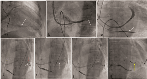
A 56-year-old male presented with a history of dyspnea on exertion New York Heart Association (NYHA) class 3 for the last 2 years. He underwent coronary angiography a year back which revealed normal coronaries. Electrocardiogram (ECG) showed normal sinus rhythm with left bundle branch block and QRS duration of 167 milliseconds. Two-dimensional echocardiography revealed an ejection fraction of 25%. He was on optimal medical treatment with beta-blockers, valsartan-sacubitril, diuretics, spironolactone, and digoxin. He was planned for CRT insertion because of refractory symptoms on optimal medical treatment. An RV lead (ENDOTAK RELIANCE™ G-59 cm, Boston Scientific) and RA lead (INGEVITY™ 52 cm, Boston Scientific) were implanted in the RV apex and right atrial appendage respectively using left subclavian venous access. A 9 Fr Coronary sinus (CS) catheter (ACUITY™ Pro, 9 FrX54 cm, Boston Scientific) was used to cannulate the CS. Selective injection of the CS showed a single large tributary which was draining the posterolateral LV wall and had acute angle orientation with CS (Figure 1a &1b). A quadripolar LV lead (ACUITY™ X4 Spiral L, Boston Scientific) was placed in the distal part of the posterolateral branch. The LV lead was not stable as the lead moved and dislodged with the respiratory movements and on withdrawing CS catheter. Repeated attempts to stabilize the LV lead were unsuccessful.

Figure 1. a. CS venogram showing a single large vein draining the posterolateral LV wall
b. Selective CS venogram showing a single tributary (arrow)
c. Deep hooking of catheter showed no further accessible veins
d. CS hooked with JR3.5 guide (yellow arrow) and CS tributary wired with SION BLUE wire (red); quadripolar LV lead is shown (white arrow)
e. Biomime 4.5X13 mm stent (arrow) placed proximal to quadripolar LV lead
f. Stent deployed at 10 atm
g. Final cine showed stent (white arrow) and intact quadripolar lead (yellow arrow)
Finally, the LV lead was stabilized by stenting the CS and jailing the LV lead, as there were no other alternative veins accessible for lead insertion (Figure 1c). Right femoral venous access was used using 6 Fr femoral sheath. A 6 Fr JR3.5 (LAUNCHER, MEDTRONIC) guiding catheter was used to access CS, and 0.014- inch coronary guidewire SION BLUE (ASAHI INTEC, Japan) was advanced into CS tributary (Figure 1d&1e). The average diameter of the CS tributary was 5 mm, so it was decided to stent with a 4.5 mm stent. Given the non-availability of a bare-metal stent, a drug-eluting stent was implanted in the CS tributary in a position 3cm away from the quadripolar lead, making sure not to damage the lead. A drug-eluting stent (DES) 4.5X13 mm (biomime™ MERIL) was deployed at 10 atm (Figure 1f&1g). The stent was deployed in the CS branch away from the main body of CS to prevent its occlusion. Post stent implantation, there was a good flow in the target vessel. Subsequently, catheters were removed without any LV lead dislodgement and there was no diaphragmatic stimulation. The procedure duration and mean fluoroscopy time were 137 min and 35 min, respectively. He was discharged after 3 days on dual antiplatelet drugs and optimal heart failure treatment. The QRS duration reduced to 110ms following CRT. He was under regular follow up with improved symptoms (NYHA class 1) and acceptable LV lead parameters of threshold and impedance of 1.5 V/0.4 ms, 900Ω respectively after 12 months.
CRT is the main modality of treatment of heart failure patients with LBBB who are refractory to optimal medical therapy. CRT improves the quality of life, reduces hospitalizations, and increases the survival of heart failure patients. The success of CRT depends on the optimal lead position and synchronous biventricular pacing. LV dislodgement results in the loss of LV stimulation resulting in failure of CRT and is reported in up to 2 to 12% of patients [1-4]. In the case of dislodgement, one may require repeat procedure or even surgical epicardial lead placement, thereby subjecting one to its attendant risks of renal failure and infections as shown in various studies [5,6]. CS interventions were done for cases of difficult LV lead placement secondary to CS occlusion, stenosis, valves, dissection, or even tortuosity. CS stenting was done earlier for the stabilization of unipolar and bipolar LV leads [7-9]. Newer quadripolar LV leads had an advantage of allowing distal placement improving lead stabilization and ability to pace proximally. In our case even though we used quadripolar lead, there was repeated dislodgement of LV lead owing to venous anatomy. Angioplasty of CS and its branches for LV lead stabilization was not widely accepted due to concerns of insulation defects, and difficulty in LV lead removal during infection and diaphragmatic stimulation. Because of these limitations, the CS stenting approach is not recommended routinely. In our case, we used this option as a last resort for stabilizing quadripolar LV lead, and a stent size of 4.5mm (CS diameter of 5 mm) was selected making sure it does not damage the LV lead but simultaneously jailing the lead. The majority of earlier reports used BMS for CS stenting, however in our case we used DES due to non-availability of BMS of appropriate size. The delivery of stents can be done via the CS catheter itself, but we chose to use femoral vein access. Our case was different from previous reports as we used quadripolar LV lead and drug-eluting stent to stabilize the LV lead.
Even though routine usage of CS stenting for LV lead stabilization is not recommended, one can use CS stenting in cases of difficult LV lead with satisfactory results on follow up.
None of the authors has a potential conflict of interest in connection with this article
- Alonso C, Leclercq C, d'Allonnes FR, Pavin D, Victor F, et al. (2001) Six-year experience of transvenous left ventricular lead implantation for permanent biventricular pacing in patients with advanced heart failure: technical aspects. Heart 86: 405-410. [PubMed]
- Salukhe TV, Francis DP, Sutton R (2003) Comparison of medical therapy, pacing and defibrillation in heart failure (COMPANION) trial terminated early; combined biventricular pacemaker-defibrillators reduce all-cause mortality and hospitalization. Int J Cardio 89: 119-120. [PubMed]
- Cazeau S, Leclercq C, Lavergne T, Walker S, Varma C, et al. (2001) Effects of multisite biventricular pacing in patients with heart failure and intraventricular conduction delay. N Engl J Med 344: 873-880. [PubMed]
- Cleland JG, Daubert JC, Erdmann E, Freemantle N, Gras D, et al. (2006) Longer-term effects of cardiac resynchronization therapy on mortality in heart failure [the CArdiac REsynchronization-Heart Failure (CARE-HF) trial extension phase]. Eur Heart J 27: 1928-1932. [PubMed]
- Ailawadi G, LaPar DJ, Swenson BR, Maxwell CD, Girotti ME, et al. (2010) Surgically placed left ventricular leads provide similar outcomes to percutaneous leads in patients with failed coronary sinus lead placement. Heart Rhythm 7: 619-625. [PubMed]
- Dekker AL, Phelps B, Dijkman B, van Der Nagel T, van Der Veen FH, et al. (2004) Epicardial left ventricular lead placement for cardiac resynchronization therapy: optimal pace site selection with pressure-volume loops. J Thorac Cardiovasc Surg 127: 1641-1647. [PubMed]
- Gellér L, Szilágyi S, Zima E, Molnár L, Széplaki G, et al. (2011) Long-term experience with coronary sinus side branch stenting to stabilize left ventricular electrode position. Heart Rhythm 8: 845-850. [PubMed]
- Biffi M, Bertini M, Ziacchi M, Diemberger I, Martignani C, et al. (2014) Left ventricular lead stabilization to retain cardiac resynchronization therapy at long term: when is it advisable?. Europace 16: 533-540. [PubMed]
- Szilágyi S, Merkely B, Molnár L, Zima E, Osztheimer I, et al. (2011) CRT implantation: Lead stabilization using coronary sinus side branch stenting. Interv Med Appl Sci 3: 142-145.

