A 57-year-old man presented to the emergency room with neck back pain for about 2 months, unresponsive to nonsteroidal anti-inflammatory drugs and progressive course of upper and lower extremity weakness with no sphincter dysfunction. The patient had no predisposing risk factors such as recent spinal surgery, trauma, instrumentation, distal site of infection, immunosupression, diabetes. He was apyrexial. Physical examination showed marked mid neck tenderness, no palpable masses were felt, no lymph nodes were felt. Neurological examination of his extremities, spasticity was positive, and power was decreased 3/5 in both lower extremities, 2/5 in both upper extremities. Bilateral Babinski signs were present and deep tendon reflexes were increased.
Full blood count and biochemistry showed white blood cell count (WBC) 10,269/L (neutrophils 71.3%; lymphocytes 21.8%; monocytes 2.2%; WBC 4.4 to 11.3/L); C-reactive protein 13.86 mg/dL (0.1 to 6 mg/dL). Magnetic Resonance imaging of the cervical spine showed the collapsed body of C4 with epidural abscess formation, complicating with spinal cord compression. He underwent urgent anterior cervical decompression and evacuation of anterior epidural abscess with fusion. The material underwent histologic examination and aerobic, anaerobic, fungal, mycobacterial cultures. A tuberculous granuloma was detected on histology. Ziehl-Neelsen stain confirmed the diagnosis. Cultures also detected Staphylococcus aureus. Treatment was started with rifampin (600 mg), Isoniazid (300 mg), ethambutol (25 mg/kg), pyrazinamide (25 mg/kg), and levofloxacin 750 mg for two months. This was followed by seven months of isoniazid and rifampin. The patient was referred to rehabilitation. One year later, the patient is able to walk independently and the back pain is gone.
Spondylodiscitis can be etiologically classified as pyogenic, granulomatous (tuberculosis, brucellosis, or fungal infection), or parasitic. Pyogenic spondylodiscitis commonly affects the lumbar column and more rarely affects the thoracic and the cervical column [1,2]. S. aureus is the predominant pathogen in pyogenic spondylodiscitis, followed in older people by enterobacteria, mainly Escherichia coli, Proteus, Klebsiella, and Enterobacter [2-4]. Mycobacterium tuberculosis is the most common cause of spondylodiscitis worldwide. Tuberculosis affects mostly the thoracic spine and involves two or more vertebral segments (Figure 1). The main contamination routes are hematogenous spread, external inoculation, or involvement from adjacent tissue [5]. Isolation of pyogenic bacteria from an abscess may guide the clinician to disregard the possibility of spine tuberculosis. It is recommended, therefore, to made mycobacterial culture and histopathological examination for all suspicious cases even when there is positive culture of pyogenic bacteria.
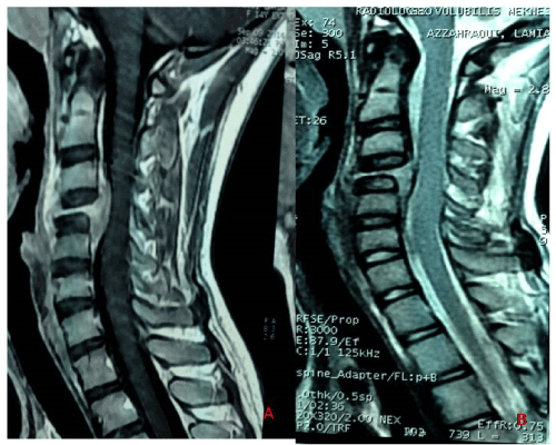
Figure 1. Preoperative MRI cervical spine showed the collapsed body of C4 with epidural abscess formation. T1-weighted (A), T2-weighted (B)
cervical; epidural abscess; Staphylococcus aureus, Mycobacterium tuberculosis
- Donnarumma P, Tarantino R, Palmarini V, De Giacomo T, Delfini R (2015) Thoracic spondylodiscitis caused by methicillin-resistant staphylococcus aureus as a superinfection of pulmonary tuberculous granuloma in an immunocompetent patient: A case report. Global Spine J 5: 144-147. [Crossref]
- Shin JH, Sung SI, Kim JK, Jung JM, Kim ES, et al. (2013) Retropharyngeal abscess coinfected with Staphylococcus aureus and Mycobacterium tuberculosis after rhinoviral infection in a 1-month-old infant. Korean J Pediatr 56: 86-89. [Crossref]
- Buchelt M, Lack W, Kutschera HP, Katterschafka T, Kiss H, et al. (1993) Comparison of tuberculous and pyogenic spondylitis. An analysis of 122 cases. Clin Orthop Relat Res 296: 192-199. [Crossref]
- Huang PY, Chen SF, Chang WN, Lu CH, Chuang YC, et al. (2012) Spinal epidural abscess in adults caused by Staphylococcus aureus: clinical characteristics and prognostic factors. Clin Neurol Neurosurg 114: 572-576. [Crossref]
- Tarantino R, Donnarumma P, Fazzolari B, Marruzzo D, Delfini R (2013) Pott’s disease: medical and surgical treatment. Clin Ter 164: 97-100. [Crossref]