--------------------------------------------------------------------- = Error percentage
Actual width of the tooth using caliper
The area of the largest diameter of voids under Motic software in micrometres at the cervical, middle and apical third using paralleling technique and mesial shift of tooth by 90⁰ was obtained.
The error was nullified by multiplying the error percentage obtained to the values obtained in Motic software. The values were converted into millimeters representing the area of the largest diameter of void
A scoring criteria to analyze the area of the largest diameter of void in mm2 using Motic software was designed in the Department by Dr Waleed K. Mukadam and Prof.Dr. Mithra N.Hegde.
SCORE |
PRESENCE OF VOID |
0 |
Absent |
>0 |
Present |
|
SCORE |
AREA OF VOID (mm2) |
1 |
0-0.01 |
2 |
0.011-0.02 |
3 |
0.021-0.03 |
4 |
0.031-0.04 |
5 |
0.041-0.05 |
6 |
>0.05 |
Scoring criteria was tabulated, where 0 is no voids and >0 is voids present.
The data obtained was statistically analysed by Kruskal Wallis test and Fisher’s exact test. Significance level was set at p-value <0.05.
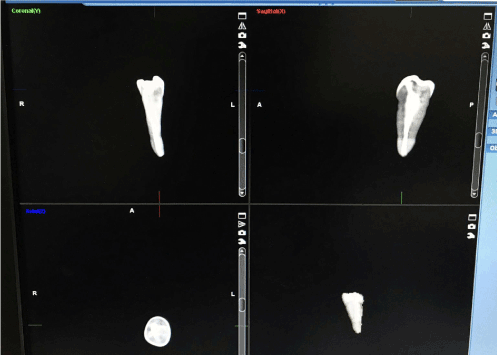
According to CBCT analysis of the obturated teeth, no voids could be detected due to presence of artefacts like noise, making it difficult to differentiate between the radiopaque GP and the teeth. High attenuation substance like GP in field of view cause bright streaks which makes it difficult to visualize the voids present in the teeth after obturation.
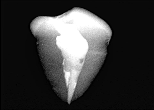
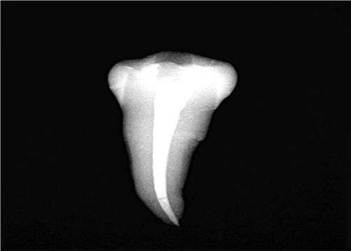
In direct digital radiography more voids could be detected with mesial shift of tooth by 90⁰ as compared to paralleling technique, hence the data obtained using paralleling technique was annulled and data obtained with mesial shift of tooth by 90⁰ was statistically analysed. (Figure 3(A), Figure 3 (B)).

Figure 3a. Mesial shift angulation in RGV.

Figure 3b. Paralleling technique in RGV.
Largest diameter of void was observed in Group B and Group C with mesial shift of tooth by 90⁰ and paralleling technique. On statistically analyzing the data using Kruskal Wallis test, there was no statistical significance between the four groups with mesial shift of tooth by 90⁰ and paralleling technique
The main objectives of Endodontic therapy are cleaning and shaping, disinfection and obturation of the root-canal system in 3D. GP is the most widely used and accepted obturation material [11]. Due to impoper obturation there could be post-operative complications resulting in failure of endodontic therapy. The quality of obturation may be influenced by various types of techniques [4].
The present study compares the quality of obturation and presence of voids using CBCT and RVG. According to the results, CBCT could not analyze voids in any of the obturating techniques.
A study done by Sogur et al, evaluation of homogeneity and length of the obturation was done using 3 different imaging methods. SPP and F-speed analogue film image exhibited superior images of obturation than LCBCT due to the presence of streaking artefacts from the sealer and GP, compromising the quality of image [12].
According to the study done by Rosen E. et al, in certain clinical situations, a more contemporary 3D imaging modality (such as CBCT imaging) may be ineffective, whereas a conventional 2D imaging modality (such as periapical radiography) may be of significant value [13].
According to Barrett J. F. et al, the main difficulty of CBCT imaging as compared to periapical radiographic images is the production of artefacts, which can be defined as ‘‘a systematic discrepancy [14].
Schulze R. et al (2011) identified the misconception that CBCT data contains fewer artefacts than their CT counterparts. CBCT imaging actually involves additional artefacts such as scatter and a generally higher noise level when a high-density material is present in the scanned volume, adversely affecting its diagnostic efficacy as proposed by Neves F. S. et al. (2014). The type of material used for root filling and its relative radiopacity as well as the CBCT acquisition parameters such as the voxel size and the field of view may sometimes influence the presence of artefacts observed in CBCT images [15,16]. In the present study also, CBCT could not be of any diagnostic value for evaluating the quality of obturation because of artefacts like noise which camouflage the voids and the obturation appears to be a dense opaque image.
The results of the current study are in contrast with Gupta et al (2015), Singh R et al (2015), Gambarini G et al (2016), B Huybrechts et al(2009) and Arora S et al (2014), where the evaluation of voids in the obturation could be assessed [4,17-19]. This could be due to the difference in CBCT hardware and software operated to visualize the voids in the obturation techniques or the voids could be calculated by evaluating the percentage of volume of root canal space before BMP and after obturation [20,21].
According to the present study area of the largest diameter of void were observed with mesial shift of tooth by 90⁰ than the paralleling technique. According to Shetty et al (2014), for accurate reproduction the image receptor (X-ray film or digital sensor) must be parallel to the long axis of the tooth and the X-ray beam should be perpendicular to the image receptor and the tooth being assessed. For better radiographic image acquisition two or three radiographs at different angles should be taken [22].
All obturations performed in the split-tooth model in the study done by Zielinski T et al (2008) also had voids; however, no quantification of the area or frequency of voids were made [23]. In this study we were able to determine the size of the void using Motic software.
In the present in vitro study, paralleling technique was done for complete analysis of voids present in the obturaion followed by mesial shift of tooth by 90⁰. Area of the largest diameter of void was observed in GuttaFlow and Calamus obturating technique (Table 1). There was no statistical significant difference (p>0.05) with presence of void between the four obturating techniques. Comparing the area of the largest diameter of void present in the coronal, middle and apical third, the four groups showed statistical significant difference(p>0.05) in the coronal third (Table 2).
Table 1. Mean, median and standard deviation values of area of the largest diameter of void present in the groups using paralleling technique and mesial shift of tooth by 90⁰.
|
Groups |
N |
Mean |
Standard deviation |
Median |
Kruskal Wallis test |
Chi square value |
p-value |
Mesial Shift of tooth by 90⁰ |
A |
10 |
0.015 |
0.010 |
0.014 |
6.76 |
0.08(NS) |
B |
10 |
0.032 |
0.017 |
0.028 |
C |
10 |
0.038 |
0.070 |
0.020 |
D |
10 |
0.020 |
0.019 |
0.016 |
Paralleling Technique |
A |
10 |
0.006 |
0.009 |
0 |
1.68 |
0.64(NS) |
B |
10 |
0.012 |
0.016 |
0.007 |
C |
10 |
0.012 |
0.027 |
0 |
D |
10 |
0.008 |
0.012 |
0 |
*p<0.05 statistically significant, p>0.05 Non significant, NS
Table 2. Fisher’s exact test comparison of area of the largest diameter of void present between the four obturating techniques at the coronal, middle and apical third
Mesial Shift of
tooth by 90⁰ |
|
Group |
Total |
Fisher’s exact test |
|
A |
B |
C |
D |
p-value |
Coronal Third |
0 |
2 |
5 |
9 |
8 |
24 |
0.006* |
20.0% |
50.0% |
90.0% |
80.0% |
60.0% |
>0 |
8 |
5 |
1 |
2 |
16 |
80.0% |
50.0% |
10.0% |
20.0% |
40.0% |
|
|
|
|
|
|
|
|
Middle Third |
0 |
3 |
1 |
6 |
5 |
15 |
0.11(NS) |
30.0% |
10.0% |
60.0% |
50.0% |
37.5% |
>0 |
7 |
9 |
4 |
5 |
25 |
70.0% |
90.0% |
40.0% |
50.0% |
62.5% |
|
|
|
|
|
|
|
|
Apical Third |
0 |
6 |
7 |
7 |
7 |
27 |
1.00(NS) |
60.0% |
70.0% |
70.0% |
70.0% |
67.5% |
>0 |
4 |
3 |
3 |
3 |
13 |
40.0% |
30.0% |
30.0% |
30.0% |
32.5% |
*p<0.05 statistically significant, p>0.05 Non significant, NS
GuttaFlow showed few area of the largest diameter of void at the apical third, 30%, whereas, more area of the largest diameter of void was seen in the middle third, 90% (Table 3). This could be due to the lack of density in cold compaction technique over thermoplasticized obturation and lateral compaction. GuttaFlow expands slightly while setting and exhibit voids and gaps because of the filling technique (Hammad M et al ., 2008).[24] 3D compaction techniques are considered to be superior over single-cone filling technique, because the volume of sealer is high relative to the volume of the cone, which promotes void formation and reduces the quality of the seal. The manufacturers of GuttaFlow recommend that it should be dispensed first in the apical part of the root canal and then a master GP cone is placed. This ensures the least amount of voids and gaps in the apical third [25].
Table 3. Fisher’s exact test comparison of area of the largest diameter of void present between apical, middle and coronal third of the four obturating techniques
Group |
Mesial Shift of
tooth by 90⁰ |
Groups |
Total |
Fisher’s exact test |
Coronal Third |
Middle Third |
Apical Third |
p-value |
A |
0 |
2 |
3 |
6 |
11 |
0.25(NS) |
20.0% |
30.0% |
60.0% |
36.7% |
>0 |
8 |
7 |
4 |
19 |
80.0% |
70.0% |
40.0% |
63.3% |
B |
0 |
5 |
1 |
7 |
13 |
0.04* |
50.0% |
10.0% |
70.0% |
43.3% |
>0 |
5 |
9 |
3 |
17 |
50.0% |
90.0% |
30.0% |
56.7% |
C |
0 |
9 |
6 |
7 |
22 |
0.45(NS) |
90.0% |
60.0% |
70.0% |
73.3% |
>0 |
1 |
4 |
3 |
8 |
10.0% |
40.0% |
30.0% |
26.7% |
D |
0 |
8 |
5 |
7 |
20 |
0.50(NS) |
80.0% |
50.0% |
70.0% |
66.7% |
>0 |
2 |
5 |
3 |
10 |
20.0% |
50.0% |
30.0% |
33.3% |
*p<0.05 statistically significant, p>0.05 Non significant, NS
Lateral compaction showed area of the largest diameter of void of 80% in the coronal third followed by GuttaFlow, Elements and Calamus (Table 2). A study conducted by Torabinejad et al, Goldman et al and Kersten et al, it was reported that “in lateral compaction pattern of voids was frequently where the fillings adapted reasonably well at the apical and coronal parts and showed longitudinal voids in the midroot section and excessive amounts of root canal sealer and open spaces between the GP cones” were observed [26, 27].
In the present study, Calamus showed the least number of area of the largest diameter of void at the coronal third 10% followed by the apical third 30% and middle third 40%. Similarly, Elements obturation technique exhibited least number of area of the largest diameter of void in the coronal third 20%, apical third 30% and middle third 50%. Calamus andElements showed superiority in obturation at the apical, middle and coronal third when compared to lateral compaction (Table 3). Calamus obturation system showed the maximum percentage of obturated volume. This system is based on the principle of warm vertical compaction where the objective is to continuously and progressively carry a wave of warm GP along the length of the master cone, starting coronally and ending in apical corkage. This method advantageously serves to initially thermosoften the master cone, maximizes the volume of GP, and effectively increases hydraulics during obturation [28]. It utilizes different sizes manual pluggers with different working end diameters to compact GP efficiently to the canal walls [7]. The consistent flow of the Calamus unit does make the obturation of curve quicker and easier. The warm GP needs to be compacted as it cools to overcome any shrinkage that will normally occur. Since the softness of the GP is mass dependent, the GP at the orifice level has the greatest mass and will stay softest for the longest time in the canal, regardless of which technique is utilized [28]. The Elements obturating System heats the GP, enabling it to flow well and allows more efficient and effective compaction as reported by Budd et al (1991) [29].
Elements obturation techniques have advantages similar to that of Calamus obturation technique. Thermoplasticized obturation techniques always have an advantage over cold compaction technique. However, for efficient and effective obturation one should master the technique for void free compaction. CBCT could not be used as a diagnostic tool for the evaluation of the quality of obturation.
Future studies with larger sample size should be conducted to evaluate the efficiency of obturation techniques.
The present study concluded that CBCT could not be used to evaluate voids present in any of the obturation techniques, due to presence of artefacts like noise and beam hardening. Owing to this result RVG was used to evaluate the quality of obturating techniques and assessments of voids in apical, middle and coronal third of the root canal system.
It was observed that there was no significant difference in any of the obturation techniques. Lateral compaction obturation showed the large diameter of voids in the apical and coronal third, whereas in the middle third GuttaFlow showed the large diameter of voids. On the other hand, Calamus followed by Elements showed superior obturation at apical, middle and the coronal third.
Future studies with larger sample size should be conducted to evaluate the efficiency of obturation techniques.
View supplementary data
- Gulsahi K, Cehreli Z, Kuraner T, Dagli F (2007). Sealer area associated with cold lateral condensation of gutta-percha and warm coated carrier filling systems in canals prepared with various rotary NiTi systems. Int Endod J 40: 275-281. [Crossref]
- Carrotte P (2004) Endodontics: Part 8 Filling the root canal system. Br Dent J 197: 667-672. [Crossref]
- Jarrett I, Marx D, Covey D, Karmazin M, Lavin M, et al. (2004) Percentage of canals filled in apical cross sections - an in vitro study of seven obturation techniques. Int Endod J 37: 392-398. [Crossref]
- Gupta R, Dhingra A, Panwar N (2015). Comparative evaluation of three different obturating techniques lateral ompaction, thermafil and calamus for filling area and voids using cone beam computed tomography: an invitro study. J Clin Diagn Res 9:15-17. [Crossref]
- Garg S, Mahajan P, Thaman D, Monga P (2015). Comparison of dentinal damage induced by different nickel-titanium rotary instruments during canal preparation: an in vitro study. J Conserv Dent. 18:302-305. [Crossref]
- Anantula K, Ganta A (2011) Evaluation and comparison of sealing ability of three different bturation techniques - Lateral condensation, Obtura II, and GuttaFlow: An in vitro study. J Conserv Dent 14: 57-61. [Crossref]
- Ruddle CJ (2010) Filling root canal systems: the Calamus 3-D obturation technique. Dent Today 29: 76, 78-81. [Crossref]
- Michael DiTolla (2013) Lauding a new endo technique. http://www.dentaleconomics.com/articles/print/volume-103/issue-12/practice/lauding-a-new-endo-technique.html
- Decurcio D, Bueno M, Alencar A, Porto O, Azevedo B, et al. (2012). Effect of root canal filling materials on dimensions of cone-beam computed tomography images. J Appl Oral Sci. 20:260-267. [Crossref]
- Kohn WG, Collins AS, Cleveland JL, Harte JA, Eklund KJ, et al. (2003) Guidelines for infection control in dental health-care settings—2003. MMWR Recomm Rep. 52: 1-61.
- Ho E, Chang J, Cheung G(2016). Quality of root canal fillings using three guttapercha obturation techniques. Restorative Dentistry and Endodontics.;41(1):22-28. [Crossref]
- Soğur E, Baksi BG, Gröndahl HG (2007). Imaging of root canal fillings: a comparison of subjective image quality between limited cone-beam CT, storage phosphor and film radiography. Int Endod J 40:179-185. [Crossref]
- Rosen E, Venezia N, Azizi H, Kamburoglu K, Meirowitz A, et al. (2016). A comparison of cone-beam computed tomography with periapical radiography in the detection of separated instruments retained in the apical third of root canal–filled teeth. J Endod 42: 1035-1039. [Crossref]
- Barrett JF, Keat N (2004) Artifacts in CT: recognition and avoidance. Radiographics 24: 1679-1691. [Crossref]
- Schulze R, Heil U, Groß D, Bruellmann D, Dranischnikow E, et al. (2011) Artefacts in CBCT: a review. Dentomaxillofac Radiol 40:265-273.
- Neves F, Freitas D, Campos P, Ekestubbe A, Lofthag-Hansen S (2014). Evaluation of cone-beam computed tomography in the diagnosis of vertical root fractures: The influence of imaging modes and root canal materials. J Endod 40: 1530-1536. [Crossref]
- Arora S, Hegde V (2014) Comparartive evaluation of novel smart-seal obturating system and its homogeneity of using cone beam computed tomography: In vitro simulated lateral canal study. J Conserv Dent 17: 364-368. [Crossref]
- Gambarini G, Piasecki L, Schianchi G, Di Nardo D, Miccoli G, et al. (2016). In vitro evaluation of carrier based obturation technique: a CBCT study. Ann Stomatol (Roma) 7:11-15. [Crossref]
- Huybrechts B, Bud M, Bergmans L, Lambrechts P, Jacobs R (2009). Void detection in root fillings using intraoral analogue, intraoral digital and cone beam CT images. Int Endod J 42:675-685. [Crossref]
- Nagaveni NB, Yadav S, Poornima P, Bharath KP, Mathew MG, et al. (2017) Volumetric evaluation of various obturation techniques in primary teeth using cone beam computed tomography an in vitro study. J Indian Soc Pedod Prev Dent. 35: 244-248. [Crossref]
- AI Qassab SJ, Hadi DA, Luke AM (2016) Evaluation of three different obturation techniques using three-dimensional cone beam computed tomography: In Vitro Study. Dentistry 6.
- Shetty A, Hegde MN, Tahiliani D, Shetty H, Bhat GT, et al. (2014). A three-dimensional study of variations in root canal morphology using cone-beam computed tomography of mandibular premolars in a South Indian Population. J Clin Diagn Res 8: ZC22-ZC24. [Crossref]
- Zielinski T, Baumgartner J, Marshall J (2008). An evaluation of guttaflow and gutta-percha in the filling of lateral grooves and depressions. J Endod 34:295-298. [Crossref]
- Hammad M, Qualtrough A, Silikas N (2008) Extended setting shrinkage behavior of Endodontic Sealers. J Endod 34: 90-99. [Crossref]
- Hammad M, Qualtrough A, Silikas N (2009). Evaluation of root canal obturation: a three dimensional in vitro study. J Endod 35: 541-544. [Crossref]
- Torabinejad M, Skobe Z, Trombly P, Krakow A, Grøn P, et al. (1978) Scanning electron microscopic study of root canal obturation using thermoplasticized gutta-percha. J Endod 4:245-250.
- Kersten H, Fransman R, VelzenS (1986). Thermomechanical compaction of gutta percha. II. A comparison with lateral condensation in curved root canals. Int Endod J. 19: 134-140. [Crossref]
- Jindal D, Sharma M, Raisingani D, Swarnkar A, Pant M, et al. (2017). Volumetric analysis of root filling with cold lateral compaction, Obtura II, Thermafil, and Calamus using spiral computerized tomography: An In vitro Study. Indian J Dent Res 28:175-180. [Crossref]
- Budd C, Weller R, Kulild J (1991) A comparison of thermoplasticized injectable gutta-percha obturation techniques. Journal of Endodontics 17:260-264.


