Abstract
Objective: It has been demonstrated that 4F child catheter could be inserted into the mother guiding catheter for back up support improvement, stent or balloon delivery, et al. Here, we aimed to determine the potential role of 4F child catheter in treating complex coronary lesions.
Methods and results: We retrospectively analyzed 40 lesions treated by 4F child catheter. The 4F child catheter was beneficial to facilitate stent or balloon delivery, to aid guidewire crossing, to capture stent, or to pick up retrograde guidewire. For CTO PCI, picking up the retrograde guidewire using the advanced greeting technique accounted for 32.1 %, indicating it was efficient for CTO revascularization. Lesion success was 95 %, device success was 95 %, and procedure success was 92.5 %. 1 patient failed because of acute thrombosis after successful stent deployment, 1 failed due to the guidewire unable to cross the target lesion. There was 1 instance of stent dislodgment and 1 instance of little pericardial effusion.
Conclusions: The 4F child catheter provided higher successful rate and it was a viable device in treating complex coronary lesions.
Key words
child catheter, angioplasty, coronary lesion, stent
Introduction
Complex coronary anatomy such as extreme vessel tortuosity, chronic total occlusions (CTOs), calcification, which often renders higher risk and difficult procedure of percutaneous coronary intervention (PCI), thus the optimal management strategy and new device have been investigated, such as larger size and deep insertion of guiding catheters, strong support wires, et al [1, 2]. The child catheter is designed to facilitate the deep insertion of the guiding catheter, which is efficient for balloons and stents delivery and serves extra backup support. In addition, it is useful to externalize the retrograde guidewire into the antegrade guiding catheter after successful reverse CART when it is impossible to do it. In this situation, the mother-child catheter serves as a connection in the mid-distal portion of the vessel, makes the re-entry into the antegrade guiding catheter possible, this technique we called it the advanced greeting technique (AGT). In this study, we retrospectively evaluated the potential role and clinical outcomes of the 4F child catheter in treating complex coronary lesions.
Materials and Methods
Study design
This retrospective study was approved by the Ethics Committee at the Bethune International Peace Hospital, and it was conducted according to the ethical principles of the Declaration of Helsinki, informed consent was obtained from all patients. The data was collected from August 2016 to April 2018 by 1 experienced operator with yearly PCI case over 500. Clinical characteristics, target lesion characteristics, ACC/AHA lesion type, indications for the use of the mother-child catheter (namely, usable to deliver the stents or balloon, poor back up support, impossible to pick up the retrograde wire, to capture the stent), lesion success (defined as percent diameter stenosis of a target lesion < 50 % achieved by any methods) rate, device success (defined as successful stent implantation in targeted lesion area) rate, and procedure success (defined as lesion success without in-hospital major adverse cardiac events) rate were analyzed. Complications (such as cardiac death, myocardial infarction, and target lesion revascularization), in-hospital outcome and 6-month follow-up were recorded and reviewed. Data were collected and analyzed by cardiologists who were experienced in quantitative coronary arteriography analysis.
Study Patients
A total of 40 patients was enrolled between August 2016 to April 2018, who were treated by the 4F child catheter. Indication for usage of this technique based on the operator's discretion.
Mother-Child Technique
The mother-child technique was used when a balloon or a stent failed to cross the target lesion, or failed to externalize the retrograde guidewire, or capture a stent. Firstly, removed these devices from the guiding catheter while placed the PCI guidewire in situ, disconnected the Y-connector from the guiding catheter and connected a small hemostatic valve. Then, inserted a 4F child catheter into the mother guiding catheter through the PCI guidewire. Next, connected the Y-connector to the 4F child catheter, and advanced the child catheter out of the mother guiding catheter, and placed the child catheter near the target lesion. In this procedure, it was important to use the anchor balloon technique with slight tension on the shaft of the balloon catheter, this step helped to avoid intimal injury by the child catheter tip. Subsequently, removed the balloon catheter and inflated/ deployed the balloon or the stent, or picked up the retrograde guidewire or captured the stent.
Follow-up
Patients treated with ACG at 6-month follow-up (e.g. death, myocardial infarction, CABG and stroke) were performed by a standardized telephone interview.
Statistical Analysis
Statistical analysis was performed using SPSS19.0 statistical software (SPSS Inc, Chicago, IL). Continuous variables were expressed as means ± standard deviation (SD), categorical variables were expressed as number and percentages (%). p < 0.05 was considered statistically significant.
Results
Patient Clinical Characteristics
40 patients who were treated with PCI using the 4F child catheter were included. The demographic data of the patients were presented in table 1. They were 63.3 ± 10.2 years of age (range, 46 to 87 years), and men accounted for 77.5 %. The left ventricular ejection fraction was 59.1 ± 11.2 %. Moreover, the majority had risk factors, such as hypertension (67.5 %), hypercholesterolemia (30 %), current smoking (22.5 %), diabetes mellitus (45 %), previous PCI (20 %), previous myocardial infarction (MI) (12.5 %), and chronic renal impairment (2.5 %). 85 % patients presented with unstable angina pectoris.
Table 1. Patient Clinical Characteristics (n = 40)
Variable |
N (%) or Mean ± SD |
Age, y |
63.33±10.22 |
Male gender |
31 (77.5%) |
Diabetes mellitus |
18 (45%) |
Hypertension |
27 (67.5%) |
Hyperlipidemia |
12 (30%) |
Smoker |
9 (22.5%) |
Previous myocardial infarction |
5 (12.5%) |
Previous PCI |
8 (20%) |
Previous bypass surgery |
0 |
Left ventricular ejection fraction, % |
59.07±11.17 |
Chronic renal impairment |
1 (2.5%) |
Unstable angina pectoris. |
34 (85%) |
Continuous variables are presented as mean ± standard deviation, whereas categorical variables are presented as frequency (percentage).
Procedural Characteristics
Procedural characteristics of the patients were provided in table 2. The target vessels were 13 (32.5 %) in left anterior descending (LAD) artery, 7 (17.5 %) in left circumflex (LCX) artery, 19 (47.5 %) in right coronary artery (RCA), and 1 (2.5 %) in left main. The morphologies of the lesions consisted of 9 (22.5 %) type B and 31 (77.5 %) type C. No type A lesions were observed. CTO accounted for 70 %. PCI carried out via the radial route accounted for 32.5 %, 2.5 % via the femoral route, 62.5 % via the bilateral route (right radial and femoral approach, or right femoral and left femoral approach), and 2.5 % via the trilateral artery (right radial, femoral together with left femoral). 40 % patients used the 6F mother guiding catheters, and 60 % patients used the 7F mother guiding catheters. The shapes of the mother guiding catheters used were EBU in 16 (40 %), SAL in 1 (2.5 %), AL in 10 (25 %), XB in 2 (5 %), BL in 1 (2.5 %), JR4 in 6 (15 %) and JL in 4 (10 %). 5 % patients treated with the mother-child technique to deliver balloon, 25 % to deliver stent, 22.5 % to externalize the retrograde guidewire, 50 % to aid the guidewire crossing the target lesion, and 2.5 % to capture the dislodged stent. Lesion success was achieved in 38 procedures (95 %), Device success was achieved in 38 procedures (95 %); procedural success was achieved in 37 procedures (92.5 %). 1 procedure failed because of acute thrombosis, 1 failed due to the guidewire unable to cross the target lesion. Stent dislodgement occurred in 1 patient when using the 4F child catheter.
Table 2. Procedural Characteristics (n = 40)
Variable |
N (%) or Mean ± SD |
Lesion vessel |
|
Left anterior descending |
13 (32.5%) |
Circumflex |
7 (17.5%) |
Right coronary |
19 (47.5%) |
Left main |
1 (2.5%) |
Lesion type |
|
A |
0 (0%) |
B |
9 (22.5%) |
C |
31 (77.5%) |
CTO |
28 (70%) |
Access site |
|
Radial artery |
13 (32.5%) |
Femoral artery |
1 (2.5%) |
Bilateral artery |
25 (62.5%) |
Trilateral artery |
1 (2.5%) |
Size of mother guiding catheter |
|
6F |
16 (40%) |
7F |
24 (60%) |
Shape of mother guiding catheter |
|
EBU |
16 (40%) |
SAL |
1 (2.5%) |
AL |
10 (25%) |
XB |
2 (5%) |
BL |
1 (2.5%) |
JR4 |
6 (15%) |
JL |
4 (10%) |
Usage of 4F child catheter (overlapped) |
|
To deliver balloon |
2 (5%) |
To deliver stent |
10 (25%) |
To externalize the retrograde guidewire |
9 (22.5%) |
To aid the guidewire cross the target lesion |
20 (50%) |
To capture the stent |
1 (2.5%) |
Lesion success |
38 (95%) |
Device success |
38 (95%) |
Procedural success |
37 (92.5%) |
Complications |
3 (7.5%) |
Thrombosis |
1 (2.5%) |
Pericardial effusion |
1 (2.5%) |
Stent dislodged |
1 (2.5%) |
Procedural Complications
1 of 40 (2.5 %) patient dead eventually due to acute thrombosis after successful stent deployment. Little pericardial effusion was observed in 1 patient due to wire perforation. Stent dislodgement occurred in 1 patient. There were no in-hospital target vessel surgical or percutaneous revascularization.
CTO Patients Characteristics
For patients with CTO table 3, the target lesion was 35.7 % in LAD, 14.3 % in LCX, and 50 % in RCA. 14.3 % of CTO PCI was carried out via the radial route, 3.6 % via the femoral route, and 82.1 % via the bilateral route. Usage of the 6F mother guiding catheter accounted for 28.6 %, while 71.4 % used the 7F mother guiding catheter. The shapes of the mother guiding catheters used were EBU in 10 (35.7 %), AL in 8 (28.6 %), XB in 1 (3.6 %), BL in 1 (23.6 %), JR4 in 6 (21.4 %) and JL in 2 (7.1 %). Delivering stent accounted for 17.9 %, externalizing the retrograde guidewire accounted for 32.1 %, aiding the guidewire crossing the occluded lesion accounted for 57.1 %. Lesion success and device success were achieved in 27 procedures (96.4 %), and procedural success was achieved in 26 procedures (92.9 %). 1 failed due to acute thrombosis after successful stent deployment, 1 failed as the guidewire unable to cross the occluded lesion.
Table 3. Characteristics of patients with CTO (n = 28)
Variable |
N (%) or Mean ± SD |
Lesion vessel |
|
Left anterior descending |
10 (35.7%) |
Circumflex |
4 (14.3%) |
Right coronary |
14 (50%) |
Access site |
|
Radial artery |
4 (14.3%) |
Femoral artery |
1 (3.6%) |
Bilateral artery |
23 (82.1%) |
Size of mother guiding catheter |
|
6F |
8 (28.6%) |
7F |
20 (71.4%) |
Shape of mother guiding catheter |
|
EBU |
10 (35.7%) |
AL |
8 (28.6%) |
XB |
1 (3.6%) |
BL |
1 (3.6%) |
JR4 |
6 (21.4%) |
JL |
2 (7.1%) |
Usage of 4F child catheter(overlapped) |
|
To deliver stent |
5 (17.9%) |
To externalize the retrograde guidewire |
9 (32.1%) |
To aid the guidewire cross the target lesion |
16 (57.1%) |
Lesion success |
27 (96.4%) |
Device success |
27 (96.4%) |
Procedural success |
26 (92.9%) |
Complications |
1 (3.6%) |
Thrombosis |
1 (3.6%) |
The advanced greeting technique
The child catheter was inserted when it was impossible to externalize the retrograde guidewire into the antegrade guiding catheter after successful reverse CART, and the mother-child catheter served as a connection in the mid-distal portion of the vessel, made the re-entry into the antegrade guiding catheter possible, this technique we called it the advanced greeting technique (AGT). AGT was used in 9 of the 28 CTO cases. As shown in table 4, all patients performed through the radial and femoral access for bilateral injection or retrograde approach. Most of the target vessel was the RCA and LAD, and all the patients had severe angulation, long lesion, or mid to severe tortuous, which predicts the need for AGT. Collateral circulation (CC) level 2 accounted for the majority. The mean estimated intubation distance of 4F child catheter into the occluded vessel was 2.5 ± 1.1 cm. Technique success was achieved in all procedures, acute thrombosis occurred in one produce after successful stent deployment. No MACCE occurred after 6-month follow-up. Representative images were taken from 1 procedure where it was impossible to cross the retrograde guidewire initially, but it did succeed with the use of the AGT (Figure 1). A 64-year-old woman with hypertension and hypercholesterolemia was referred to our hospital for coronary angiogram due to unstable angina. She performed LAD stent delivery 8 years ago in other hospital. The coronary angiogram here showed total occlusion of LAD after D1 receiving collaterals from RCA. RCA proximal had 85 % stenosis and 70 % stenosis at the first turning point, proximal and middle of LCX with 40-50 % stenosis, proximal OM1 with 70 % stenosis (Figure 1A-B). Bilateral PCI was performed with 6F SAL1.0 guiding catheter for RCA (Medtronic, Minneapolis, USA), and 7F EBU 3.5 guiding catheter for LAD (Medtronic, Minneapolis, USA). As the occluded stent showed severe angulation and long lesion, it was hard to use the antegrade approach, so the retrograde approach was attempted first. A 3.0X18mm stent was deployed in RCA lesion, and the result was excellent. Then the Sion was passed to the septal branch via a Corsair 150, retrograde wire penetration into the occluded segment were tried using different guidewires (pilot150, run-through NS), however, these wires entered the subintimal space repeatedly (Figure 1C). Next, we attempted the antegrade approach, the Miracle6 was knuckled into the occluded stent via a Corsair 150 (Figure 1D), and a 4F child catheter was inserted into the mother guiding catheter along the PCI guide wire after withdrawn the Corsair 150 (Figure 1E). Then, the retrograde guidewire XT-R and Corsair 150 were advanced into the antegrade guiding catheter (Figure 1F). The XT-R was exchanged with a RG3 wire (Asahi Intecc, Aichi, Japan). The RG3 tip was introduced into antegrade catheter lumen and externalized, then the child catheter was retrieved and the miracle6 was advanced to the distal of LAD via the Crusade (TERUMO, Tokyo, Japan) (Figure 1G). Next, balloon dilatation with a 2.75 × 15mm Boston balloon and deployment of one 3.0 × 18 mm XienceV stent (Abbott Vascular, Santa Clara, USA) proceeded without difficulties, the final angiographic result was excellent (Figure 1H).
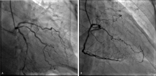
Figure 1 A,B. LAD with severely angulation occlusion (arrow).
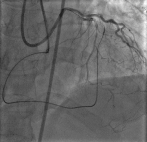
Figure 1C. The retrograde guidewire passed to the subintimal at the proximal of LAD (arrow).
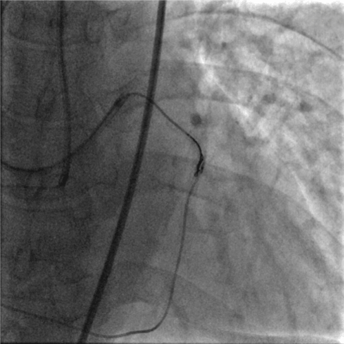
Figure 1D. The antegrade guidewire knuckled into the occluded stent together with a Corsair.
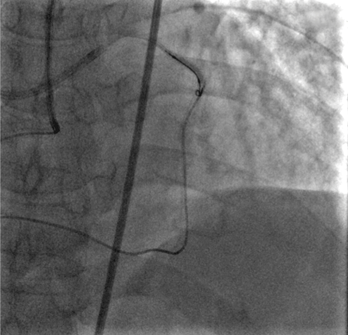
Figure 1E. The 4F child catheter was advanced to the distal of the occluded stent using an anchor balloon technique. The arrow indicates the tip of the child catheter.
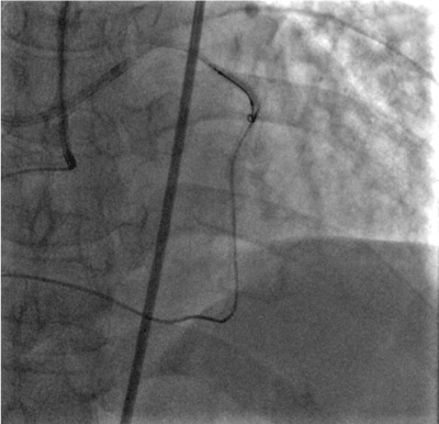
Figure 1F. The retrograde guidewire and Corsair were advanced into the antegrade guiding catheter.
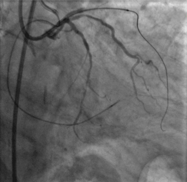
Figure 1G. The antegrade guidewire passed to the distal of LAD
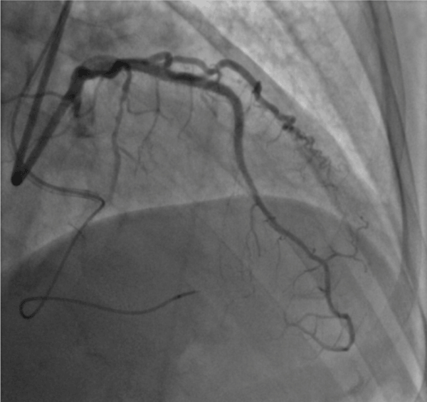
Figure 1H. The result was excellent.
Table 4. Summary of cases performed using AGT. GC = Guiding Catheter; CC: Collateral Circulation.
Case |
Access site |
Lesion vessel |
GC |
Challenge |
CC |
Intubation
Depth (cm) |
Complication |
6-month
MACCE |
1 |
R/F |
RCA |
7F AL |
Severe angulation/long lesion/mid tortuous |
1 |
1.5 |
None |
None |
2 |
R/F |
LAD |
6F XB |
Severe angulation/long lesion/mid tortuous |
2 |
2.6 |
Acute thrombosis |
|
3 |
R/F |
RCA |
7F AL |
Long lesion |
2 |
2.5 |
None |
None |
4 |
R/F |
RCA |
7F AL |
Severe angulation/long lesion/mid tortuous |
1 |
3.0 |
None |
None |
5 |
R/F |
LAD |
7F EBU |
Severe angulation |
2 |
5.2 |
None |
None |
6 |
R/F |
LAD |
7F EBU |
Long lesion |
2 |
2 |
None |
None |
7 |
R/F |
LAD |
7F EBU |
Severe angulation/long lesion/mid tortuous |
2 |
2 |
None |
None |
8 |
R/F |
LCX |
7F EBU |
Long lesion/severe tortuous |
2 |
1.5 |
None |
None |
9 |
R/F |
RCA |
7F AL |
Severe angulation/long lesion/mid tortuous |
1 |
1.8 |
None |
None |
Discussion
6F mother guiding catheter for PCI is the current standard. Nevertheless, it may be difficult with a 6F guiding catheter when treating complex coronary lesions, like delivering balloon/stent, or picking up the retrograde guidewire, due to severe calcification, poor back-up support, long lesion, CTO, et al. New devices and techniques have been proposed to overcome these disadvantages. The mother-child technique has been demonstrated to be one of the most effective techniques to deliver balloon/ stent into complex lesions and to externalize the retrograde guidewire [3, 4]. In the present study, we inserted a 4F child catheter into a 6F or larger guiding catheter to access the potential usage of a 4F child catheter in PCI of complex coronary lesions, only type B2/C lesions were included, with 77.5 % of type C lesions. The usage of the 4F child catheter leaded to favorable clinical outcomes even with such complexity of the target lesions.
Failure of advancing the guidewire or delivering a stent/balloon across the target lesion during PCI is often due to poor backup support, especially in complex lesions. When the back up support of a guiding catheter is limited, introduction of the 4F/5F child catheter into the coronary artery through the 6F or larger mother guiding catheter provides increased backup support [4, 5]. Compared with the 5F child catheter, the 4F child catheter has smaller outer diameter, so it could insert deeper into the distal coronary artery without compromise the coronary flow [3]. In this study, we discovered that 50 % of the included patients needed the 4F child catheter to provide improvement in the backup support in order to aid the guidewire crossing the target lesion and the percentage was higher in patients with CTO.
In CTO PCI, the balloon or stent inability to cross the occluded lesion accounts for 10 % of the failed intervention [6]. Our study showed 25 % and 2 % of all included patients needed the 4F child catheter to deliver stent and balloon respectively. The mother-child catheter is an efficacious device to deliver balloon or microcatheter crossing the occluded segment [7]. A body of studies have discovered that the mother-child catheter results in increased back-up and facilitates stent delivery in complex lesions [8-10]. Considering the small outer diameter of 4F child catheter, the deeper insertion makes it serves as a sheath to deliver a balloon/stent to the coronary lesions.
Both the antegrade and retrograde skillsets are required for the hybrid approach to treatment of CTO PCI. For the retrograde approach, it is difficult to externalize the retrograde guidewire to the antegrade guide catheter sometimes due to the long collateral, severe calcification, angulation or long occlusion lesion, et al, child catheter often required for this procedure [11]. In this study, 32.1 % of the CTO patients needed this device to externalize the retrograde guidewire. Here, we presented a case where the retrograde approach was used to revascularize a LAD CTO, however, the retrograde guidewire entered the subintimal space of proximal LAD. To solve this, a 4F child catheter was introduced via an antegrade wire and was advanced over successfully, this insertion made the distance between the antegrade guiding catheter and the reentry site of retrograde guidewire shorter, thus facilitated the pickup of the retrograde guidewire and microcatheter. Then the externalization was completed, and LAD CTO was revascularized. This case illustrates a new technique using the child catheter to complete wire externalization and indicates the value of child catheter in treating CTOs. This technique was performed in 9 out of 28 CTO patients, these target lesions were extremely challenging due to severe angulation, long lesion, or tortuosity, bilateral angiography and CC level 1 or 2 were need for AGT. The estimated intubation distance was shorter than the those using Guidezilla, and it has been reported that the intubation distance of 4F child catheter < 5 cm failed to increase the back-up support, suggesting the 4F child catheter in patients performed AGT played a role in making the distance between the antegrade guiding catheter and retrograde guidewire shorter and serving as a connection in the mid-distal portion of the vessel, thus facilitated the re-entry of the retrograde guidewire into the antegrade guiding catheter possible. In addition, the shorter intubation distance reduced the subintimal space, and decreased the incidence of complications.
Acute thrombosis occurred in one produce after successful stent deployment, the angiography showed severe thrombus burden within vessel lesions and led to poor prognosis. This situation was unpredictable and associated with multi-factors [12-14], including patient, balloon predilation, stent, poor responders to antiplanet or anticoagulation drugs, as well as related procedure. A combination of these factors was the most likely cause for acute thrombosis. Therefore, we must take care to prevent thrombus formation during PCI procedure that would be detrimental to clinical outcomes. Inhibiting generation of thrombin or thrombin using anticoagulants represents standard therapy in PCI procedure. Efficient and safe anticoagulation to prevent thrombosis from forming on the device is crucial in the setting of PCI [14-17]. As CTO procedure takes longer time, the antegrade mother guiding catheter may be leaved unused for some time when trying the retrograde approach, or the 4F child catheter is inserted through the antegrade mother guiding catheter, et al, so preventing catheter thrombus formation is extremely necessary during this condition. No MACCE occurred after 6-month follow-up.
We had 1 instance of stent dislodgement during delivering the stent when using the 4F child catheter, as the coronary lesions was excessive tortuosity, the stent delivery was failed. In this case, the stent was dislodged in the 4F child catheter, so the 4F child catheter was withdrawn and a 5F child catheter was inserted to aid stent delivery, and the stent was successfully delivered. If the stent dislodged in the coronary artery during the PCI procedure, retrieving the stent appears difficult after failed delivery, and the risk of stent dislodgment is high, so deploying or crushing the stent is the recommended action in this course.
In conclusion, the 4F child catheter can be advanced into the mother guiding catheter for back up support improvement, stent or balloon delivery, guidewire crossing and retrograde guidewire externalization, et al. For CTO PCI, the retrograde guide wire externalization using 4F child catheter is efficient and fast. Attention must be paid to adequate anticoagulation and to use exchanging-length guide wire when removing the child catheter while the guidewire remained in situ.
References
- Saeed B, Banerjee S, Brilakis ES (2008) Percutaneous coronary intervention in tortuous coronary arteries: associated complications and strategies to improve success. J Interv Cardiol 21: 504-511. [Crossref]
- Di Mario C, Ramasami N (2a008) Techniques to enhance guide catheter support. Catheter Cardiovasc Interv 72: 505-512. [Crossref]
- Takeshita S, Shishido K, Sugitatsu K, Okamura N, Mizuno S, et al. (2011) In vitro and human studies of a 4F double-coaxial technique ("mother-child" configuration) to facilitate stent implantation in resistant coronary vessels. Circ Cardiovasc Interv 4: 155-161. [Crossref]
- Takeshita S, Takagi A, Saito S (2012) Backup support of the mother-child technique: technical considerations for the size of the mother guiding catheter. Catheter Cardiovasc Interv 80: 292-297. [Crossref]
- Takahashi S, Saito S, Tanaka S, Miyashita Y, Shiono T, et al (2004) New method to increase a backup support of a 6 French guiding coronary catheter. Catheter Cardiovasc Interv 63: 452-456. [Crossref]
- Hoebers LP, Claessen BE, Dangas GD, Ramunddal T, Mehran R, et al. (2014) Contemporary overview and clinical perspectives of chronic total occlusions. Nat Rev Cardiol 11: 458-69. [Crossref]
- Kovacic JC, Sharma AB, Roy S, Li JR, Narayan R, et al. (2013) GuideLiner mother-and-child guide catheter extension: a simple adjunctive tool in PCI for balloon uncrossable chronic total occlusions. J Interv Cardiol 26: 343-350. [Crossref]
- de Man FH, Tandjung K, Hartmann M, van Houwelingen KG, Stoel MG, et al. (2012) Usefulness and safety of the GuideLiner catheter to enhance intubation and support of guide catheters: insights from the Twente GuideLiner registry. EuroIntervention 8: 336-344. [Crossref]
- Ali M, Yagoub H, Ibrahim A, Ahmed M, Ibrahim M, et al. (2018) Anchor-balloon technique to facilitate stent delivery via the GuideLiner catheter in percutaneous coronary intervention. Future Cardiol 14: 291-299. [Crossref]
- Mamas MA, Fath-Ordoubadi F, Fraser DG (2010) Distal stent delivery with Guideliner catheter: first in man experience. Catheter Cardiovasc Interv 76: 102-111. [Crossref]
- Mozid AM, Davies JR, Spratt JC (2014) The utility of a guideliner catheter in retrograde percutaneous coronary intervention of a chronic total occlusion with reverse cart-the "capture" technique. Catheter Cardiovasc Interv 83: 929-932. [Crossref]
- Nakano M, Yahagi K, Otsuka F, Sakakura K, Finn AV, et al. (2014) Causes of early stent thrombosis in patients presenting with acute coronary syndrome: an ex vivo human autopsy study. J Am Coll Cardiol 63: 2510-2520. [Crossref]
- Montalescot G, Hulot JS, Collet JP (2009) Stent thrombosis: who's guilty? Eur Heart J 30: 2685-2688. [Crossref]
- Gurbel PA, Tantry US (2007) Stent thrombosis: role of compliance and nonresponsiveness to antiplatelet therapy. Rev Cardiovasc Med 1: S19-26. [Crossref]
- Grayburn PA, Willard JE, Brickner ME, Eichhorn EJ (1991) In vivo thrombus formation on a guidewire during intravascular ultrasound imaging: evidence for inadequate heparinization. Cathet Cardiovasc Diagn 23: 141-143. [Crossref]
- Kaeberich A, Raaz U, Vogt A, Maedgefessel L, Neuhart E, Krezel C, et al (2014) In vitro comparison of the novel, dual-acting FIIa/FXa-inhibitor EP217609C101, unfractionated heparin, enoxaparin, and fondaparinux in preventing cardiac catheter thrombosis. J Thromb Thrombolysis 37: 118-130. [Crossref]
- Schlitt A, Rupprecht HJ, Reindl I, Schubert S, Hauroeder B, et al. (2008) In-vitro comparison of fondaparinux, unfractionated heparin, and enoxaparin in preventing cardiac catheter-associated thrombus. Coron Artery Dis 19: 279-284. [Crossref]


