We present the clinical case of a 62-year-old male who went to the hospital emergency department in September 2008 due to hypertensive crisis, finding advanced chronic kidney disease and initiating haemodialysis by means of a central venous catheter (CVC).
Native of Mauritania and with language barrier. Since its beginning in haemodialysis, the diagnosis of secondary hyperparathyroidism stands out, as well as multiple vascular access problems that involve the placement of multiple central venous catheters. He presented several episodes of bacteremia with bacterial endocarditis on tricuspid valve and thrombosis of superior and inferior vena cava requiring angioplasties and stents at both levels. Valued by the Haematology service that rules out autoimmune or associated coagulation disorder except weakly positive lupus anticoaugulante.
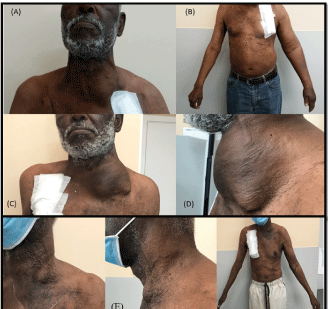
In June/2017, a cervical tumour of firm consistency in the inferolateral and left anterior region of the neck, associated with signs of venous hypertension in the left upper limb (MSI) was detected on physical examination (Figure 1A, 1B) where he carried an immature humerus-cephalic suckrus-cephalic fistula (FAV) and CVC in the left internal jugular vein.

Figure 1. (A and B): initial tumor conditioning venous hypertension MSI (C and D): cervical tumor 2 years after and before surgery (E): after excéresis of the tumor that included total thyroidectomy
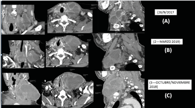
By cervical ultrasound is an occupant lesion of space of indeterminate nature may be vascular lineage. CT was performed with contrast, highlighting a laterocervical mass of 6.6 x 5 cm of diameter in axial plane and 5 cm of craniocaudal diameter of polycystic morphology with walls and hypercaptant thick septa. It is also described a solid pole of cranial location of 1.8 x 1.5 cm hyperdense with dystrophic calcifications that displaces the airway and larynx and produces compression and thrombosis of the left jugular vein requesting cytological study (Figure 2A).

Figure 2. (A): laterocervical mass of 6,6 x 5 cm of diameter in axial plane and 5 cm of craniocaudal diameter of polycystic morphology. (B and C): progressive increase in tumor size with displacement of the airway and larynx as well as compression and thrombosis of the left jugular vein
We decided to change CVC to the right internal jugular vein and perform cytological and microbiological studies by means of Eco-PAAF of several of the lesions, all of which were negative for malignancy and without microbial growth.
Clinically the patient so far remained stable without odinophagia, dysphonia or dyspnea. Analytically, it presented tumour markers and proteinogram in blood without significant alterations except B2 microglobulin: 39.1 μg/ml and an elevation in the gammaglobulin fraction of heterogeneous type (polyclonal). In terms of thyroid function, TSH was always kept within normal and only in October 2019 was a discretely decreased free T4 hormone (0.66 ng/dl) found.
In the absence of diagnosis and the progressive growth of the cervical tumour, interconsultations are requested to the specialties of Haematology, Otolaryngology (ENT) and Infectious Service, ruling out haematological neoplasms, hydatidosis, mycobacterial infections, mycoplasmas, rickettsias, Q fever and CMV.
A new CT with contrast and PET-CT is also requested in which the same multiloculate mass with cystic content is continued to be described, but with an increase in size with respect to the previous study of 7.6 cm x 5.8 cm respectively: without finding other neoplastic lesions or metastases to another level. Despite the diagnostic presumption of colloid cyst, no connection with the thyroid gland can be seen (Figure 2B). Again, at Eco-PAAF we again obtained negative cytological and microbiological studies.
Evolutionarily, the patient begins with dysphonia and dysphagia coinciding with the even greater increase in the size of the tumour (Figure 1C-1E), finally going in October/2019 to the emergency department for cervical bleeding in the context of tumour physitulysis to the skin.
On this occasion, a new cervical CT scan was performed, which shows a marked radiological worsening of the tumour with approximate diameters of 7 x 13 x 11 cm in its AP, T and CC axes respectively (previous 5 x 5.8 x 7.6 cm) as well as an important expansive effect, displacing the airway laterally to a greater extent and partially collapsing its light at the supraglottic level (Figure 2C).
For the third time, Eco-PAAF was performed on several of the lesions, this time being positive for malignancy and suggestive of papillary thyroid carcinoma [CATEGORY VI (Bethesda)].
The patient underwent surgery and underwent a total thyroidectomy. The anatomopathological report describes the existence of a classic variant papillary thyroid carcinoma, present both in the tumour mass dependent on the left hemitiroid without lymphovascular or perineural invasion. In the case of the isthmus and the right thyroid lobe, no evidence of malignancy was found.
Evolutionarily, he required surgical reintervention by left supraclavicular collection with good posterior evolution presenting as the only initial sequel a mild dysphonia.
After months of an apparent good evolution is in imageological control tumour recurrence in situ associated with cervical and systemic ganglion metastases. Given these findings, surgical reoperation was decided with recurrent mass excisis and left ganglion emptying without complications.
With more than 3 years of evolution the patient remains alive, with acceptable quality of life at the moment and in follow-up by the services of END, Endocrinology and Nuclear Medicine pending treatment with radioactive iodine I-13.
Patients with chronic kidney disease on dialysis (CDDS) have a higher risk of developing cancer when compared to the general population [1] and in the case of thyroid carcinoma its frequency is higher in the first group of patients [2].
The diagnosis of thyroid cancer in the dialysis patient is usually rapid and is often found as a chance finding in the context of the study of secondary hyperparathyroidism or during the pre-kidney transplant study [2].
In our case, the difficulty of the etiological diagnosis despite the large size of the tumour, the prolonged time of evolution, as well as the histological studies, imaging tests and multidisciplinary interconsultations carried out, are striking.
Papillary thyroid cancer is most common between the fourth and fifth decades of life with a higher incidence in women, with the ratio being approximately 2.5: 1.
A recent study conducted in the haemodialysis population found that the mean time between the onset of haemodialysis and the diagnosis of cancer was 64.40 ± 41.81 months and the risk factors that were significantly related to its onset were: advanced age (especially in ≥ 70 years), male sex and liver diseases [3].
These data contrast with our male patient who has been on haemodialysis for 12 years, the history of liver disease is not collected, and who was 61 years old at the time of diagnosis.
These data contrast with our male patient who has been on haemodialysis for 12 years, the history of liver disease is not collected, and who was 61 years old at the time of diagnosis. Papillary and follicular cancers are considered differentiated cancers, and, in these patients, surgery is the treatment of choice. There are other malignant thyroid entities such as medullary thyroid cancer (isolated or familial, forming part of multiple endocrine neoplasia syndrome type 2 [MEN2]) and primary thyroid lymphoma.
Thyroid metastases can also be found from other cancers, the most common being breast, colon, kidney cancer and melanoma [4].
The histology of our clinical case was compatible with a classical variant papillary thyroid carcinoma and given the size and complications presented, surgical treatment with total thyroidectomy was decided in doubt of tumor extension to the right hemitiroid.
Most thyroid cancers have an excellent prognosis so most patients with papillary cancer do not die from this disease [4]. In a number of patients with a median follow-up of 16 years, mortality related to this type of cancer in patients without metastases at diagnosis was 6% [5].
According to the criteria of the ATA (American Thyroid Association), our patient initially, despite the tumor size, met criteria of low risk of persistent/recurrent disease (thyroid carcinoma confined to the thyroid gland) [4].
In the case of patients at risk of recurrence, ablative therapy with radioactive iodine I-13 may be necessary [2]. Radio iodine is administered after thyroidectomy in patients with differentiated thyroid cancer to remove residual normal thyroid tissue by providing adjuvant therapy for subclinical micro metastatic disease and/or to provide treatment for clinically apparent residual or metastatic thyroid cancer [6].
In patients who develop distant metastases, radioactive iodine therapy is sometimes less effective so other treatments are preferred, such as: systemic chemotherapy (kinase inhibitors), external radiation therapy, percutaneous injection of ethanol into cervical lymph node metastases, radiofrequency ablation of cervical, bone and lung metastases as well as palliative embolization of bone metastases [4].
Given the tumour recurrence in our patient has been decided treatment with radioactive iodine with palliative character given the systemic extension found, not being considered a candidate for treatment more aggressive.
With this clinical case we want to highlight how difficult and prolonged it can be to diagnose such a frequent pathology that can become fatal in a patient on haemodialysis, despite assessing these patients at least 2 or 3 times a week and subjecting them to multiple complementary examinations.
Cancer is the second leading cause of death and morbidity in Europe (3.7 million new cases each year) and the median age of patients at diagnosis is 65 years [7].
If we take into account the increase in the average age of our patients on dialysis, and the improvement of the techniques that lead to a survival each time of these patients; the close cancer-kidney connection explains the need, today more than ever, to develop Onconephrology as a subspecialty with faster and more effective diagnostic and therapeutic protocols.
- Maisonneuve P, Agodoa L, Gellert R, Stewart JH, Buccianti G, et al. (1999) Cancer in patients on dialysis for end-stage renal disease: an international collaborative study. Lancet 354: 93-99. [Crossref]
- Gallegos-Villalobos A, García López F, Escalada C (2014) Uso de yodo radiactivo I-131 y monitorización de radiactividad en pacientes con enfermedad renal crónica en hemodiálisis. Nefrologia 34: 317-322.
- Myung J, Choi JH, Yi JH (2020) Cancer incidence according to the National Health Information Database in Korean patients with end-stage renal disease receiving hemodialysis. Korean J Intern Med 35: 1210-1219. [Crossref]
- Tuttle RM. Differentiated thyroid cancer: Overview of management. 2020.
- Mazzaferri EL, Jhiang SM (1994) Long-term impact of initial surgical and medical therapy on papillary and follicular thyroid cancer. Am J Med 97: 418-428. [Crossref]
- Tuttle RM. Papillary thyroid cancer: Clinical features and prognosis. 2020.
- de Francisco Ángel LM, Macia M, Alonso F. En: Lorenzo V, López Gómez JM (Eds). http://www.revistanefrologia.com/es-monografias-nefrologia-dia-articulo-Onconefrología


