Urachal pathologies are rare to be encountered and can varies from benign conditions to malignant lesions. Here we report a 35 years old lady newly diagnosed diabetic patient, complaining of abdominal pain and urine frequency for 3 months, abdominal CT scan showed 3.5 cm Urachal cyst with internal calcification, patient underwent Robotic assisted laparoscopic Urachal excision with partial cystectomy. pathologic examination of the cystic mass was consistent with mucinous cystadenoma, patient post-operative course was unremarkable.
urachal mass, mucinous cystadenoma, urachus
The urachus is a 3-10 cm tubular pre-peritoneal structure, that is situated between bladder dome and umbilicus, it is the remnant of obliterated umbilical artery, however, it can abnormally remain open completely, form a cystic structure or to be open in one end forming a sinus or bladder diverticulum, urachus is surrounded by umbilicovesical fascia so the disease process is contained to this space.
Urachal cyst manifest usually in adults and it contribute to 30 % of urachal anomalies, it can give raise to certain pathologies, like abscess formation, sinus or diverticulum that could harbor adenocarcinoma. One extremely rare pathology to be reported is mucinous cystadenoma of the urachus, with few cases reported in literature, it’s a benign condition but the possibility that it could have a low malignant potential is uncertain. here we report a 35 years old female patient with Urachal mucinous cystadenoma, that could add to the existing literature about this rare condition.
This is a 35 years old Asian female, newly diagnosed by diabetes Meletus type 2, complaining of mild abdominal pain and urinary frequency that started 3 months ago, no other urinary symptoms, surgical and other medical history were unremarkable, family history was insignificant, physical examination showed mild periumbilical tenderness otherwise it was unremarkable.
Labs: Renal function test, Urinalysis and Urine culture were negative.
Imaging: (Bedside US showed a cystic lesion that is adjacent to the bladder dome, so CT scan was requested for better characterization. Abdomen and pelvis CT scan with contrast: showed a 3.5 cm non enhancing cystic lesion with mural calcification in the midline of prevesical space at the bladder dome with clear separation between the bladder and the cystic lesion wall (Figure 1).
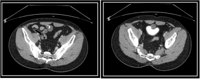
Figure 1. Abdomen and pelvis CT scan with contrast
No additional lesion within midline of anterior abdomen and bladder anterior wall along urachal course.
No focal lesion or wall thickening in distended urinary bladder.
No lymph node enlargement, ascites or other abnormalities in pelvic cavity (Figure 2).
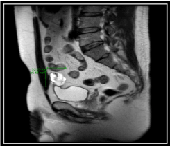
Figure 2. MRI-T2 weighted images: showed 3.5cm sized septated cyst with internal mural calcification, separable from the bladder wall
The diagnosis of urachal cyst was made and decision of urachal excision with partial cystectomy is taken due to uncertainty of the cyst behavior.
Robotic assisted laparoscopic urachal cyst excision using Da Vinci Si surgical robotic system, Urachal cystic mass was dissected away from the bladder without being separated from the urachus, then urachus was excised and specimen extracted by EndoBag (Figure 3).
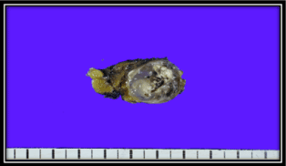
Figure 3. Urachal cyst excision
Goss description: The specimen submitted fresh "Urachal tumor", measuring 6.0 x 3.0 x 2.5cm. There is an encapsulated well-circumscribed mass (3.5 x 2.3 x 1.5cm), which is filled with gelatinous material and there is focal area with calcification (25%). The mass is grossly abutting on circumferential resection margin.
Microscopic description: Urachal mucinous cystadenoma arising from the urachal remnant, with ossifying metaplasia
Follow up at 9 months: CT scan is unremarkable; patient is symptoms free.
The urachus is a tubular structure that is obliterated after birth, however urachal remnant can be an origin to many anomalies or lesions that varies from benign to malignant conditions [1].
Wu, et al. [2] reviewed 10 reported cases of urachal cystadenoma , patients were aged between 29 to 72 years old with median age of 54, 8 patients were male and 2 were female , majority of the patients presented with abdominal pain and periumbilical swelling felt on examination and one patient presented with urinary symptoms in the form of hematuria and mucusuria. Rarely, in the case of a cyst rupture, the patient might present with severe and acute abdominal pain. On the other hand, a portion of the patients were asymptomatic, and the tumor was found incidentally by palpation or imaging for other causes, their biochemistry profile was within normal range , in our patient however there was no abdominal pain , urinary symptoms in form of frequency but no hematuria or mucusuria, there was no palpable periumbilical mass on examination, on CT scan calcification within the cyst was a common finding among most of the reported cases including our patients [2,3].
Urachal cystic masses are rare condition with uncertain behavior they can vary from being benign to malignant, but transformation from benign mucinous urachal cystadenoma to malignant cystadenocarcinoma was not observed among all the reported patients follow up [2], concurrent mass or neoplasm rarely occur with urachal mucinous cystadenoma, Kelly, et al. reported a case of urachal mucinous cyst that was found incidentally during CT scan for a patient with sigmoid colon adenocarcinoma, first it was thought of as a metastasis from other site of adenocarcinoma (sigmoid colon cancer) but on histopathological examination after resection it turned to lacked nuclear beta-catenin expression which is rare in the spectrum of urachal mucinous tumors, and it is suggested to be used to distinguish the urachal mucinous neoplasm from metastatic colorectal cancer [4-6].
Development of Pseudomyxoma peritonei was reported to be caused rarely by mucinous cystadenocarcinoma of the urachus but not reported or linked to the benign urachal cystadenoma, and fortunately no risk of malignant transformation was observed in the reported cases [7] mucinous cystadenoma of the urachus is a benign condition and carries a good prognosis as per reported cases, we believe that resection is the appropriate management since differentiating it from urachal cystadenocarcinoma is uncertain and cannot be done without histopathological examination, care should be taken while resecting the tumor as tumor spillage in case of malignant lesion can give raise to Pseudomyxoma peritonei at peritoneal spread which have a poor prognosis even with aggressive treatment [8].
Mucinous cystadenoma is a rare urachal pathology that could be symptomatic or incidentally found while investigating for other clinical conditions, on CT and MR imaging, non-enhancing mass with mural calcification was a common finding in the reported cases. mucinous cystadenoma is a benign condition that has no malignant transformation potential as seen in reported cases, we believe that resection of the urachal cyst with partial cystectomy if needed, is the most appropriate management as definite diagnosis by histopathological examination is needed. However, care should be taken while resecting he mass not to violate the cyst wall as the tumor behavior is unknown at this point, and Pseudomyxoma peritonei was reported after tumor spillage in case of malignant cystadenocarcinoma.
We hope that our reported case will add to the existing literature.
- Liang L, Zhou N, Xu H, Liu D, Lu Y (2017) Urachal mucinous adenocarcinoma with pseudomyxoma peritonei: A case report. Medicine 96: e7548. [Crossref]
- Wu J, Liu A, Chen A, Zhang P (2017) Urachal borderline mucinous cystadenoma: A rare case report and literature review. Medicine 96: e8740. [Crossref]
- Wang D, Kauffman E, Sule N (2016) Mucinous Cystadenoma of the Urachus: A Case Report and Review of the Literature. Am J Clin Pathol 146: 57.
- Brennan K, Johnson P, Curtis H, Arnason T (2019) Urachal Mucinous Cystic Tumor of Low Malignant Potential with Concurrent Sigmoid Colon Adenocarcinoma. Case Rep Gastrointest Med p. 9.
- Brabletz T, Jung A, Hermann K, Günther K, Hohenberger W, et al. (1998) Nuclear overexpression of the oncoprotein beta-catenin in colorectal cancer is localized predominantly at the invasion front. Pathol Res Pract 194: 701-704. [Crossref]
- Paner GP, McKenney JK, Barkan GA, Yao JL, Frankel WL, et al. (2011) Immunohistochemical analysis in a morphologic spectrum of urachal epithelial neoplasms: Diagnostic implications and pitfalls. Am J Surg Pathol 35: 787-798. [Crossref]
- Shinohara T, Misawa K, Sano H, Okawa Y, Takada A (2006) Pseudomyxoma peritonei due to mucinous cystadenocarcinoma in situ of the urachus presenting as an inguinal hernia. Int J Clin Oncol 11: 416-419. [Crossref]
- Amin MB, Smith SC, Eble JN, Rao P, Choi WW, et al. (2014) Glandular neoplasms of the urachus: a report of 55 cases emphasizing mucinous cystic tumors with proposed classification. Am J Surg Pathol 38: 1033-1045. [Crossref]