Abstract
Background: Currently there is lack of consensus amongst physicians regarding the management of a mobile floating atheromatous plaque (MFAP). Our case report contributes to the on-going body of literature in an attempt to establish precedent on current management of MFAP.
Case report: 70-year-old man experienced a transient ischemic attack (TIA) with weakness in his right arm. Carotid duplex scan showed a mobile plaque in the left proximal internal carotid artery (ICA) with ICA/CCA ratio of 0.55 (<50% stenosis). The patient remained asymptomatic during his 10-day admission and was conservatively managed; with no complications post one-year follow up.
Conclusion: Mobile carotid lesions are a rare pathology with its management being supported by a limited but growing body of literature. We further demonstrate the efficacy of medical therapy in treating an atheromatous carotid plaque that has <50% stenosis. Our review further demonstrates medical therapy as being current preferred treatment modality by the majority of clinicians, and sheds light on the need for further investigation and characterizing and categorizing these mobile carotid lesions.
Key words
carotid occlusive disease, mobile plaque, vascular surgery
Introduction
A mobile atheromatous floating carotid plaque is an infrequent pathology, which occurs due to unknown mechanisms, and is often responsible for drastic outcomes [1]. Diagnostic imaging demonstrating mobile atheromatous plaque (MAP) at the carotid bifurcation or at the origin of the internal carotid artery (ICA) is a rare finding [2]. Currently, there is insufficient research and evidence regarding the management of this pathology. The current management of patients with mobile atheromatous floating plaques remains undetermined. Some authors proposed medical management using anti-platelet and/or anticoagulation therapy, whereas others favour surgical management.
Case report
A 70-year-old man presented to the emergency department with chief complaints of right arm weakness. His past medical history was significant for hypertension and hypercholesterolemia. A carotid duplex ultrasound showed an inhomogeneous soft appearing mobile plaque at the proximal left internal carotid artery (ICA) Figure 1, with peak systolic velocity of 49 cm/sec and an internal carotid artery/common carotid artery (ICA/CCA) ratio of 0.55 (<50% stenosis as per Carotid Consensus Criteria). Patients’ blood pressure maintained controlled with atenolol. He was started on pravastatin 20 mg daily and therapeutically anti-coagulated with a heparin infusion. Over the next 48 hours, he remained asymptomatic with a normal neurological examination. Heparin was then discontinued, and dual anti-platelet therapy was started (aspirin (ASA) 81 mg daily and clopidogrel 75 mg daily). Twenty-four hours later, a carotid CT angiogram (CTA) was conducted which showed no signs of intraluminal filling defects in the left CCA or ICA. A repeat U/S was done 72 hours later and showed an unchanged calcified inhomogeneous atheromatous plaque within the left carotid bulb extending into the origin of the ICA; however, the mobile plaque in the proximal ICA was no longer present. MRI of the brain nine days after admission to hospital showed small focal areas of acute infarction involving the left parietal lobe presumably secondary to thromboembolism from the left ICA. The patient remained asymptomatic during his length of stay of 10 days and was discharged home on ASA 81 mg daily, Clopidogrel 75 mg daily, Atenolol 50 mg BID and Pravastatin 20 mg daily.
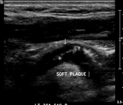
Figure 1. There is inhomogenous soft appearing mobile plaque in the internal carotid artery.
One year later, the pa2021 Copyright OAT. All rights reservf TIA or neurological dysfunctions. A repeat US showed a residual calcified plaque with no evidence of stenosis or mobility (Figure 2).
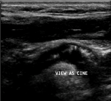
Figure 2. 1-year follow-up. Homogenous calcified atheromatous plaque noted within the distal CCA, carotid bulb, and extending into the proximal ICA. No soft/mobile plaque is noted.
Discussion
The incidence of an MAFP is one in every 2000 patient, as a recent study showed [3-5]. Despite the epidemiology, current management of this condition is limited with no clinical trials conducted [1]. Bhatti et al. ultimately demonstrated both medical and surgical management as being used in the treatment of MAPs, with neither being deemed as superior [1]. Meanwhile, for symptomatic lesions specifically described as mobile plaques, Ko et al, reported two cases with symptomatic MAP that were treated with ticlodipine 200mg daily [6,7]. Similar to the case that is presented here, follow up post one year in Ko et al, showed disappearance of floating plaque [7,8]. Granted that our patient can be classified as symptomatic, it is evident that medical management was both logical and sufficient. Similar presentation and management has been used in the past, as seen with Ko et al. Furthermore, two additional cases of asymptomatic MAPs reported by Szendro et al were also treated with medical therapy alone, and showed complete resolution of the MAPs on follow-up imaging [9]. Additional study conducted by Kim, showed two cases, which were also managed by anti-coagulation with no adverse effects post one-year, follow up [6].
The use of anti-coagulation allows the demolishment of floating plaques through plasmin, which will dissolve the clot by degrading fibrin [3]. Furthermore, it prevents the formation of future clots by inhibiting thrombin. Both physiological functions of anti-coagulation allow it to be a safe and logical action in patients with MFAP. Conducting an emergent endartectomy may be beneficial, but carries a high risk of neurological complications [7]. These complications can occur due to an emboli from the plaque, or by possible ischemic damage due to clamping of the carotid [4]. In the case of this patient, surgical intervention would in turn be considered in the case of worsening severity of the lesion, or if the patient had worsening symptomatology with inability to tolerate medical management.
Conclusion
In conclusion, the current body of literature on management of mobile carotid plaques has no consensus. Both medical and surgical methods are deemed appropriate. There are various cases, which argue for either side, with neither course of management showing superiority. Considering this, anti-coagulation alone was enough to prevent another stroke in our patient without the need of a surgical procedure.
References
- Bhatti AF, Leon LR, Labropoulos N, Rubinas T, Rodriguez H, et al. (2007) Free-floating thrombus of the carotid artery: Literature reiew and case reports. J Vasc Surg 45: 199-205.
- Cho YP, Kwon TW, Kim GE (2002) Sonographic appearance of a free-floating atheromatous plaque in a patient with acute stroke. J Clin Ultrasound 30: 317-321.
- Ezihe-Ejiofor JA, Hutchinson N (2013) Anticlotting mechanisms 1: physiology and pathology. Contin Educ Anaesth Crit Care Pain 13: 87-92.
- Moncayo KE, Vidal JJ, García R, Pereira D (2015) Surgical management of a mobile floating carotid plaque. Interact CardioVasc Thorac Surg 20: 443-444.
- Kim JG, Lee SJ (2015) Two Cases with Free-Floating Thrombus in the Common Carotid Artery. J Cardiol Clin Res 3: 1047.
- Khurana D, Saini M, Laldinpuii J, Khandelwal N, Prabhakar S (2009) Mobile plaque in the internal carotid artery: A case report and review. Ann Indian Acad Neurol 12: 185-187.
- Ko PT, Lin SK, Chang YJ, Ryu SJ, Chu CC (1997) Carotid floating plaques associated with multiple cerebral embolic strokes: Case reports. Angiol 48: 255-261.
- Lukic S, Prokin AL, Zivanovic Z, Zekic TK, Sekaric J, et al. (2013) Mobile floating carotid plaque in a young woman. Neurol India 61: 700-701. [Crossref]
- Szendro G, Sabetai MM, Tegos TJ, Dhanjil S, Lennox AF, et al. (1999) Mobile carotid plaques: the natural history of two asymptomatic and non-operated cases. J Vasc Surg 30: 357-362.