Key words
Cardiac Tumors, Echocardiography
Case study
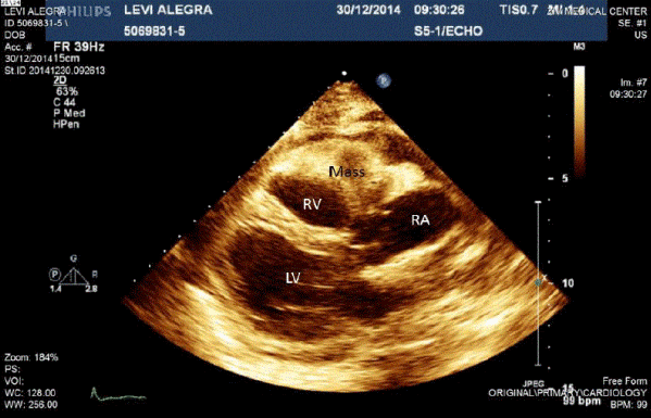
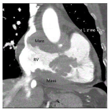
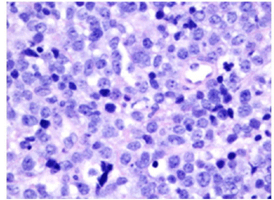
A 63-year-old woman with a medical history of smoking and epilepsy was hospitalized after a two-week period of shortness of breath. Her physical examination was notable for tachycardia and distant heart sounds. She underwent a transesophageal echocardiogram which showed a large mass engulfing the right ventricle and atrium, infiltrating the free wall of the ventricle into the tricuspid valve and compressing the superior vena cave (Figure 1). A chest CT revealed a large mass infiltrating the right ventricle and atrium and intruding the SVC (Figure 2). She was hemodynamically stable. A multidisciplinary team decided that a biopsy was necessary. The post-operative course was uneventful. The biopsy specimen showed a diffuse large B-cell lymphoma (Figure 3). Chemotherapy was immediately started.

Figure 1. Transesophageal echocardiogram - Parasternal long axis view with the mass attached to the RV and RA

Figure 2. ECG gated contrast enhanced CT - coronal reconstruction demonstrating the mass engulfing the RVOT and the aortic root

Figure 3. H&E staining (X40) showing diffuse sheets of lymphoma cells
Primary cardiac lymphomas comprise only 1% of all primary cardiac tumors, which are rarer than metastatic tumors of the heart [1]. B-cell lymphoma is the most common type, typically involving the right cardiac chambers. Presentation usually includes signs of obstruction or emboli. Treatment is based on chemotherapy and radiotherapy, with better prognosis than that of other cardiac malignancies [2].
References
- Reynen K (1996) Frequency of primary tumors of the heart. Am J Cardiol 77: 107. [Crossref]
- Nascimento AF, Winters GL, Pinkus GS (2007) Primary cardiac lymphoma: clinical, histologic, immunophenotypic, and genotypic features of 5 cases of a rare disorder. Am J Surg Pathol 31: 1344-1350. [Crossref]
Editorial Information
Editor-in-Chief
Article Type
Case report
Publication History
Received: June 01, 2019
Accepted: June 10, 2019
Published: June 13, 2019
Copyright
©2019 Saute M. This is an open-access article distributed under the terms of the Creative Commons Attribution License, which permits unrestricted use, distribution, and reproduction in any medium, provided the original author and source are credited.
Citation
Saute M (2019) Cardiac lymphoma involving the right heart. Surg Case Rep Rev, 3. DOI: 10.15761/SCRR.1000137
Corresponding author
Milton Saute
Department of Cardiothoracic Surgery, Rabin Medical Center, Beilinson Campus, Petach Tiqva 49100, Israel
E-mail : bhuvaneswari.bibleraaj@uhsm.nhs.uk


