Bowen’s disease, also known as intraepithelial neoplasia, is a very slow-growing carcinoma in situ. In 3-5% of patients, it can progress to an invasive squamous cell cancer. The anal localization of this disease is very rare. In Bowen’s disease, surgical resection is the standard of treatment. Radiotherapy keeps its place in recurrent or unresectable cases. However, surgical resection with sufficient margin is sometimes difficult or may damage the corresponding organ-function. We report a case of Bowen’s anal canal, in whom radiotherapy showed a good outcome. A 59-years-old male patient diagnosed had Bowen’s anal canal disease. On examination, we considered that performing large excision with a negative margin is uncertain because of the anatomical localization, and also surgery may destroy the anal sphincter function. After discussing this issue with the patient, we decided to employ an exclusive radiotherapy at a dose of 60 Gray in 30 fractions over 49 days. The lesion disappeared with a complete clinical and radiological response and preservation of the anal sphincter with an 18 months follow-up. We believe that radiotherapy might be an option for Bowen’s disease depending on the location and topological situation.
Bowen’s disease, anal canal, radiotherapy
Bowen’s disease, first described by Johan Bowen in 1912 [1], is high-risk intraepithelial neoplasia that appears primarily on the neck, face, genital organs and perianal region. Anal localization is quite rare. The most important risk factor is infection with HPV 16 and 18 [2,3]. Biopsy with pathological examination provides the definitive diagnosis [4,5]. The curative treatment is surgical excision with negative margins. Other less aggressive therapeutic methods can be discussed if the surgery is invasive with unacceptable morbidity or if the patient refuses, such as radiotherapy. We propose to evaluate the therapeutic efficacy of radiotherapy on this localized disease at the anal canal. We report the case of a patient diagnosed with Bowen’s anal canal disease treated with exclusive radiotherapy.
This is a 59-years-old male patient with a non-significant medical history, followed for three years for recurrent anal margin condylomas after three surgical resections. The clinical examination revealed a tumor of the anal margin encompassing the anal orifice and at the rectal touch a rough appearance of the anterior wall of the anal canal (Figure1). The anoscopy showed a budding and friable lesion of the anal margin extended into the anal canal. The biopsy of the anal margin lesion shows a low-grade dysplasia on an acuminate condyloma with signs of HPV impregnation, while the biopsy of the anal canal lesion showed carcinoma in situ. The colonoscopy showed no other lesions.
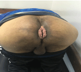
Figure 1. Condyloma of the anal margin encompassing the anal orifice
Because of the recurrent nature and, the localization of the disease performing a large excision with negative margins without causing sphincter dysfunction has proved impossible. On the other hand, radiotherapy could deliver a curative dose of radiation to the entire anal canal with margin. Our patient agreed and began treatment.
Our patient was treated with conformal therapy via a posterior field with an energy of 06 MV, a right anterior oblique and a left anterior oblique with an energy of 18 MV each. A 5 mm bolus was used to increase surface dose. Our patient received 30 fractions at a 2 Gy fraction per day, with a total of 60 Gy. During the course of the radiotherapy, physical exam revealed radiation dermatitis grade I, treated symptomatically.
The follow-up consisted of a quarterly clinical examination and annual MRI. The evolution was marked by a good clinical and radiological response. A pelvic MRI performed at six months after completion of treatment concluded to a complete clinical response (Figure2).
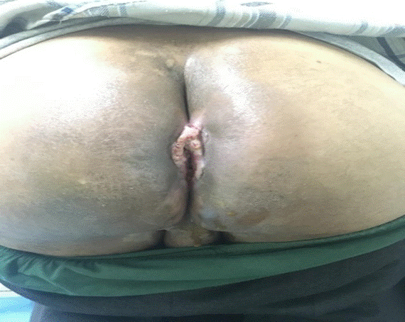
Figure 2. Total clinical response in the anal canal and regression of the condyloma of the anal
18 months after the radiation treatment, our patient is disease free with good anal sphincter anal.
Bowen’s disease, also known as intraepithelial neoplasia, is carcinoma in situ, and rarely invades adjacent organs or metastases [6]. It is linked to several factors, including ionizing radiation, sun exposure, arsenic exposure [7], immunosuppression [8], human papillomavirus infection [9], genetic factors, trauma, chemical carcinogens and, X-rays.
HPV infection is most commonly responsible for etiology. Up to 80 percent of AIN cases are believed to develop from infections from the Human Papilloma Virus (HPV) 16 and 18 [10]. HPV E6 and E7 gene products inactivate the mitotic regulator proteins of tumor suppressor genes (p53 and Rb), thereby permitting malignant transformation [11]. Histologically, anal intraepithelial neoplasia (AIN) I and AIN II involve less than two-thirds of mucosal thickness and are considered low-grade lesions [12]. AIN III involves more than two-thirds of mucosal thickness, and is considered high-grade (Bowen’s disease), with malignant potential that warrants treatment [13].
This disease can occur at any age, much higher in people aged 20-45 years old, with an equal incidence rate between the two sexes [14]. Bowen’s disease of the anal canal is characterized by mild and infrequent clinical manifestations, such as burning or itching [15], with the appearance of perianal mass or bleeding in more than one-third of patients [16]. Forty percent of cases are asymptomatic and they are often detected incidentally in hemorrhoidectomy specimens [17]. Dermatological diseases such as Paget’s disease, psoriasis, nummural dermatitis, seborrheic dermatitis, lichen planus, lupus vulgaris, secondary syphilis should be considered in the differential diagnosis [18,19]. The definite diagnosis is made by histopathological examination of biopsy material. The histopathology shows an epidermis replaced by grossly disordered keratinocytes with nuclear atypia, bizarre mitotic figures, a relatively intact basal layer, and often involvement of the pilosebaceous apparatus [20].
A therapeutic strategy was not recommended [21]. The decisions for the treatment are influenced by several factors such as lesion size and thickness, the equipment available and the perceived for poor wound healing. The therapy of the choice is surgical excision. It is necessary to resect the entire lesion with negative margins to reduce the recurrence rate [22]. Nerve injuries, damage to the anal sphincter and, disease recurrence are the main complications following surgical excision of anal tumors. According to a study by Marchesa et al., the incidence of local recurrence of Bowen disease after surgical treatment depends on the surgical used technique. In the case of large excision surgery, this recurrence is at 23.1% with a margin greater than 1cm, 53.3% after local excision with a margin less than 1 cm and, 80% after laser therapy [23]. It has been found that the wider was the surgery, the lower the recurrence was. Forthermore, with HPV-16 and HPV-18, the risk of developing recurrent disease with surgery alone is high. Even with negative margins, Bowen’s disease can recur in HPV positive regions. But the surgical technique also depends on the tumor localization. In our case, performing large excision surgery and having a negative margin is uncertain because of the anatomical localization with the possibility of preserving the sphincter tonicity of the anal canal.
Because of the morbidity and the high recurrence rates associated with surgical resection of Bowen’s disease, several treatment modalities have been applied, including imiquimod [24], external fluorouracil [25], laser therapy, radiotherapy and, photodynamic therapy [26].
Of these, radiation therapy is appropriate for larger and recurrent lesions [21] with good functional and cosmetic results. A study by Herat et al. had shown the efficacy of concomitant radio-chemotherapy in Bowen disease of the anal canal in an immunodeficient patient with good therapeutic tolerance and clinical response after 10 months of treatment [27]. Troicki et al. reported a recurrence of perianal Bowen disease initially treated with surgery. During this recurrence, the patient was treated with exclusive radiotherapy at 45Gy in 25 fractions with good tolerance and control at two years from the end of irradiation [28].
Radiation therapy is also advantageous in patients who refuse surgery, for large or multiples lesions, for lesions in cosmetically sensitive areas, and in patients who are predisposed to formation keloids. The purpose of radiation treatment is to minimize the risk of recurrent cancer in the treated area with as little toxicity as possible do that good cosmesis and function are maintained.
To achieve this type of outcome, radiotherapy planning and implementation must be tailored to the anatomic site, tumor volume, histology, previous treatment and patient’s age. The treatment planning and implementation are usually simple, because the cancer is on the surface and can be visualized and palpated readily and observed directly during each set-up.
As for our patient, he is not immunodeficient, he was operated on three times for a condyloma of the anal canal. This time, Bowen’s disease was diagnosed and he received exclusive radiation therapy of 60Gy in 30 fractions over 49 days. There was a marked clinical improvement and regression of the lesion during treatment.
There is no standard protocol for follow-up of anal Bowen’s disease. Beck recommends annual physical examination, flexible sigmoidoscopy, also punch biopsy and random biopsy from the margins of the skin grafts if new lesion emerges [29]. Relapse was reported nine years after wide 20 surgical excision in the literature [30]. Therefore, follow-up period may need to encompass a period of time longer than 5 years.
At 18 months of control, the patient had no visible lesion of the anal canal, and without palpable mass on the rectal touch. Sphincter preservation was also noted. Not only did this patient report a good quality of life with preservation of the anal sphincter but the most important is that the disease did not recur at 18 months of follow-up after irradiation. Radiotherapy is a therapeutic option in Bowen’s anal canal disease that has shown its efficacy.
Bowen’s disease localized at the anal canal is a rare pathology. Different therapeutic methods have been evaluated. Radiotherapy might be a considered option of treatment based on the topological conditions of the lesion, with good local control.
We thank the Radiotherapy department and our radiotherapist-‘s colleagues at the National Institute of Oncology of Rabat who provided care and support for the patient.
The authors report no conflict of interest concerning the case in this paper.
Written informed consent was obtained from our patient for publication of this case report and accompanying images.
- Bowen JT (1912) Precancerous dermatoses: A study of two cases of chronic atypical epithelial proliferation. J Cutan D 30: 241-255.
- Cleary RK, Schaldenbrand JD, Fowler JJ, Schuler JM, Lampman RM (1999) Perianal Bowen’s disease and anal intraepithelial neoplasia: review of the literature. Dis Colon Rectum 42: 945-951. [Crossref]
- Cox NH, Eedy DJ, Morton CA (2007) Therapy guidelines and audit subcommittee, British association of dermatologists. Guidelines for management of Bowen’s disease: 2006 update. Br J Dermatol 156: 11-21.
- Skinner PP, Ogunbiyi OA, Scholefield JH, Start RD, Smith JH (1997) Skin appendage involvement in anal intraepithelial neoplasia. Br J Surg 84: 675-678.
- Northfett DW, Swift PS, Palefsky JM (1996) Anal neoplasia. Pathogenesis, diagnosis, and management. Hematol Oncol Clin North Am 10: 1177-1187.
- Heidi Nelson Anus (2012) Chapter 53, In Sabiston Textbook of Surgery, 19th ed. Elsevier, Philadelphia.
- Shannon RL, Strayer DS (1989) Arsenic-induced skin toxicity. Hum Toxicol 8: 99-104.
- Drake AL, Walling HW (2008) Variations in presentation of squamous cell carcinoma in situ (Bowen’s disease) in immunocompromised patients. J Am Acad Dermatol 59: 6871.
- Derancourt C, Mougin C, Chopard Lallier M, Coumes-Marquet S, Drobacheff C (2001) Oncogenic human papillomaviruses in extra-genital Bowen disease revealed by in situ hybridization. Ann Dermatol Venereol 128: 715-718.
- Siegel JF, Mellinger BC (1992) Human papillomavirus in the male patient. Urol Clin North Am 19: 83-91.
- Steenbergen RD, de Wilde J, Wilting SM, Brink AA, Snijders PJ (2005) HPV-mediated transformation of the anogenital tract. J Clin Virol 32: S25-833.
- Colquhoun P, Nogueras JJ, Dipasquale B, Petras R, Wexner SD (2003) Interobserver and intraobserver bias exists in the interpretation of anal dysplasia. Dis Colon Rectum 46: 332-338.
- Skinner PP, Ogunbiyi OA, Scholefield JH, Start RD, Smith JH (1997) Skin appendage involvement in anal intraepithelial neoplasia. Br J Surg 84: 675-678.
- Bertagni A, Vagliasindi A, Ascari Raccagni A, Valmori L, Verdecchia GM (2003) Perianal Bowen’s disease: a case report and review of the literature. Tumori 89: 16-18.
- Leonard D, Beddy D, Dozois EJ (2011) Neoplasms of anal canal and perianal skin. Clin Colon Rectal Surg 24: 54-63. [Crossref]
- Margenthaler JA, Dietz DW, Mutch MG, Birnbaum EH, Kodner IJ et al. (2004) Outcomes, risk of other malignancies, and need for formal mapping procedures in patients with perianal Bowen’s disease. Dis Colon Rectum 47: 1655-1660.
- Thaler K, Wexner SD (2001) Cancers of the anal margin and anal canal, Bowen’s disease, and paget’s disease. Operative Techniques in General Surgery 3: 171-182.
- Cox NH, Eedy DJ, Morton CA (2007) Therapy guidelines and audit subcommittee, British association of dermatologists. Guidelines for management of Bowen’s disease: 2006 update. Br J Dermatol 156: 11-21.
- Marchesa P, Fazio VW, Oliart S, Goldblum JR, Lavery IC (1997) Perianal Bowen’s disease: a clinico pathologic study of 47 patients. Dis Colon Rectum 40: 1286-1293.
- Dupree MT, Kiteley RA, Weismantle K (2001) Radiation therapy for Bowen’s disease: Lessons for lesions of the lower extremity. J Am Acad Dermatol 45: 401- 404.
- Morton CA, Birnie AJ, Eedy DJ (2014) British association of dermatologists’ guidelines for the management of squamous cell carcinoma in situ (Bowen’s disease) 2014. Br J Dermatol 170: 245-260.
- Westers-Attema A, van den Heijkant F, Lohman BG, Nelemans PJ, Winnepenninckx V (2014) Bowen’s disease: A six-year retrospective study of treatment with emphasis on resection margins. Acta Derm Venereol 94: 431-435.
- Marchesa P, Fazio VW, Oliart S, Goldblum JR, Lavery IC (1997) Perianal bowen’s disease: a clinicopathologic study of 47 patients. Dis Colon Rectum 40: 1286-1293.
- Patel GK, Goodwin R, Chawla M, Laidler P, Price PE, et al. (2006) Imiquimod 5% cream monotherapy for cutaneous squamous cell carcinoma in situ (Bowen’s disease): a randomized, double-blind, placebo-controlled trial. J Am Acad Dermato 54: 1025-1032.
- Morton C, Horn M, Leman J, Tack B, Bedane C (2006) Comparison of topical methyl aminolevulinate photodynamic therapy with cryotherapy or Fluorouracil for treatment of squamous cell carcinoma in situ: Results of a multicenter randomized trial. Arch Dermatol 142: 729735.
- Neubert T, Lehmann P (2008) Bowen’s disease-a review of newer treatment options. Ther Clin Risk Manag 4: 1085-1095.
- Asoka Herat, Kyoko Shirato, Diona L Damian, Robert Finlayson, Margot Whitfeld (2006) Invasive squamous cell carcinoma arising in refractory perianal Bowen’s disease in a HIV-positive individual. Australasian Journal of Dermatology 47: 120-123.
- Filip Troicki, Alexandros Pappas, Robert Noone, Albert DeNittis (2010) Radiation therapy of recurrent anal squamous cell carcinoma in-situ: a case report. Journal of Medical Case Reports 4: 67.
- Beck DE (1995) Paget’s disease and Bowen’s disease of the anus. Semin Colon Rectal Surg 6: 143-149.
- Northfett DW, Swift PS, Palefsky JM (1996) Anal neoplasia. Pathogenesis, diagnosis, and management. Hematol Oncol Clin North Am 10: 1177-1187. [Crossref]