Introduction
Most common causes of large bowel obstruction (LBO) are adenocarcinoma of the colon and rectum, volvulus, and benign diverticular stricture [1]. This case represents a unique cause of large bowel obstruction and signifies importance of connecting urologic complaints to bowel obstruction and classic signs on basic imaging that can lead to a rapid diagnosis.
An 80 year-old-man with a history of HTN, BPH, Hartman’s procedure in 2007 for perforated diverticulitis with reversal in 2009, and three subsequent incisional hernia repairs, presented to the outpatient clinic for four months of persistent constipation. The patient reported minimal watery bowel movements daily, with the sense of incomplete defecation. He denied abdominal pain, nausea, vomiting or fevers. In addition, he also noted increased urinary frequency and urgency. Most concerning to the patient was a dramatic increase in abdominal distention. He tried several over the counter laxatives including bisacodyl, milk of magnesia, polyethylene glycol 3350, Senna, and lactulose without any relief.
Additionally, an X-ray from three months prior at an outside facility revealed a remarkably distended colon, particularly the transverse colon with possible obstruction, making the presentation suspicious for an anastomotic stricture. A CT scan was recommended at that time, however the patient refused. He returned four months later without any interval improvement.
Case report
On physical exam, All the vitals were within normal limit. His abdomen was significantly distended with a healed midline surgical scar. He had normal bowel sounds, was diffusely tympanic on percussion, on palpation no tenderness or guarding. Baseline lab work identified elevated creatinine - 2.45 mg/dL, BUN - 30 mg/dL and decreased GFR - 24 mL/min/1.73M2. He was sent to the Emergency Department for urgent CT scan and further management. A CT without IV contrast was performed in the ED which revealed a gaseous distention of the sigmoid colon to 14.7 cm with moderate right hydroureteronephrosis (Figure 1). A 1.8 × 3.2 cm obstructive calcified stone was seen at the right UVJ with marked urinary bladder distention (Figure 2).
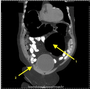
Figure 1. Computed Tomography of abdomen and pelvis without oral contrast. 1: Gaseous distention of the sigmoid colon to 14.7 cm. 2: Moderate right hydroureteronephrosis with an obstructing 1.8 × 3.2 cm (506 Hounsfield units) ovoid calcific density at the right UVJ
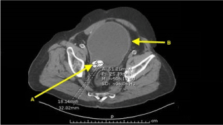
Figure 2. A: Large obstructing stone and B: Distended bladder seen
Discussion and conclusion
This patient was diagnosed with an LBO due to a distended urinary bladder secondary to renal calculus.
A large bowel obstruction occurs when there is a luminal occlusion of the colon, with dilatation of the large bowel proximal to the site of the obstruction [1]. An LBO most often occurs in the elderly; however, it is 2-5 times less frequent than a small bowel obstruction [1]. Patients with LBO often present with abdominal pain, distention and constipation; however, this can vary among patient populations [1]. Although rare, causes of extrinsic compression include post-operative fibrous adhesions, abdominal hernias, extrinsic neoplasms, intra-abdominal abscesses, aneurysms, hematomas and urological causes [2].
The most common urological cause for LBO in the literature was benign prostatic hypertrophy [BPH] causing massive bladder dilation in elderly males. However, bladder compression due to renal stone causing LBO has not previously been reported in the literature [3].
Kleinhaus et al. [4] described four cases of recto sigmoid compression due to bladder distention. The radiological findings of narrowing of the recto sigmoid by a distended bladder are quite characteristic. A large pelvic homogeneous soft-tissue mass seen on plain x-ray of the abdomen with or without gaseous distension of the colon, should arouse suspicion leading to evaluation with CT scan or bladder decompression [4,5].
Buntzen et al. [6] showed that anal pressure was markedly increased, and rectal motility was absent as bladder distention worsened. This was attributed to the excitatory 'vesico-anal' reflex produced at the spinal cord level. In our patient, there was enormous bladder distention with a volume of 6.1 L. His bladder was large enough to compress the colon, presenting as abdominal distention and constipation likely due to physiological reflex fecal stasis [6].
Urethral catheterization was performed with a total of 6.1 L of urine evacuated from the bladder. The patient noted instant relief from abdominal distention. To rule out an alternative etiology for large bowel obstruction, the patient underwent colonoscopy. The colonoscopy revealed a colon with no intraluminal abnormalities, and a healthy appearing surgical anastomosis. On the day of discharge, his creatinine improved to 1.59, and his abdominal distention had fully resolved.
Acknowledgement
We are thankful to our patient, for giving us informed consent to use relevant clinical data, to prepare and publish this manuscript.
References
- Feldman M, Friedman L, Brandt L (2016) Sleisinger and Fordtran’s Gastrointestinal and Liver Disease: Pathophysiology/Diagnosis/Management. (10th edn), PA: Elsevier Inc. Philadelphia: 2.
- Somwaru A, Phillips S (2017) Imaging of uncommon causes of large-bowel obstruction. AJR Am J Roentgenol 209: W277-W286. [Crossref]
- Mac Giobuin S, Kavanagh DO, Ryan R, Kinsella A, Myers E, et al. (2009) Acute colonic obstruction due to benign prostatic hypertrophy. Ir Med J 102: 52-53. [Crossref]
2021 Copyright OAT. All rights reserv
- Kleinhaus U, Kaftori J (1978) Rectosigmoid pseudostenosis due to urinary retention. Radiology 127: 645-647. [Crossref]
- Jaffe T, Thompson WM (2015) Large-bowel obstruction in the adult: classic radiographic and CT findings, etiology and mimics. Radiology 275: 651-663. [Crossref]
- Buntzen S, Nordgren S, Delbro D, Hultén L (1995) Anal and rectal motility responses to distension of the urinary bladder in man. Int J Colorectal Dis 10: 148-151. [Crossref]